Zol 3
advertisement

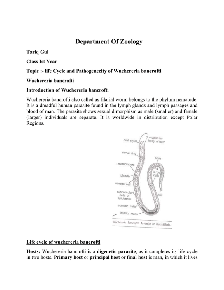
Department Of Zoology Tariq Gul Class Ist Year Topic :- life Cycle and Pathogenecity of Wuchereria bancrofti Wuchereria bancrofti Introduction of Wuchereria bancrofti Wuchereria bancrofti also called as filarial worm belongs to the phylum nematode. It is a dreadful human parasite found in the lymph glands and lymph passages and blood of man. The parasite shows sexual dimorphism as male (smaller) and female (larger) individuals are separate. It is worldwide in distribution except Polar Regions. Life cycle of wuchereria bancrofti Hosts: Wuchereria bancrofti is a digenetic parasite, as it completes its life cycle in two hosts. Primary host or principal host or final host is man, in which it lives in lymph glands and lymph passages and blood. The secondary host or intermediate host or vector host is an invertebrate or blood sucking insect usually a mosquito. (A) Life cycle in man: (1) Infection of man: When an infected mosquito bites a man, it liberates the parasites in the skin wound caused by its proboscis. The mosquito liberates both male and female Juveniles (young ones) in the human body. (2) Copulation: Copulation takes place when individuals of both the sexes (male and female) are present in the same lymph gland. (3) Larval development in Man: Female is viviparous (probably ovoviviparous), which releases numerous Juveniles called microfilariae. They are born in an immature state, they are in fact embryos rather than Juveniles. They are microscopic, about 0.2 to 0.3 mm long and are surrounded by a delicate circular sheath (membrane) and containing rudiments of various adult structures. The outer body surface of microfilariae is covered by flattened epidermal cells and on inner side a column of cytoplasm is present which contains many nuclei. Important structures from anterior end to posterior end are, future mouth or oral stylet, nerve ring band, nephridiopore, renette cell, darkly staining inner mass, four large cells and future anus. Microfilariae which are discharged into the lymph vessels soon enter blood vessels and circulate with blood showing active movements. They migrate to reside ultimately in deeper blood vessels of thorax. But they do not undergo further development until sucked by the intermediate host, i.e. mosquito. Microfilariae in human blood finally die unless ingested by the intermediate host. (4) Development in Mosquito: In the stomach of mosquito microfilariae lose their sheath and penetrate the stomach wall and migrate to thoracic muscles or wing muscles, where they undergo metamorphosis and grow. During growth they first change into a plump sausage-shaped organism (fatty meat piece) of about 150250µ long. Then they change into a more elongated form, and finally to form a long slender shaped juvenile of third infective stage. Microfilariae undergo two moults in about 10 days to reach the third stage larvae which are about 1.5 mm long. Infective juveniles now migrate into mosquito’s labium (proboscis). (5) Infection of new human host: When this infected mosquito bites a potential human host by its proboscis, the infective juveniles come out of its labium and get entered into the skin wound made by mosquito. In new human host, juveniles pass into lymph glands and lymph passages, where they coil up and develop into adult forms. Adults copulate and the females deliver (give birth) microfilariae. PATHOGENECITY OR DISEASE OF Wuchereria bancrofti Filariasis or Elephantiasis (1) Occurrence: Filariasis disease is found throughout the World. Its causative agent is a nematode called Wuchereria bancrofti. The nematode lives in the lymphatic system of man, where they obstruct the flow of lymph, causing a severe condition known as elephantiasis. In this disease, the limbs or other body parts grow to enormous size. (2) Symptoms: The incubation period of this disease is long and the symptoms may appear after 8-16 months. Light infection produces no serious symptoms. It causes filarial fever, mental depression, headache, etc. In heavy infection, accumulation of living or dead filarial worms finally blocks the lymphatic vessels and glands, resulting in various pathological conditions. Most spectacular is the immense swelling of the affected body parts known as elephantiasis or filariasis. Due to lymphatic obstruction, lymph cannot move back into the circulatory system and accumulates into organs and causes these organs to swell or enlarge to a greater extent (lymphedema). Generally lower limbs, scrotum in males, legs and mammary glands are affected. There occurs inflammation of lymphatic vessels (lymphangitis) and lymphatic glands (lymmphadentis). (3) Diagnosis: The disease can be diagnosed by the study of microfilariae after staining. Microfilariae of different species are identified after their specific shape and morphological characters. (4) Treatment (therapy): For the treatment of filariasis, some drugs are used. The effective drugs used are heterazan, compounds of antimony and arsenic, cyanine dyes, diethylcarbamazine citrate, etc. These drugs destroy microfilariae from the circulation. Large swellings can be removed by surgery. (5) Prevention (Prophylaxis or control): Preventive measures are as under: (a) Destruction or Eradication of insect vector i.e. mosquito. In endemic areas, small trees and bushes should be cleared off. (b) Use mosquito repellent cream on exposed parts of the body. (c) Periodic fumigation and spray of insecticides of sleeping rooms should be done. (d) The mosquito nets or screens should be used for preventing ourselves from the bite of mosquito and avoid sleeping on ground floors. (e) Do not keep stagnant water in the surroundings as these pools of water are their breeding places. (f) Wear clothes with long sleeves, full length pants and socks for proper protection. CANAL SYSTEM IN SPONGES A distinguishing feature (important feature) of all sponges is that their body surface is perforated by numerous apertures for the entrance and exit of water current. Inside the body, the water current flows through certain system of spaces (canals) collectively forming the canal system. TYPES OF CANAL SYSTEM The arrangement and complexity of internal channels (spaces) vary considerably in different sponges. Accordingly, the canal system has been divided into three types, I.e. Ascon type, Sycon type and Leucon type. (1) ASCON TYPE (ASCONOID CANAL SYSTEM) It is the simplest type of canal system which is found in Leucosolenia (asconoid sponge). Its (sponge) body surface is perforated by a large number of minute pores called incurrent pores or ostia or dermal ostia. These pores are intracellular spaces within tube like cells called the porocytes. These pores (ostia) open directly into the spongocoel (large central cavity). The spongocoel is the single, large central cavity in the sponge body and is lined by the flagellated collar cells or choanocytes. Spongocoel opens to outside through a narrow circular opening called the osculum, which is located at the free distal end. Surrounding sea water enters the canal system through ostia. Flow of water is maintained by the beating of flagella of collar cells or choanocytes. Passage of water current in the body of sponge may be shown as under: Outside water through Ostia Spongocoel through Osculum Ascon type To Outside (2) SYCON TYPE (SYCONOID CANAL SYSTEM) Sycon type of canal system is more complex than Ascon type. It is found in sycon and Grantia (syconoid sponge). It can be theoretically derived from the ascon type by horizontal folding of its body wall. Body wall of syconoid sponges includes two types of canals, incurrent canal and radial canal. Both types of canals are interconnected by minute pores. Ostia found on the outer surface of the body open into the incurrent canals. These incurrent canals are non-flagellated, as they are lined by pinacocytes and these incurrent canals opens into radial canals through minute openings (pores) called prosopyles. Radial canals are flagellated channels as they are lined by choanocytes (flagellated collar cells). These radial canals open into the spongocoel by small pores called apopyles. The spongocoel is a narrow, non-flagellated cavity lined by pinacocytes and it opens to the exterior through an excurrent pore called the osculum. Passage of water current in the body of sponge may be shown as under: Outside water Ostia Incurrent canals Prosopyle Radial canals Sycon type Apopyle Spongocoel Osculum To Outside (3) LEUCON TYPE (LEUCONOID CANAL SYSTEM) This canal system is most complex type of canal system and is found in leuconoid sponges such as Spongilla. It is formed due to further folding of body wall of Sycon type, especially radial canals of sycon type, which form flagellated chambers. This type of canal system has become irregular and often branched. Flagellated chambers are small, spherical and are lined by choanocytes (flagellated collar cells) and these flagellated chambers are formed due to folding of radial canals. All other spaces are lined by pinacocytes. Incurrent canals open into flagellated chambers through small pores called prosopyles. Flagellated chambers open into excurrent canals through another small pore called apopyles. These excurrent canals are developed as a result of shrinkage and division of spongocoel which has disappeared and these excurrent canals open to the outside through an osculum. Passage of water current in the body of sponge may be shown as under: Outside water Ostia Incurrent canals prosopyle Flagellated chambers canals Apopyle Excurrent Osculum To Outside Leucon type Leucon type of canal system has three grades such as Eurypylous type, Aphodal type and Diplodal type. (a) Eurypylous type: It is the simplest and most primitive Leucon type of canal system. In this type, the flagellated chambers opens directly into the excurrent canals by broad apertures called the apopyles e.g. Plakina. (b) Aphodal type: In this type, the flagellated chambers opens into the excurrent canals by a narrow canal (pore) called the aphodus e.g. Geodia. (c) Diplodal type: In this type, besides aphodus, another tube (canal) called as prosodus is present between incurrent canal and flagellated chamber e.g. Spongilla. FUNCTIONS OF CANAL SYSTEM The canal system helps the sponge in nutrition, respiration, excretion and reproduction. The water current flowing through the canal system brings food and oxygen and carries away faeces, nitrogenous wastes and carbon-dioxide. It also carries spermatozoa from one sponge to another sponge for the fertilization of ova there.


