Assisted Ventilation of the Neonate
advertisement
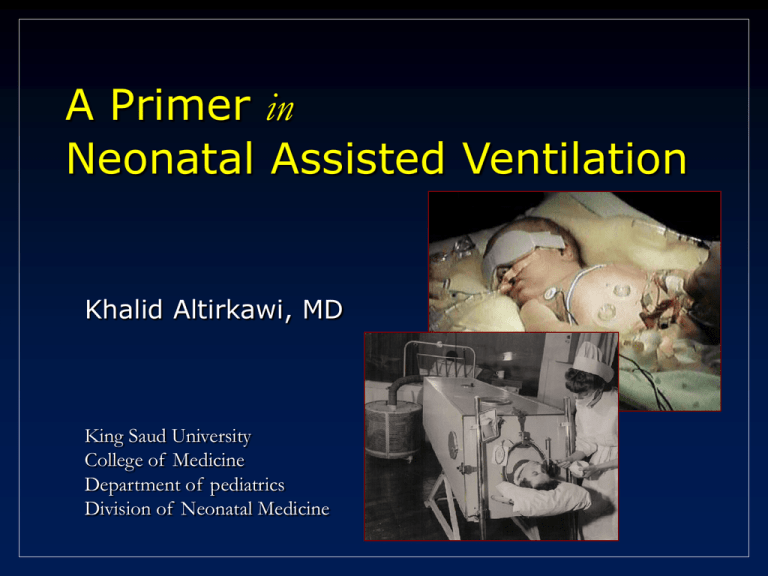
A Primer in Neonatal Assisted Ventilation Khalid Altirkawi, MD King Saud University College of Medicine Department of pediatrics Division of Neonatal Medicine Introduction • • • • Control of breathing Pulmonary mechanics Gas exchange mechanisms Historical synopsis • Assisted Ventilation Strategies • Strategies for preventing lung damage Control of breathing • Receptors • Chemoreceptors • Mechanoreceptors • Reflexes Control of Breathing • Ventilation (CO2 elemination) is maintained by fine adjustments in tidal volume (VT ) and respiratory rate (RR) that minimize the work of breathing. • Motoneurons in the CNS regulate inspiratory and expiratory muscles activity. • Chemoreceptors and mechanoreceptors provide feedback to these neurons to adjust ventilation continuously Chemoreceptors • Central (in the brain stem) • Affected by changes in PaCO2 • And changes in pH (independently of PaCO2 values) • Peripheral (carotid and aortic bodies) • Stimulated by hypoxia • In neonates, acute hypoxia produces a transient increase in ventilation that disappears quickly Mechanoreceptors • Stretch receptors: • Located in airway smooth muscles • Respond to changes in tidal volume (VT) • Mediate: • Hering-Breuer inflation reflex _ a brief period of decreased or absent respiratory effort following a good lung inflation • Head paradoxical reflex _ At slow ventilator rates, large VT will stimulate augmented inspirations (may be one of the mechanisms by which caffeine helps weaning from ventilator) Mechanoreceptors • Juxtamedullary (J) receptors: • Located in the interstitium of the alveolar wall • Stimulated by interstitial edema, fibrosis and by pulmonary capillary engorgement (CHF). • Its stimulation increases respiratory rate Mechanoreceptors • Baroceptors: • Located in aortic and carotid sinuses • Mediate: • The baroreflex _ Hypertension hypoventilation or apnea Hypotension hyperventilation. Pulmonary Mechanics pulmonary mechanics are dynamic, frequently changing over time Airway Pressure Gradient • Gas flows down pressure gradients between airway opening and alveoli • Pressure gradient depends on: • Compliance of lung parenchyma and chest wall • Resistance to airflow Compliance • The elasticity or distensibility of the system (lungs, chest wall): Compliance = ∆ volume/∆ pressure • The chest wall is compliant in neonates and does not impose a substantial elastic load compared with the lungs. Resistance • The capacity of the air conducting system (airways, ETT) and tissues to oppose airflow Resistance = ∆ pressure/∆ flow • Resistance depends on: • • • • Total cross-sectional area of the airways (including ETT) Length of airways Flow rate Density and viscosity of gas breathed Time constant • A measure of the time necessary for the alveolar pressure to reach 63% of the change in airway pressure. Time constant = resistance x compliance Delivery of pressure and volume is complete (95% to 99%) after three to five time constants • Different lung regions may have different time constants due to varying compliance and resistance Change in pressure (%) 100 95 99 86 80 60 98 63 40 20 1 2 3 4 Time constant (X) 5 Incomplete inspiration (short Ti) ↓TV Hypercapnea ↓MAP hypoxemia Incomplete expiration (short TE) Gas trapping ↓Compliance ↓TV ↑MAP ↓TV ↓Cardiac output Hypercapnia hyperoxemia Gas exchange Hypoxemia and Hypercapnia may coexist, although some disorders may affect gas exchange differentially Gas exchange • Hypercapnia usually is caused by: • Hypoventilation • Severe V/Q mismatch • Hypoxemia is usually due to: • • • • V/Q mismatching (RDS) R to L shunting (PPHN) Hypoventilation (apnea) Diffusion abnormalities Gas exchange: CO2 • In CMV, elimination of CO2 is directly proportional to alveolar minute ventilation. minute ventilation = (tidal volume - dead space) X frequency • In high frequency ventilation: CO2 elimination = (tidal volume)2 x frequency Gas exchange: O2 • Oxygenation is determined by FiO2 and MAP MAP = K (PIP - PEEP) (TI/TI + TE) + PEEP • ↑MAP ↑lung volume and improved V/Q matching ↑oxygenation • Within the physiologic range, MAP is independent of rate Assisted Ventilation Strategies • The principles • Historical synopsis • Continuous Positive Airway Pressure (CPAP) • Conventional Mechanical Ventilation (CMV) • High-Frequency Ventilation (HFV) • Extracorporeal Membrane Oxygenation (ECMO) CMV Principles • Gas flows along a pressure gradient • In spontaneous ventilation, negative intrathoracic pressure opposes “neutral” atmospheric pressure • Some mechanical ventilators attempt to mimic nature (negative extrathoracic pressure) “iron lung” • In most mechanical ventilators, positive (supraatmospheric) machine-generated pressure vs. intrathoracic pressure Characteristics of Positive Pressure CMV Cycle to expiration Pressure Flow determines rate of rise and peak pressure Pressure limited = “PIP” Begin inspiration Time Ti Te In order to prevent uncontrolled over-distention, the PIP is time-limited, pressure-limited The Oxygenation story: the North American experience • Focus on enhancing oxygenation during spontaneous breathing • Continuous Positive Airway Pressure (CPAP) • Continuous Negative Airway Pressure (CNAP) • Continuous Respiratory Airway Pressure (CRAP) • Addition to mechanical ventilation a Positive End Expiratory Pressure (PEEP) Pressure Adding PEEP PEEP Ti Time Te The Oxygenation story: Across “The Pond” • Focus on enhancing oxygenation during mechanical ventilation • Systematically explored the “reversed I:E ratio” • Spontaneous breathing is characterized by a shorter inspiratory time compared to expiratory time • Improved oxygenation as one moved from 1:2 to 1:1 to 2:1 to 4:1 Reversed I:E ratio Pressure Reversed I:E Ti Time Te Eureka! Pressure Reversed I:E Raise PIP Increase flow Increased PEEP Ti Time Te Conventional mechanical ventilation • The variables • The modes • The modalities CMV variables • PIP • Changes in PIP affect both PaO2 (by altering the MAP) and PaCO2 (by its effects on VT) • A high PIP may increase the risk of volutrauma, with resultant air leaks and BPD • Larger infants tend to have more compliant lungs, therefore requiring a lower PIP CMV variables • PEEP • Adequate PEEP prevents alveolar collapse, maintains lung volume at end expiration, and improves V/Q matching. • ↑ PEEP MAP and FRC improving oxygenation. • Vey high PEEP may decrease venous return, cardiac output, and increase pulmonary vascular resistance • Increases in both PIP and PEEP have opposite effects on CO2 elimination CMV variables • Rate: • In large RCTs, relatively high rates (60 breaths/min) resulted in a decreased incidence of pneumothorax in preterm infants who had RDS • Generally, a high rate, low VT strategy is preferred CMV variables • I:E ratio: • The major effect of an increase in the I:E ratio is to increase MAP and improve oxygenation. • Changes in the I:E ratio are not as effective in increasing oxygenation as are changes in PIP or PEEP • Changes in the I:E ratio usually do not alter VT unless TI and TE become relatively too short CMV variables • TI & TE • A long TI increases the risk of pneumothorax • Shortening TI is helpful during weaning • In RCT: limiting TI to 0.5 seconds resulted in a significantly shorter duration of weaning • In patients who have CLD a longer TI (around 0.8 sec) may result in improved VT and better CO2 elimination CMV variables • FiO2 • During increasing support, increase FiO2 first until it reaches about 0.6 to 0.7, then increase MAP • During weaning, decrease FiO2 initially (~ 0.4 to 0.7) before you decrease MAP maintaining an appropriate MAP may allow substantial reduction in FiO2 • Reduce MAP before a very low FiO2 is reached a higher incidence of air leaks has been observed if distending pressures are not weaned earlier CMV variables • Flow: • Changes in flow have not been well studied in infants, but they probably affect arterial blood gases minimally as long as a sufficient flow is used. • In general, flows of 8 to 12 L/min are sufficient in most neonates. • High flows are needed when inspiratory time is shortened to maintain an adequate tidal volume. CMV Modalities (Target Variable) • Volume controlled: • Set tidal volume is delivered, VT is pressure-limited • Pressure controlled: • Constant inspiratory pressure, ie. decelerating variable flow • Time or flow cycled: method by which inspiration is started/ended • Volumes vary with lung compliance Modes of CMV Untriggered Triggered • IPPV: • SIMV • Machine rate faster than spontaneous • IMV: • Slower than spontaneous and asynchronous • SIPPV or Assist/Control • Pressure Supported Ventilation (PSV) • Volume Guaranteed (VG) Problems in CMV • Problem: • Asynchrony: • Ineffective ventilation and oxygenation • Fluctuation in BP (associated with IVH) • Solutions: • Paralysis and sedation • Synchronized ventilation Synchronizing Means of detecting the initiation of a breath • Thoracic impedance • Abdominal movement • Change in esophageal pressure • Change in airway pressure • Change in airway flow Problems • • • • • Artifact “auto triggering” Antiphasic triggering Delayed response time Lack of response Triggered (Synchronous) Ventilation • SIMV • Only “X” breaths per minute are supported with “machine breaths” in synchrony • SIPPV • Every sensed breath is supported by “machine breath” • PSV • Every sensed breath is supported by fixed pressure, but patient controls flow and TI • VG • Preset volume delivered every time Volume Guarantee (VG) • The ventilator delivers a specified expired volume (VT) with the lowest possible pressure • The preferred mode in which to use VG is PSV • Best use is when mechanics are rapidly changing • Do NOT use VG if the leak is > 40% Volume Guarantee (VG) • The Problem • Preset volume delivered may be excessive or inadequate in a rapidly changing clinical setting • The Solution • Over several breaths, records volume achieved and pressure generated to achieve that volume • Target “new” pressure computed to achieve predefined set volume Characteristics of Triggered CMV “Support” in untriggered phase Machine’s Support Frequency of support A/C Preset flow, Ti, PIP, PEEP All sensed breaths Set rate “guaranteed” SIMV Preset flow, Ti, PIP, PEEP All sensed breaths in rated-defined “window” CPAP Set rate “guaranteed” Preset PIP, PEEP Patient’s flow, Ti All sensed breaths +/- SIMV or CPAP PSV Clinical Use of Triggered CMV Typical Setting Advantage Disadvantage Critically ill, unstable, paralyzed Control Control is an illusion SIMV Stable, spontaneously breathing but not with consistent rate or MV Increased dependence on patient = “wean” Increased dependence on patient = “wean” PSV Actively attempting to decrease ventilatory support Greatest synchronization A/C Draeger: no SIMV A/C and PSV vs. SIMV High frequency ventilation HFV Characteristics of HFV • Continuous distending pressure • Small VT (less than anatomic dead space) • Rapid ventilator rates HFV: Examples in Nature • Humming bird • ~250 bpm while at rest • One-way flow via parabronchi and air sacs (NOT tidal) • Panting dog • VT less than deadspace • Very high respiratory rate HFV: Impediment to Gas Flow Impedance Airway frequency impedance HFV: Impediment to Gas Flow Alveolae frequency HFV: Impediment to Gas Flow impedance Airway Airway + Alveolae Alveolae frequency impedance HFV: Resonant Frequency “Sweet spot” frequency So… In High Frequency Ventilation • Ventilation is controlled by frequency and (VT)2 • Frequency and VT are inversely related frequency = ETT resistance, VT HFV: The Design HFOV • • • • • Diaphragm or piston Active expiration Single control of Paw Single device Optimum volume, low pressure • Air trapping • Fixed (rigid) circuit HFJV • • • • • High pressure jet Passive expiration Multiple controls of Paw CMV in series Low volume, low pressure • Air trapping • Triple lumen ET tube HFV: The Strategy HFOV • Severe homogeneous lung disease • RDS • Early-onset pneumonia • PPHN (in concert with iNO) Target: RDS HFJV • Severe heterogeneous lung disease • MAS • Late-onset pneumonia • Airleak syndromes • Pneumothorax • PIE Target: barotrauma Comparative Pressure Profiles Common Starting Points Starting CMV • Choose mode (SIMV, PSV, ….) • Select PEEP based upon lung disease to achieve optimal inflation • Select PIP or volume to generate VT = 4-6 ml/kg • Start with rate 30-40 • Blood gases in 30 minutes to determine baseline Starting HFOV • Frequency • 10 to 12 Hz for infants >1500g, 15 Hz if <1500 g • IT • 33% of cycle • Amplitude (P) • Perceptible vibration movement down to groin • Paw • Equal to CMV if restrictive disease • 2-3 cm H2O above CMV if atelectatic disease Starting HFJV • Frequency (jet) • 420 (LBW), 240 - 360 (term or long time constant) • 3-5 CMV breaths • IT • 0.02 seconds • PIP (Jet) • To vibrate chest (~PIP on CMV) • PEEP (CMV) • To maintain Paw for optimal inflation HFV: Therapeutic strategies • Blood gas 20-30 minutes after initiation • Chest radiograph within 4 hrs after initiation, and whenever lung over-inflation is suspected • Adjust Paw to maintain optimal lung volume • After improvement • Decrease Paw to maintain FiO2 0.3-0.4 • Decrease VT if PaCO2 lower than target HFV: Therapeutic strategies • Atelectasis? • Increase Paw by 1-2 cm H2O until O2 requirement • Hypercarbia with high lung volumes? • Consider air trapping/inadvertent PEEP HFV: Pitfalls and Complications • HFV is more effective at ventilation, so the risk of hypocarbia is greater. Hypocarbia is clearly correlated with PVL • Lung overdistention airleak • Air trapping Hypercarbia • Increased intrathoracic pressure Reduced systemic venous return Hypotension HFV vs. CMV in prophylaxis • There is no evidence that elective use of HFV ( HFOV or HFFI) provides any greater benefit to premature infants who have RDS than CMV • Data are limited and results are mixed as to whether HFJV may reduce the incidence of CLD • Preferential use of HFV as the initial mode of ventilation to treat RDS in premature infants is not supported HFV vs. CMV in rescue therapy • There is no evidence that HFV provides any longterm benefit over CMV in patients who have respiratory failure • No RCTs to support the use of HFV over CMV in the treatment bronchopleural or tracheo-esophageal fistula • HFV in this population appears to diminish the amount of continuous air leak and improve patient stabilization Take Home Generalizations • “A high frequency ventilator can ventilate a stone.” • Each underlying disease has a pathophysiology that suggests the ventilator-of-choice • Diffuse, homogeneous vs. dishomogenous • Presence or absence of barotrauma • Underlying cardiovascular compromise Strategies to Prevent Lung Injury Strategies to Prevent Lung Injury • Permissive Hypercapnia • Low Tidal Volume Ventilation • Alternative Modes of Ventilation: • • • • • • Patient-triggered Ventilation Synchronization High-frequency Ventilation Liquid ventilation Proportional Assist Ventilation Tracheal Gas Insufflation Ventilator-associated lung injury • Biotrauma: • Barotrauma (high pressure, low volume) • Volutrauma (high volume, low pressure) • Markers of lung injury are present with the use of high volume and low pressure, but not with the low volume and high pressure • The heterogeneity of lung tissue involvement in many diseases predisposes some parts of the lung to volutrauma • Oxidant injury Permissive hypercapnia • Priority is given to the prevention or limitation of over-ventilation rather than to maintenance of normal blood gases • Two large retrospective studies: • Hypocapnia during the early neonatal course resulted in an increased risk of lung injury Permissive hypercapnia • RCT: • • • • Surfactant-treated infants BW = 854+163 g, GA = 26+1.4 wks Assissted ventilation during the first 24 hours Two groups; Permissive hypercapnia (PaCO2 = 45 - 55 mm Hg) or normocapnia (PaCO2 = 35 - 45 mm Hg) • Results: the number of patients receiving assisted ventilation during the intervention period was lower in the permissive hypercapnia group (P =0.005) When to avoid hypercapnia • IVH risk starts to rise when PaCO2 ~ 53-57 • Since IVH is an early phenomenon, we need to change upper limit of PaCO2 target range after risk period has ended i.e. for Premature infants in first two weeks of life, PCO2 goal is 45 – 55 mm Hg. after two weeks of life, PCO2 goal is 55 – 65 mm Hg as long as pH > 7.20 Proportional Assist Ventilation • PAV matches the onset and duration of both inspiratory and expiratory support • Support is in proportion to the volume and flow of the spontaneous breath (decrease the elastic or resistive work of breathing selectively) • RCTs are needed Tracheal gas insufflation • The added dead space of the ETT ↑anatomic dead space ↓minute ventilation ↑ PaCO2 • Gas delivered to the distal part of ETT during exhalation washes out this dead space and the accompanying CO2 Continuous positive airway pressure CPAP CPAP PROS • ↑ alveolar volume & FRC • Alveolar recruitment • Alveolar stability • Improved V/Q matching • Redistribution of lung water CONS • Increased risk for air leaks • Overdistention • CO2 retention • CVS impairment • Decreased compliance • Potential to increase PVR Early CPAP • Rescue therapy of established RDS • Decreases O2 requirements • Decreases the need for mechanical ventilation • May reduce mortality • The optimal time to start CPAP depends on the severity of RDS (PaO2~ 50 torr, FiO2 ~ 0.4) Prophylactic CPAP • Does not decrease the incidence or severity of RDS • Does not reduce the rate of complications or death Non-invasive ventilation NIV NIV – the rationale • One of the initial forms of respiratory support used in preterm infants with RDS • Recently it has been reintroduced for initial management of RDS and indications such as apnea and to improve extubation success after invasive ventilation NIV – the mechanism • The intermittently increased nasal pressure: • Is transmitted to the lower airways enhancing VT • Via the nose may act as a stimulus and reduce apnea episodes • Increases MAP better alveolar recruitment and higher lung volume • Clears the exhaled gas from the upper airway and thus reducing the anatomical dead space. NIV – the Modalities • Initially, NIV was accomplished using conventional IMV mode (N-IMV) • By adding triggering, other modalities became available: N-A/C (aka N-SIPPV), N-PSV, NSIMV • There are no data on what are the best settings during NIV NIV and apnea • Effects of NIV on apnea: • Not consistent • Greater among infants who present with a more frequent apnea while on N-CPAP • May be more effective in those infants with poorer lung function and on N-CPAP • May not always lead to improved ventilation and gas exchange, but can partially reduce work of breathing The End Suggested Reading • Goldsmith and Karotkin. Assisted Ventilation of the Neonate 2003: 183-202 • Froese and Kinsella. High Frequency Oscillatory Ventilation: Lessons from the neonatal/pediatric experience. Critial Care Medicine 2005;33:S115-121 • Keszler. High Frequency Ventilation: Evidence-based Practice and Specific Clinical Indications NeoReviews 2006;7:e234-241 Optimizing Lung Volume • Lung hysteresis refers to the fact that lung volume and compliance at a given transpulmonary pressure is higher in deflation than in inflation Froese AB: Neonatal and Pediatric Ventilation: Physiological and clinical perspectives. In Marini JJ, Slutsky AS (eds): Physiological Basis of Ventilatory Support. New York, Marcel Dekker, 1998. P. 1346

