Functional Organization of Nervous Tissue
advertisement
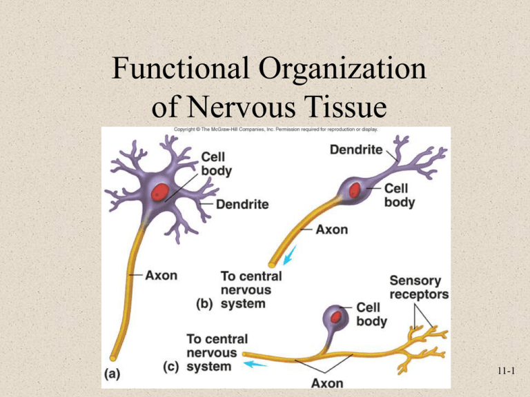
Functional Organization of Nervous Tissue 11-1 The Nervous System • Components – Brain, spinal cord, nerves, sensory receptors • Responsible for – Sensory perceptions, mental activities, stimulating muscle movements, secretions of many glands • Subdivisions – Central nervous system (CNS) – Peripheral nervous system (PNS) 11-2 Central Nervous System • Consists of – Brain • Located in cranial vault of skull – Spinal cord • Located in vertebral canal • Brain and spinal cord – Continuous with each other at foramen magnum 11-3 Peripheral Nervous System • Two subcategories – Sensory or afferent – Motor or efferent • Divisions – Somatic nervous system – Autonomic nervous system (ANS) » Sympathetic » Parasympathetic » Enteric 11-4 Nervous System Organization 11-5 Cells of Nervous System • Neurons or nerve cells – Receive stimuli and transmit action potentials – Organization • Cell body or soma • Dendrites: Input • Axons: Output • Neuroglia or glial cells – Support and protect neurons 11-6 Types of Neurons • Functional classification – Sensory or afferent: Action potentials toward CNS – Motor or efferent: Action potentials away from CNS – Interneurons or association neurons: Within CNS from one neuron to another • Structural classification – Multipolar, bipolar, unipolar 11-7 Neuroglia of CNS • Astrocytes – Regulate extracellular brain fluid composition – Promote tight junctions to form blood-brain barrier • Ependymal Cells – Line brain ventricles and spinal cord central canal – Help form choroid plexuses that secrete CSF 11-8 Neuroglia of CNS • Microglia – Specialized macrophages • Oligodendrocytes – Form myelin sheaths if surround axon 11-9 Neuroglia of PNS • Schwann cells or neurolemmocytes – Wrap around portion of only one axon to form myelin sheath • Satellite cells – Surround neuron cell bodies in ganglia, provide support and nutrients 11-10 Myelinated and Unmyelinated Axons • Myelinated axons – Myelin protects and insulates axons from one another – Not continuous • Nodes of Ranvier • Unmyelinated axons 11-11 Electrical Signals • Cells produce electrical signals called action potentials • Transfer of information from one part of body to another • Electrical properties result from ionic concentration differences across plasma membrane and permeability of membrane 11-12 Sodium-Potassium Exchange Pump 11-13 Membrane Permeability 11-14 Ion Channels • Nongated or leak channels – Always open and responsible for permeability – Specific for one type of ion although not absolute • Gated ion channels – Ligand-gated • Open or close in response to ligand binding to receptor as ACh – Voltage-gated • Open or close in response to small voltage changes 11-15 Resting Membrane Potential • Characteristics – Number of charged molecules and ions inside and outside cell nearly equal – Concentration of K+ higher inside than outside cell, Na+ higher outside than inside – At equilibrium there is very little movement of K+ or other ions across plasma membrane 11-16 Changes in Resting Membrane Potential • K+ concentration gradient alterations • K+ membrane permeability changes – Depolarization or hyperpolarization: Potential difference across membrane becomes smaller or less polar – Hyperpolarization: Potential difference becomes greater or more polar • Na+ membrane permeability changes • Changes in Extracellular Ca2+ concentrations 11-17 Local Potentials • Result from – Ligands binding to receptors – Changes in charge across membrane – Mechanical stimulation – Temperature or changes – Spontaneous change in permeability • Graded – Magnitude varies from small to large depending on stimulus strength or frequency • Can summate or add onto each other 11-18 Action Potentials • Series of permeability changes when a local potential causes depolarization of membrane • Phases – Depolarization • More positive – Repolarization • More negative • All-or-none principle – Camera flash system 11-19 Action Potential 11-20 Refractory Period • Sensitivity of area to further stimulation decreases for a time • Parts – Absolute • Complete insensitivity exists to another stimulus • From beginning of action potential until near end of repolarization – Relative • A stronger-than-threshold stimulus can initiate another action potential 11-21 Action Potential Frequency I n s e r • Number of potentials produced per unit of time to a stimulus • Threshold stimulus – Cause an action potential • Maximal stimulus • Submaximal stimulus • Supramaximal stimulus 11-22 Action Potential Propagation 11-23 Saltatory Conduction 11-24 The Synapse • Junction between two cells • Site where action potentials in one cell cause action potentials in another cell • Types – Presynaptic – Postsynaptic 11-25 Chemical Synapses • Components – Presynaptic terminal – Synaptic cleft – Postsynaptic membrane • Neurotransmitters released by action potentials in presynaptic terminal – Synaptic vesicles – Diffusion – Postsynaptic membrane • Neurotransmitter removal 11-26 Neurotransmitter Removal 11-27 Summation 11-28

