The Nervous System
advertisement
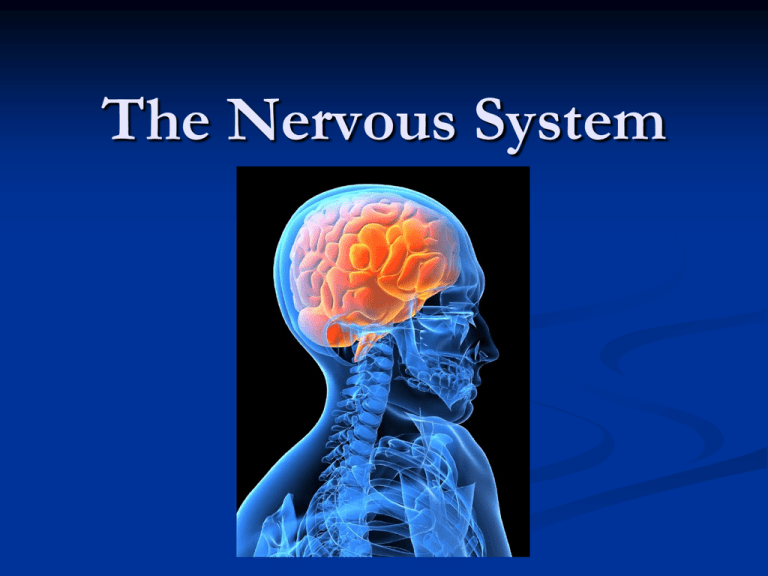
The Nervous System The Nervous System The master controlling and communication system of the body Three Overlapping Functions Sensory Input Monitors internal and external stimuli (Changes) Afferent pathway to the brain receptors Integration Processes and interprets information Decides the appropriate response Motor Output Efferent pathway to effector organs (muscles or glands), effects a response Organization of the Nervous System Central Nervous System (CNS) Brain and spinal cord Dorsal body cavity Integrating command center Peripheral Nervous System (PNS) Nerves to and from brain and spinal cord Communication links to the CNS Spinal nerves and cranial nerves Peripheral Nervous System Sensory Afferent Division Impulses to the brain and spinal cord (CNS) Monitors internal and external changes Somatic afferent Skin, skeletal muscle, joints Visceral afferent Organs within ventral body cavity Peripheral Nervous System Motor Efferent Division From CNS to effector muscle, organs, and glands Somatic efferent nervous system Voluntary nervous system Autonomic nervous system Involuntary Visceral motor nerve fibers (to smooth and cardiac muscle and glands) Sympathetic (emergency situations) Parasympathetic (conserves energy, nonemergency functions) Classification P. 224 Nervous System THE CELLS OF THE NERVOUS SYSTEM Nervous Tissue-Types of Cells Two Principal Cell Types Neurons: excitable nerve cells Supporting cells: Neuroglia “Nerve Glue” Support, insulate, and protect neurons by surrounding and wrapping neurons. Central nervous system supporting cells Astrocytes Microglia Ependymal cells Oligodendrocytes Peripheral nervous system supporting cells Satellite cells Schwann cells Neuroglia-Supporting Cells of CNS Astrocytes Most abundant *Star-shaped Radiating processes cover neurons and anchor them to adjacent blood vessels Controls exchange between blood vessels and neurons Determines capillary permeability Astrocytes (Cont) Controls chemical environment around neurons Cleans up leaked potassium and recycling released neurotransmitter Guide migration of young neurons Microglia Small, ovoid cells, with thorny processes (spider-like) Branches monitor health of neurons Migrate toward injured neuron Transform into a macrophage when invading microorganisms or dead neurons are present and dispose of debris Protective role because cells of immune system are denied access to CNS Ependymal cells Simple squamous to columnar in shape Line the cavities of the brain and spinal cord Beating of their cilia circulates cerebrospinal fluid that cushions brain and spinal cord Oligodendrocytes Wrap cytoplasmic extensions around neurons and form myelin sheaths in CNS Insulates and protects Supporting Cells of the PNS Satellite cells Surround neuron cell bodies within ganglia Protective and cushioning cells Schwann cells Surround and form myelin sheaths around larger nerve fibers Functionally similar to oligodendrocytes Neurons Highly specialized to transmit nerve impulses Neurons Nerve cells Structural units of the nervous system Special characteristics of neurons: 1. 2. 3. 4. Conduct nerve impulses Extreme longevity Amiotic (exceptions: olfactory and hippocampus) (become injured and do not regenerate) High metabolic rate: requires abundant oxygen and glucose All Neurons have cell bodies and processes, plus three functional components: receptive or input region, conducting component, and secretory or output component. Cell Body Metabolic center No centrioles Ribosomes and rough ER (Nissl Substance)most active of any cell in the body Plasma membrane acts as part of the receptive surface Most located within the CNS (called nuclei) Cell body collections in the PNS are called ganglia Cell Processes Brain and spinal cord (CNS) contain both cell bodies and their processes The PNS consists chiefly of neuron processes Bundles of neuron processes are called tracts (CNS) and nerves (PNS). Dendrites Short, tapering, hundreds/cell Receptive (input) region; convey incoming messages(electrical signals) toward the cell body Large surface area Cell Processes Axon (conducting component) One Axon (Single process) per neuron Axon arises from axon hillock of cell body Generates nerve impulses and transmits them away from the cell body Long axons called nerve fibers Axon terminals –created where the axon branches at the terminal end 100-1000+ terminal branches per axon Axon terminals are the secretory portion—secretes neurotransmitters Myelin Sheath (neurilemma) Myelin: whitish, fatty material covering long nerve fibers; has waxy appearance Protects and provides an electrical insulation covering for large and long nerve fibers Increases speed of transmission of nerve impulses Unmyelinated fibers conduct impulses slowly Associated only with axons; Dendrites always unmyelinated Formed by Schwann cells in PNS—made of concentric layers of cell membranes (no channel or carrier proteins) called the neurilemma Myelin (Cont) Nodes of Ranvier are the gaps between Schwann cells— regularly spaced Oligodendrocytes form CNS myelin sheaths—coils around 60 fibers at the same time—no neurilemma because of the absence of coiling of cells Regions of the brain and spinal cord with myelinated fibers called white matter. Gray matter contains mostly cell bodies and unmyelinated fibers Classification of Neurons Structural Classification Multipolar neurons Bipolar neurons Unipolar Neurons Functional Classification Sensory (afferent) neurons Motor (efferent) neurons Association (interneurons) neurons Nervous Tissue Structures p. 227 CELL BODY DENDRITE Nervous Tissue Structures AXON AXON TERMINAL Nervous Tissue Structures SCHWANN CELLS NODES OF RANVIER
