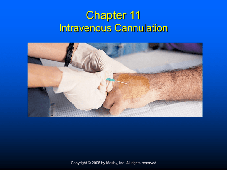
Chapter 11
Intravenous Cannulation
Copyright © 2006 by Mosby, Inc. All rights reserved.
Objectives
Define the term intravenous cannulation
Recall the indications and contraindications of
intravenous cannulation
Identify the equipment used to perform intravenous
cannulation
Select preferred solutions for use in management of
trauma and medical emergencies
Copyright © 2006 by Mosby, Inc. All rights reserved.
Objectives
Recall the recommended ratio of IV replacement to
blood loss in patients experiencing hypovolemic
shock
Describe the methods used to determine the proper
IV flow rate
List the advantages, disadvantages, and
complications associated with the use of peripheral
veins
Copyright © 2006 by Mosby, Inc. All rights reserved.
Objectives
Identify the veins that are commonly used for
peripheral intravenous cannulation
Recall the steps used to perform peripheral
intravenous cannulation
Use problem-solving skills with IV lifelines that are
not functioning properly to determine the cause and
correct the problem
Copyright © 2006 by Mosby, Inc. All rights reserved.
Objectives
List complications associated with IV therapy
Demonstrate the steps for discontinuing an IV
lifeline
Copyright © 2006 by Mosby, Inc. All rights reserved.
Introduction
Intravenous cannulation
Placement of a catheter in a vein
Administer blood, fluids, medications
Obtain blood specimens
Copyright © 2006 by Mosby, Inc. All rights reserved.
Introduction
Intravenous cannulation
Indications
• Cardiac disease
• Hypoglycemia
• Seizures
• Shock
• Precautionary measure
Contraindications
• Sclerotic veins
• Burned extremities
• Critical patients
Copyright © 2006 by Mosby, Inc. All rights reserved.
Body Substance Isolation Precautions
When performing IV therapy
Hepatitis B virus (HBV)
Human immunodeficiency virus (HIV)
Copyright © 2006 by Mosby, Inc. All rights reserved.
Body Substance Isolation Precautions
Gloves
Barrier protections
Proper disposal
Copyright © 2006 by Mosby, Inc. All rights reserved.
IV Cannulation—Equipment
IV solution
Administration set
Extension set
Needles/catheters
Tourniquet
Protective gloves
Gown and goggles
Tape
Antibiotic swabs/ointment
Gauze dressings
10ml-35mL syringes
Vacutainer holder
Assorted blood collection
tubes
Padded arm boards
Copyright © 2006 by Mosby, Inc. All rights reserved.
Equipment
Intravenous solutions
25 – 1000 mL
Clear plastic bag
Two ports
Copyright © 2006 by Mosby, Inc. All rights reserved.
Equipment
Colloids and crystalloids
Normal saline and
lactated Ringer’s
3 L IV crystalloid/1 L
blood lost (3:1)
Copyright © 2006 by Mosby, Inc. All rights reserved.
Equipment
Administration set
Piercing spike
Drip chamber
Flow clamp
Drug administration port
Connector end
Copyright © 2006 by Mosby, Inc. All rights reserved.
Equipment
Microdrip chamber
Precise amounts
Copyright © 2006 by Mosby, Inc. All rights reserved.
Equipment
Macrodrip chamber
Large amounts
Copyright © 2006 by Mosby, Inc. All rights reserved.
Equipment
IV flow rates
Medical emergencies
• “To keep open” (TKO)
• Precautionary measure
• Administer medications
Copyright © 2006 by Mosby, Inc. All rights reserved.
Equipment
IV flow rates
Trauma
• Patient’s response
•
•
•
•
Pulse
B/P
Capillary refill <6 y/o
Cerebral function
Severe hypovolemic patient
Wide open rate
2-3 L limit
3-4 times normal flow
Copyright © 2006 by Mosby, Inc. All rights reserved.
Equipment
Volutrol chamber
Specific amounts
Pediatric infusions
Copyright © 2006 by Mosby, Inc. All rights reserved.
Equipment
Over-the-needle catheter
Through-the-needle catheter
Copyright © 2006 by Mosby, Inc. All rights reserved.
Equipment
Hollow needle (butterfly
type)
Copyright © 2006 by Mosby, Inc. All rights reserved.
Equipment
IV catheter with attached syringe
Copyright © 2006 by Mosby, Inc. All rights reserved.
Differences Between Arteries and Veins
Copyright © 2006 by Mosby, Inc. All rights reserved.
Sites for Peripheral Venous Cannulation
Sites used in routine
situations
Copyright © 2006 by Mosby, Inc. All rights reserved.
Sites for Peripheral Venous Cannulation—
Other Sites
Copyright © 2006 by Mosby, Inc. All rights reserved.
Procedure for Performing
IV Cannulation
Insert spiked piercing
into IV bag
Drip chamber filled to
half way
Copyright © 2006 by Mosby, Inc. All rights reserved.
Procedure for Performing
IV Cannulation
Place tourniquet
Make slip knot
Copyright © 2006 by Mosby, Inc. All rights reserved.
Procedure for Performing
IV Cannulation
Complete band placement
Cleanse site
Copyright © 2006 by Mosby, Inc. All rights reserved.
Procedure for Performing
IV Cannulation
Pull skin taut—needle
bevel facing up
Penetrate at juncture
Copyright © 2006 by Mosby, Inc. All rights reserved.
Procedure for Performing
IV Cannulation
Enter vein from either top
or side
Watch for blood flashback
Copyright © 2006 by Mosby, Inc. All rights reserved.
Procedure for Performing
IV Cannulation
Advance needle
Slide catheter
Copyright © 2006 by Mosby, Inc. All rights reserved.
Procedure for Performing
IV Cannulation
Remove needle
Draw blood sample
Copyright © 2006 by Mosby, Inc. All rights reserved.
Procedure for Performing
IV Cannulation
Release tourniquet
Connect IV
Copyright © 2006 by Mosby, Inc. All rights reserved.
Procedure for Performing
IV Cannulation
Secure in place
Commercial device
Copyright © 2006 by Mosby, Inc. All rights reserved.
Procedure for Performing
IV Cannulation
Proper disposal of used
needle
Copyright © 2006 by Mosby, Inc. All rights reserved.
Procedure for Performing
IV Cannulation
Documentation
Date/time
Type/amount
Device used
Site
Number of attempts
Flow rate
Adverse reactions
Name of EMT-I
Copyright © 2006 by Mosby, Inc. All rights reserved.
Procedure for Performing
IV Cannulation
Common puncture sites that use an arm board
Copyright © 2006 by Mosby, Inc. All rights reserved.
Procedure for Performing
IV Cannulation
Complications
Pain
Catheter shear
Circulatory overload
Cannulation of an artery
Hematoma or infiltration
Local infection
Air embolism
Pyrogenic reaction
Copyright © 2006 by Mosby, Inc. All rights reserved.
Drawing Blood
Equipment
Copyright © 2006 by Mosby, Inc. All rights reserved.
Discontinuing the IV Lifeline
Equipment
Protective gloves
Sterile gauze pads
Adhesive bandage
Close control valve
Carefully untape and remove dressing
Copyright © 2006 by Mosby, Inc. All rights reserved.
Discontinuing the IV Lifeline
Stabilize tissue with dressing
Withdraw catheter
Cover puncture site with dressing and tape
Copyright © 2006 by Mosby, Inc. All rights reserved.
External Jugular Vein Cannulation
Copyright © 2006 by Mosby, Inc. All rights reserved.
Elderly Patients
Copyright © 2006 by Mosby, Inc. All rights reserved.
Summary
IV cannulation is used for administering
blood, fluids, or medications into circulatory
system and for obtaining blood samples
IV placement should never delay patient
transport
Normal saline and lactated Ringer's solution
are recommended for prehospital therapy;
they are isotonic crystalloid solutions
Copyright © 2006 by Mosby, Inc. All rights reserved.
Summary
Crystalloid solutions diffuse out of circulatory system
quickly; at least 3 L must be administered for every
liter of blood lost
Two most common administration sets are micro
(delivers 60 drops/mL) and macro (delivers 10 to 20
drops/mL)
Plastic over-the-needle catheters are most commonly
used because they can be better anchored and
permit freer patient movement
Copyright © 2006 by Mosby, Inc. All rights reserved.
Summary
In noncritical patients, use distal veins on dorsal
aspect of hand as IV sites
During cardiac arrest, preferred sites are veins of
antecubital fossa
Difficult cannulation can occur in obese persons,
during shock and cardiac arrest, in chronic mainline
drug users, in elderly patients with "fragile" or "rolling
veins," and in small children
Copyright © 2006 by Mosby, Inc. All rights reserved.
Summary
When selecting equipment for IV therapy, ensure fluid
is clear, not outdated, and bag has no leaks
Employ infection control procedures when
assembling administration set, cannulating vein, and
attaching IV tubing to catheter
Discard needles appropriately
Copyright © 2006 by Mosby, Inc. All rights reserved.
Summary
Release tourniquet once IV is connected to catheter
Carefully monitor patient for signs of improvement
and for signs of circulatory overload
Some complications of IV therapy are pain, catheter
shear, circulatory overload, cannulation of artery,
infiltration/hematoma, local infection, and air
embolism
Copyright © 2006 by Mosby, Inc. All rights reserved.
Questions?
Copyright © 2006 by Mosby, Inc. All rights reserved.


