Fetal Pig Dissection
advertisement
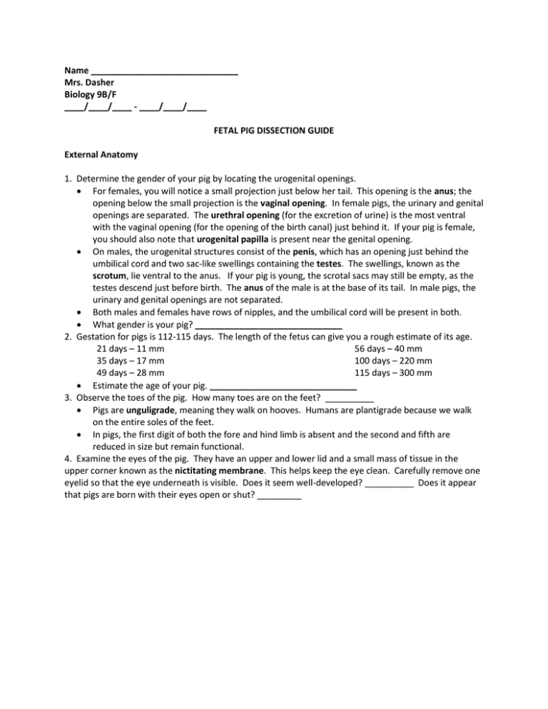
Name ______________________________ Mrs. Dasher Biology 9B/F ____/____/____ - ____/____/____ FETAL PIG DISSECTION GUIDE External Anatomy 1. Determine the gender of your pig by locating the urogenital openings. For females, you will notice a small projection just below her tail. This opening is the anus; the opening below the small projection is the vaginal opening. In female pigs, the urinary and genital openings are separated. The urethral opening (for the excretion of urine) is the most ventral with the vaginal opening (for the opening of the birth canal) just behind it. If your pig is female, you should also note that urogenital papilla is present near the genital opening. On males, the urogenital structures consist of the penis, which has an opening just behind the umbilical cord and two sac-like swellings containing the testes. The swellings, known as the scrotum, lie ventral to the anus. If your pig is young, the scrotal sacs may still be empty, as the testes descend just before birth. The anus of the male is at the base of its tail. In male pigs, the urinary and genital openings are not separated. Both males and females have rows of nipples, and the umbilical cord will be present in both. What gender is your pig? ______________________________ 2. Gestation for pigs is 112-115 days. The length of the fetus can give you a rough estimate of its age. 21 days – 11 mm 56 days – 40 mm 35 days – 17 mm 100 days – 220 mm 49 days – 28 mm 115 days – 300 mm Estimate the age of your pig. ______________________________ 3. Observe the toes of the pig. How many toes are on the feet? __________ Pigs are unguligrade, meaning they walk on hooves. Humans are plantigrade because we walk on the entire soles of the feet. In pigs, the first digit of both the fore and hind limb is absent and the second and fifth are reduced in size but remain functional. 4. Examine the eyes of the pig. They have an upper and lower lid and a small mass of tissue in the upper corner known as the nictitating membrane. This helps keep the eye clean. Carefully remove one eyelid so that the eye underneath is visible. Does it seem well-developed? __________ Does it appear that pigs are born with their eyes open or shut? _________ Internal Anatomy 1. Examination of the oral cavity. With a pair of scissors cut deeply into both corners of the mouth. This may be difficult as you must cut through both tissue and home. Open the mouth. Be sure to follow the curvature of the throat and do not cut straight back into the neck tissue. Examine the oral cavity containing the tongue and teeth. Notice the ridged roof of the mouth called the hard palate. The soft palate is the fleshy portion of the roof of the mouth and lies caudal to the hard palate. Locate the tongue with all its taste buds (also known as sensory papillae). Pigs have two types of teeth – incisors located in the front of the oral cavity and cheek teeth located toward the back of the oral cavity. To find the next few structures, you will have to cut through the bone of the jaw. Gentle pressure will be required for opening the mouth. o Far back in the oral cavity is the pharynx, a common passage for food going to the esophagus and air going into the lungs. Locate the tear-shaped epiglottis, a flap-like structure at the top of the trachea. The esophageal opening, which is the entrance to the esophagus (food tube), can also be found at the back of the throat. The esophagus is located behind the trachea. 2. Preparing the specimen. Use two pieces of strong twine and tie one around a wrist and one around an ankle of the pig. Pull each under the dissecting pan and tightly tie the twine to the opposite wrist or ankle. Making incisions – use upward pressure whenever possible, by turning the blade over. Make a shallow incision along midline of body, starting at neck. o use light pressure cutting through neck muscles. o Cut through until you see a whitish tube the size of a pencil: the trachea. o Observe the thymus on either side of the trachea. o Observe the brown oval structure above the trachea: the thyroid. o Remove both the thyroid and thymus at this time. Continue the incision to include the thoracic cavity. Ribs and sternum will create resistance. o Use scissors to cut through the sternum, being careful of the organs beneath. Expose the organs of the chest cavity. o heart: enclosed by whitish membrane: the pericardium o Lungs: lateral to the heart. Cut along the body wall edge to free the diaphragm. Make a shallow incision all the way to the umbilical cord o Observe pg. 19 to complete this incision. make a U-shaped cut for males make a circle for females Continue the cut to the pubic region and anus. o cut about 1/8” until reaching the pubis (cartilage) o Be careful with abdominal cuts due to organs underneath. o Stop when you can see the peritoneum – white, shiny, semitransparent membrane. At the pubic region, the muscles are deeper. Cut slightly off-center and deep. Pin back skin, muscles and membranes to reveal the internal organs. Cut the skin flaps back close to the back bone so they will remain open. Be careful not to injure the kidneys. 3. Examination of the digestive system. The large, reddish-brown organ that occupies much of the abdominal space is the liver. Gently lift it up and probe it to locate the gall bladder which is on the pig’s right side. The diaphragm, a thin brown muscular tissue, is the tough muscle which separates the thoracic and abdominal cavities. The esophagus goes through it to the stomach. The esophagus carries the food from the pharynx to the stomach. Locate the stomach on the upper left side of the abdominal cavity, underneath the liver. The stomach resembles a pouch in appearance and is connected to the esophagus on its anterior end. Slit open the stomach to see the longitudinal ridges. The constricted caudal portion of the stomach leads to the small intestine. Observe that the small intestine is not loose in the abdominal cavity but is held in place by the mesentery. Check for veins and arteries in the clear mesentery – these carry absorbed nutrients to the liver through the hepatic-portal vein. Cut open the small intestine. Inside the small intestine are finger-like projections called villi. The villi increase the surface area of the small intestine for absorption. These villi are microscopic. The large intestine appears as a compact coil and is larger in diameter than the small intestine. Locate the junction of the small and large intestine. Below this junction you may be able to find a small pouch-like structure called the caecum. This structure is similar to the appendix in humans. It helps in the slow digestion of plant materials in other animals. Follow the large intestine to the rectum. This lies in the dorsal wall of the abdominal cavity and is the straight end portion of the large intestine. Water is absorbed by the body in the large intestine. Waste material stored in the rectum leaves the body through the anus. Locate the pancreas which is a large white granular organ located below the stomach. The pancreas makes a variety of digestive enzymes that travel to the small intestine. The red elongated organ extending around the outer curvature of the stomach is the spleen. It resembles a tongue. It helps to destroy old red blood cells. 4. Examination of the circulatory system. The circulatory system of the pig consists of the heart, arteries, veins, and capillaries. There are two major parts of this system. Pulmonary circulation supplies the lungs with blood. The systemic circulatory system supplies all parts of the body except the lungs. You will need to cut through the sternum to open the thoracic cavity. Covering the heart is a thin, tough membrane called the pericardium. Partially covering the heart is the thymus gland (globular structure). The thymus is largest in young individuals. It is part of the immune system. The heart is composed of four chambers. Locate the two atria and two ventricles. Your pig has been injected with colored latex which makes it easy to locate the veins (blue latex) and the arteries (red latex). Locate the superior/anterior and inferior/posterior vena cava. These carry blood from the cranial and caudal portions of the body, respectively. Find the pulmonary veins which carry blood from the lungs to the left atrium. This carries oxygenated blood from the lungs back to the heart. The most noticeable artery is the aorta. The aorta curves to the left and passes cranially along the dorsal side of the thoracic and abdominal wall. The next largest artery is the pulmonary artery. It arises from the anterior portion of the right ventricle and soon divides into the right and left pulmonary arteries. Other arteries are named for the body part they serve. The gastric artery leads to the stomach, the hepatic artery leads to the liver, the renal artery leads to the kidney, and the carotid artery leads to the head. Locate the carotid artery and jugular vein. 5. 6. 7. 8. Remove the heart by carefully cutting the arteries and veins leading to and from the heart as far away from the heart as possible. DO NOT damage any lung tissue. Cut the heart in half through the frontal plane using a sharp blade. Ask Mrs. Dasher for assistance if at any time you are uncertain of the procedure. Identify the right atrium, right ventricle, left atrium, and left ventricle. The structure between the two ventricles is the septum. You may be able to visualize the valves. Remember that the atrioventricular valves occur between the atria and ventricles, the semilunar valves are found between the ventricles and the pulmonary artery and vein. On the surface of the heart are the coronary artery and veins. A unique feature of the fetal mammal heart is the ductus arteriosus. This short vessel allows blood to bypass pulmonary circulation until birth, at which time there is a complete closure of the vessel. Examination of the respiratory system The respiratory system is responsible for the exchange of gases. Most mammals must take in oxygen to burn food and must rid itself of carbon dioxide waste one it is born. Air enters through the external nares. Air is drawn into the nasopharynx or nose chambers where sensory nerve cells detect smell. Here, also, is where the glottis, the opening of the trachea, may be found. The trachea is a tube that extends from the neck to the chest. It is white and lined with cartilage. The enlargement at the anterior end of the trachea is the larynx which contains the vocal cords. The trachea splits in the chest cavity into two bronchi. Each of these air tubes extends into the lungs and splits into smaller tubes called bronchioles. The lungs are located on either side of the heart. The lungs are made up of tiny air sacs called alveoli (microscopic) where gas exchange occurs. Locate the thin muscular diaphragm just above the liver. This muscle is responsible for drawing air into the chest cavity. Spasms of this muscle result in hiccups. Examination of the urogenital system. “Uro” of this term represents the urinary system. “Genital” represents the reproductive system. These systems are structurally related. Remember that you are dealing with paired structures, meaning what is observed on one side can also be observed on the other. To find the kidneys, look for two lumps low in the abdominal cavity. They are behind a membrane called the peritoneum. You will need to carefully remove the peritoneum to see the bean-shaped kidneys. Locate the ureter originating from the hilus. Follow the ureter posteriorly until it joins the urinary bladder. Do not remove any of these organs. The renal artery and vein also come out of the kidney. The artery carries blood to the kidney; the vein carries blood out of the kidney. Prepare for observation of reproductive system(s). Pull the hind legs apart. With scissors, cut anteriorly a little to one side of the mid-ventral line to avoid cutting the penis in the male. Press firmly on the tissue between the legs to feel the cartilaginous structure of the pubic symphysis. This is part of the pelvic girdle. Continue the incision anteriorly and cut through the pubic symphysis. Expose the urethra. This tube leads from the bladder to the outside world. Examination of the male reproductive system. Examine the scrotal sac (scrotum) at the posterior end of the male pig. Open one sac and determine the presence of a testis. If not found in the scrotum, they may be found in a tubelike structure called the inguinal canal; they will appear as small oval organs. In either case, locate one of the testes and note the coiled tubule making up the epididymis. Follow this tube forward as it passes through the inguinal canal as the vas deferens. 9. Examination of the female reproductive system. Spread the legs to separate the pubic symphysis and thereby expose the female reproductive system. Locate the oval-shaped ovaries which are found caudal to the kidneys. Leading from the ovaries are the twisted oviducts or Fallopian tubes. The oviducts continue posteriorly and are soon supported by broad ligaments. Further on, the oviducts join to form the common uterus. You will notice a slight constriction of the common uterus marking the location of the cervix. Posterior to the cervix, the remainder of the tube forms the vagina.
