Here is the Original File
advertisement
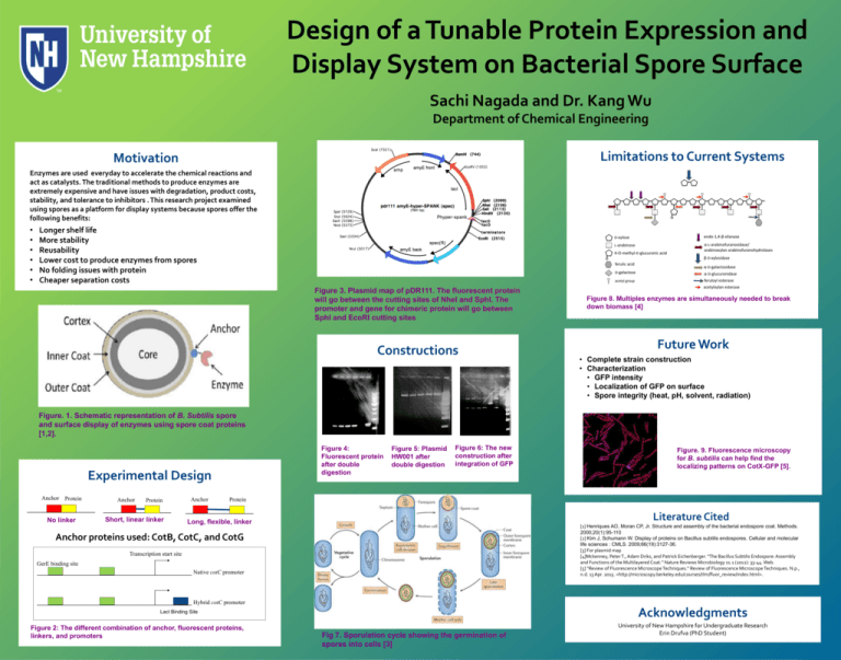
Design of a Tunable Protein Expression and Display System on Bacterial Spore Surface Sachi Nagada and Dr. Kang Wu Department of Chemical Engineering Limitations to Current Systems Motivation Enzymes are used everyday to accelerate the chemical reactions and act as catalysts. The traditional methods to produce enzymes are extremely expensive and have issues with degradation, product costs, stability, and tolerance to inhibitors . This research project examined using spores as a platform for display systems because spores offer the following benefits: • • • • • • Longer shelf life More stability Reusability Lower cost to produce enzymes from spores No folding issues with protein Cheaper separation costs Figure 3. Plasmid map of pDR111. The fluorescent protein will go between the cutting sites of NheI and SphI. The promoter and gene for chimeric protein will go between SphI and EcoRI cutting sites Constructions Figure 8. Multiples enzymes are simultaneously needed to break down biomass [4] Future Work • Complete strain construction • Characterization • GFP intensity • Localization of GFP on surface • Spore integrity (heat, pH, solvent, radiation) Figure. 1. Schematic representation of B. Subtilis spore and surface display of enzymes using spore coat proteins [1,2]. Figure 4: Fluorescent protein after double digestion Experimental Design Anchor Protein No linker Anchor Protein Short, linear linker Anchor Figure 5: Plasmid HW001 after double digestion Figure 6: The new construction after integration of GFP Figure. 9. Fluorescence microscopy for B. subtilis can help find the localizing patterns on CotX-GFP [5]. Protein Literature Cited Long, flexible, linker [1] Henriques AO, Moran CP, Jr. Structure and assembly of the bacterial endospore coat. Methods. 2000;20(1):95-110 [2] Kim J, Schumann W. Display of proteins on Bacillus subtilis endospores. Cellular and molecular life sciences : CMLS. 2009;66(19):3127-36. [3] For plasmid map [4]Mckenney, Peter T., Adam Driks, and Patrick Eichenberger. "The Bacillus Subtilis Endospore: Assembly and Functions of the Multilayered Coat." Nature Reviews Microbiology 11.1 (2012): 33-44. Web. [5] "Review of Fluorescence Microscope Techniques." Review of Fluorescence Microscope Techniques. N.p., n.d. 13 Apr. 2015. <http://microscopy.berkeley.edu/courses/tlm/fluor_review/index.html>. Anchor proteins used: CotB, CotC, and CotG Transcription start site GerE binding site Native cotC promoter Hybrid cotC promoter Acknowledgments LacI Binding Site Figure 2: The different combination of anchor, fluorescent proteins, linkers, and promoters Fig 7. Sporulation cycle showing the germination of spores into cells [3] University of New Hampshire for Undergraduate Research Erin Drufva (PhD Student)
