Lecture 7a
advertisement
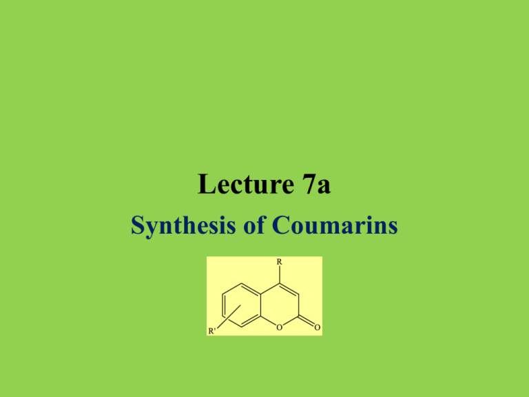
Lecture 7a
Synthesis of Coumarins
Introduction I
• Coumarin (2H-chromen-2-one) was first isolated from the tonka bean
(coumarou) or sweet clover in 1820 by A. Vogel. It has a sweet odor
that is recognized as the scent of a new-mown hay and a bitter taste.
• Since 1882, it has been used in perfumes and as aroma enhancer in
pipe tobacco and some alcoholic drinks (i.e., mulled wines).
However, it is banned as food additive in the United States due to the
concerns of its hepatotoxicity (chemical-driven liver damage).
• Many coumarin derivatives like dicumarol, warfarin and
acenocoumarol act as anticoagulant and vitamin K antagonist.
• Umbelliferone (7-hydroxyl-coumarin), found in carrots, coriander
and garden angelica.
• Hymecromone (7-hydroxy-4-methylcoumarin, 4-methylumbelliferone), the compound synthesized in the lab, is used in bile
therapy. It is also used in the fluorometric determination of enzyme
activity.
Introduction II
• Coumarin itself has been used as an edema modifier. Like other benzopyrones,
it is known to stimulate macrophages to degrade extracellular albumin,
allowing faster resorption of edematous fluids.
• Umbelliferone absorbs ultraviolet light strongly at several wavelengths,
which led to its use as a sunscreen and an optical brightener for textiles
as well as gain medium for dye laser. It can also be used as a fluorescence
indicator for metal ions such as copper and calcium.
• W. H. Perkin, the discoverer of the aniline dyes, first synthesized coumarin
in 1868 by the reaction of salicylic aldehyde and acetic acid anhydride in
the presence of a base.
Mechanism I
• In 1884, H. v. Pechmann developed a condensation reaction using a phenol and
a carboxylic acid or ester containing a b-carbonyl group to afford a coumarin
• The reaction can be regarded as an esterification or transesterification followed
by the attack of the activated carbonyl group in ortho position to the oxygen atom
attached to the ring.
• The final step is a dehydration that leads to the formation of an additional double
bond in the newly formed six-membered ring.
transesterification
R’=m-OH
enolization
electrophilic attack
dehydration
• Alternatively, a mechanism that starts with an attack of the ring to the keto group, which
is followed by a transesterification to form the six-membered ring has been proposed.
Mechanism II
• The reaction is usually catalyzed by sulfuric acid, phosphoric acid,
aluminum trichloride or trifluoroacetic acid. Unfortunately, many of
these reactions require large quantities of the catalyst and elevated
temperatures.
• More recently, bismuth chloride (5 % mol, 75 oC), iron(III) chloride
in ionic liquids, montmorillonite K-10 or iron(III) fluoride under
solvent-free conditions have been used successfully. In addition,
Keggin-type heteropolycompounds (i.e., H3PW12O40) have been
used under solvent-free conditions.
• The reaction works better for donor-substituted benzene ring
(i.e., polyhydroxy, amino, etc.). In some cases, a polycondensation
is observed particularly if an excess of ethyl acetoacetate was used.
• If amines are reacted with ethyl acetoacetate, 4-methyl-2-hydroxylquinolines are formed via acetoacetanilides (using p-TSA/MW) or
2-methyl-4-hydroxyquinolines via ethyl b-anilinocrotonates.
Experimental I
• In a 50 mL Erlenmeyer flask,
resorcinol, ethyl acetoacetate
and Amberlyst A15 are mixed
thoroughly
• Why is a small container used
here?
• The flask is covered with a
watch glass and is then heated
in the microwave
• After removing the reaction
mixture, it is allowed to cool
slowly for 30 minutes
• Which setting is used here?
• A minimal amount of hot
solvent (methanol:water=5:1)
is added
• Why is the mixture used here?
The reaction is run in micro-scale,
which means that only little liquid
is formed upon heating
The reaction should be run at 30 %
power, which is equal to ~210 W
• How can the cooling process
slowed down?
The flask is isolated with paper towels
and aluminum foil
The compound dissolves well in hot
methanol but less it the cold solvent
mixture
Experimental II
• The catalyst is removed from
the hot solution
• Upon cooling, the product
precipitates as a white solid
• The crystals are isolated by
vacuum filtration in a Hirsch
funnel and rinsed
• After sucking air through the
solid for 5-10 minutes, the
compound is dried under the
heating lamp (or in an open
container until next lab
meeting).
• A sample for GC/MS analysis
is submitted
• How is this accomplished?
The bulk of the solution can be decanted
and the rest be transferred with a pipette
• What is used to rinse the
crystals?
First, the ice-cold solvent mixture is used
and then some ice-cold methanol
• What is the proper concentration
here? 2 mg/mL in ethyl acetate
Characterization I
• Fluorescence
• Stage 1: Excitation
• A photon of energy hνEX is supplied by an external source such as an
incandescent lamp or a laser and absorbed by the fluorophore, creating
an excited electronic singlet state (S1‘).
• Stage 2: Excited-State Lifetime
• The excited state exists for a finite time (typically 1–10 ns). During this
time, the fluorophore undergoes conformational changes and is also subject
to a multitude of possible interactions with its molecular environment.
• The energy of S1‘ is partially dissipated, yielding a relaxed singlet excited
state (S1) from which fluorescence emission originates.
• Not all the molecules initially excited by absorption return to the ground state
(S0) by fluorescence emission. Other processes may also depopulate S1.
Characterization II
• Stage 3: Fluorescence Emission
•
A photon of energy hνEM is emitted, returning the fluorophore to its ground state S0. Due to energy dissipation during
the excited-state lifetime, the energy of this photon is lower, and therefore of longer wavelength, than the excitation
photon hνEX.
•
The difference in energy or wavelength represented by (hνEX – hνEM) is called the Stokes shift (dashed line:
excitation, solid line: emission). The Stokes shift is fundamental to the sensitivity of fluorescence techniques
because it allows emission photons to be detected against a low background, isolated from excitation photons.
In contrast, absorption spectrophotometry requires measurement of transmitted light relative to high incident light
levels at the same wavelength.
•
• The beverage contains quinine, which absorbs at l=350 nm and emits at l=460 nm (bright blue/cyan hue)
350000 Fluorescence Spectrum of Hymechromone in MeOH
300000
250000
200000
150000
100000
50000
0
300
350
400
450
500
Characterization III
• Fluorescence Test
•
•
•
•
•
•
Some of the product is dissolved in 95 % ethanol
Tube A: 1 mL of 1 M sodium hydroxide solution is added
Tube B: 1 mL of 2 M HCl is added
Tube C: add nothing to this test tube
How do the solutions look like at this point?
Place all three tubes in the UV-light (long wavelength,
365 nm). How the solutions look like now?
• Warning: Do not look straight into the UV-lamp!
Characterization IV
• Crystal Structure (7-hydroxy-4-methylcoumarin)
• 7-hydroxy-4-methylcoumarin is almost planar but the hydroxyl derivative
displays a more than 100 oC higher melting point than coumarin.
• The higher melting point is due to the formation of hydrogen bonds
between the phenolic hydroxyl group and the oxygen atom of the
carbonyl group of a neighboring molecule (d(O-H…O=C=189 pm),
which is similar in length compared to water.
Characterization V
• Infrared Spectrum (7-hydroxy-4-methylcoumarin)
•
•
•
•
•
n(OH)=2400-3400 cm-1
n(C=O)=1673 cm-1
n(C=C)=1596 cm-1
n(CO)=1067, 1134 and 1267 cm-1
d(oop, tri-subst.alkene)=838 cm-1
n(OH)
d(oop)
n(C=O)
n(C=C)
n(CO)
Characterization VI
• 1H-NMR Spectrum (in DMSO-d6)
•
•
•
•
d=10.5 ppm (OH, H1)
d=6.8 ppm (d, H2) and 7.5 ppm (d, H3) (aromatic protons)
d=6.7 ppm (s, H6) and 6.1 ppm (s, H5) (aromatic and alkene proton)
d=2.3 ppm (CH3, H4)
H2O in
DMSO
H3 H2
H6 H5
H1
H4
DMSO
Characterization VII
•
13C{1H}-Spectrum
• Ten signals total
• Five quaternary carbons
• One carbonyl function (C9, 161.6)
• Three ipso-carbons (C1 (160.7),
C4 (112.5), C5 (154.0))
• One disubstituted alkene (C7, 155.3)
• Four methine carbons
• Three CH (C2 (113.3), C3 (127.1),
C6 (102.6))
• One CH (C8, 110.7)
• One methyl carbon (C10, 18.6)
Characterization VIII
• Mass Spectrum
• m/z=176 ([M]+)
• m/z=148 ([M-28]+): loss of one carbon monoxide molecule
leading to a benzofuran
• m/z=147 ([M-28-1]+): loss of one carbon monoxide molecule
and a proton
• m/z=120 ([M-28-28]+): loss of two carbon monoxide molecules
