Adaptations in dopamine neurochemistry in
advertisement

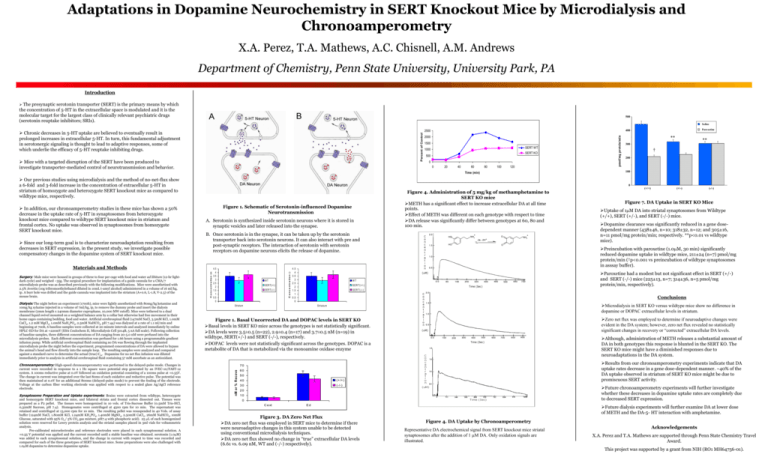
Adaptations in Dopamine Neurochemistry in SERT Knockout Mice by Microdialysis and Chronoamperometry X.A. Perez, T.A. Mathews, A.C. Chisnell, A.M. Andrews Department of Chemistry, Penn State University, University Park, PA Introduction The presynaptic serotonin transporter (SERT) is the primary means by which the concentration of 5-HT in the extracellular space is modulated and it is the molecular target for the largest class of clinically relevant psychiatric drugs (serotonin reuptake inhibitors; SRIs). 500 Saline Mice with a targeted disruption of the SERT have been produced to investigate transporter-mediated control of neurotransmission and behavior. 2000 1500 SERT WT 1000 SERT KO 500 0 0 20 40 60 80 100 120 Surgery: Male mice were housed in groups of three to four per cage with food and water ad libitum (12-hr lightdark cycle) and weighed ~35g. The surgical procedure for implantation of a guide cannula for a CMA/7 microdialysis probe was as described previously with the following modifications. Mice were anesthetized with 2.5% Avertin (10g tribromoethylethanol diluted in 10mL t-amyl alcohol) administered in a volume of 16 ml/kg, ip. A burr hole was drilled and the guide cannula was implanted into the striatum (A+0.6, L-1.8, V-2.5) of the mouse brain. Dialysis: The night before an experiment (1700h), mice were lightly anesthetized with 80mg/kg ketamine and 10mg/kg xylazine injected in a volume of 7ml/kg, ip, to remove the dummy probe and insert the dialysis membrane (2mm length x 240mm diameter cuprophane, 10,000 MW cutoff). Mice were tethered to a dual channel liquid swivel mounted on a weighted balance arm by a collar but otherwise had free movement in their home cages containing bedding, food and water. Artificial cerebrospinal fluid (147mM NaCl, 3.5mM KCl, 1.0mM CaCl2, 1.2 mM MgCl2, 1.0mM NaH2PO4, 2.5mM NaHCO3, pH 7.44) was dialyzed at a rate of 1.1 ml/min and beginning at 700h, 6 baseline samples were collected at 20 minute intervals and analyzed immediately by online HPLC-ED for DA at +220mV (ESA Coulochem II, Microdialysis Cell 5014B, 5 nA full scale). Following collection of baseline samples, three different concentrations of DA ranging from 20-5.0 nM were perfused into the microdialysis probes. Each different concentration was perfused for 1.66 hours using a programmable gradient infusion pump. While artificial cerebrospinal fluid containing no DA was flowing through the implanted microdialysis probe the night before the experiment, programmed concentrations of DA were allowed to bypass the animal’s head and flow directly into the sample loop. The resulting samples were analyzed and compared against a standard curve to determine the actual (true) Cin. Dopamine for no net flux infusion was diluted immediately prior to analysis in artificial cerebrospinal fluid containing 57 mM ascorbate as an antioxidant. Chronoamperometry: High-speed chronoamperometry was performed in the delayed-pulse mode. Changes in current were recorded in response to a 1 Hz square wave potential step generated by an IVEC-10/FAST-12 system. A 100ms reductive pulse at 0.0V followed an oxidation potential consisting of a 100ms pulse at +0.55V. The change in current was integrated over the last 80ms of each oxidative and reductive pulse. The potential was then maintained at 0.0V for an additional 800ms (delayed-pulse mode) to prevent the fouling of the electrode. Voltage at the carbon fiber working electrode was applied with respect to a sealed glass Ag/AgCl reference electrode. Synaptosome Preparation and Uptake experiments: Brains were extracted from wildtype, heterozygote and homozygote SERT knockout mice, and bilateral striata and frontal cortex dissected out. Tissues were prepared as a P2 pellet. The tissues were homogenized in 10 vols. of Tris-Sucrose Buffer (0.5mM Tris-HCl, 0,32mM Sucrose, pH 7.4). Homogenates were centrifuged at 4500 rpm for 10 min. The supernatant was retained and centrifuged at 13,000 rpm for 10 min. The resulting pellet was resuspended in 40 Vols. of assay buffer (124mM NaCl, 1.80mM KCl, 1.24mM KH2PO4, 1.40mM MgSO4, 2.50mM CaCl2, 26mM NaHCO3, 10mM Glucose, saturated with 95% O2 / 5% CO2 gas mixture, pH7.4 with phosphoric acid). 25 L of each homogenized solution were reserved for Lowry protein analysis and the striatal samples placed in 3ml vials for voltammetric analysis. Pre-calibrated microelectrodes and reference electrodes were placed in each synaptosomal solution. A +0.55 V potential was applied and the current recorded until a stable baseline was obtained. serotonin (1.0M) was added to each synaptosomal solution, and the change in current with respect to time was recorded and compared for each of the three genotypes of SERT knockout mice. Some preparations were also challenged with 1.0M dopamine to determine dopamine uptake. A. Serotonin is synthesized inside serotonin neurons where it is stored in synaptic vesicles and later released into the synapse. B. Once serotonin is in the synapse, it can be taken up by the serotonin transporter back into serotonin neurons. It can also interact with pre and post-synaptic receptors. The interaction of serotonin with serotonin receptors on dopamine neurons elicits the release of dopamine. 4 .5 4 3 .5 3 2 .5 2 1 .5 1 0 .5 0 WT SER T(+/-) SER T(-/-) C o n c e n t r a t io n ( n M ) Materials and Methods Figure 1. Schematic of Serotonin-influenced Dopamine Neurotransmission 4 .5 4 3 .5 3 2 .5 2 1 .5 1 0 .5 0 Figure 4. Administration of 5 mg/kg of methamphetamine to SERT KO mice METH has a significant effect to increase extracellular DA at all time points. Effect of METH was different on each genotype with respect to time DA release was significantly differ between genotypes at 60, 80 and 100 min. HO HO + NH3 O -2e, -2H+ + NH3 300 † 200 3-Hydroxytyramine (+/-) (-/-) Figure 7. DA Uptake in SERT KO Mice Uptake of 1M DA into striatal synaptosomes from Wildtype (+/+), SERT (+/-), and SERT (-/-) mice. Dopamine clearance was significantly reduced in a gene dosedependent manner (438±46, n=10; 318±32, n=12; and 305±16, n=11 pmol/mg protein/min; respectively. **p<0.01 vs wildtype mice). Preincubation with paroxetine (1.0M, 30 min) significantly reduced dopamine uptake in wildtype mice, 211±24 (n=7) pmol/mg protein/min (†p<0.001 vs preincubation of wildtype synaptosomes in asssay buffer). Paroxetine had a modest but not significant effect in SERT (+/-) and SERT (-/-) mice (225±13, n=7; 314±36, n=5 pmol/mg protein/min, respectively). S E R T(+ /-) S E R T(-/-) Conclusions Microdialysis in SERT KO versus wildtype mice show no difference in dopamine or DOPAC extracellular levels in striatum. S tria tu m Zero net flux was employed to determine if neuroadaptive changes were evident in the DA system; however, zero net flux revealed no statistically significant changes in recovery or “corrected” extracellular DA levels. Figure 1. Basal Uncorrected DA and DOPAC levels in SERT KO Basal levels in SERT KO mice across the genotypes is not statistically significant. DA levels were 3.5±0.5 (n=22), 2.9±0.4 (n=17) and 3.7±0.5 nM (n=19) in wildtype, SERT(+/-) and SERT (-/-), respectively. DOPAC levels were not statistically significant across the genotypes. DOPAC is a metabolite of DA that is metabolized via the monoamine oxidase enzyme Although, administration of METH releases a substantial amount of DA in both genotypes this response is blunted in the SERT KO. The SERT KO mice might have a diminished responses due to neuroadaptations in the DA system. Results from our chronoamperometry experiments indicate that DA uptake rates decrease in a gene dose-dependent manner. ~40% of the DA uptake observed in striatum of SERT KO mice might be due to promiscuous SERT activity. 70 60 50 40 (+/+) 30 (-/-) Future chronoamperometry experiments will further investigate whether these decreases in dopamine uptake rates are completely due to decreased SERT expression. 20 10 0 C e xt (+/+) O WT Stria tu m nM or % R ecovery Since our long-term goal is to characterize neuroadaptation resulting from decreases in SERT expression, in the present study, we investigate possible compensatory changes in the dopamine system of SERT knockout mice. ** 0 C o n c e n t r a t io n ( n M ) In addition, our chronoamperometry studies in these mice has shown a 50% decrease in the uptake rate of 5-HT in synaptosomes from heterozygote knockout mice compared to wildtype SERT knockout mice in striatum and frontal cortex. No uptake was observed in synaptosomes from homozygote SERT knockout mice. ** 100 Time (min) Our previous studies using microdialysis and the method of no-net-flux show a 6-fold and 3-fold increase in the concentration of extracellular 5-HT in striatum of homozygote and heterozygote SERT knockout mice as compared to wildtype mice, respectively. pmol/mg protein/min Percent of Control Chronic decreases in 5-HT uptake are believed to eventually result in prolonged increases in extracellular 5-HT. In turn, this fundamental adjustment in serotonergic signaling is thought to lead to adaptive responses, some of which underlie the efficacy of 5-HT reuptake inhibiting drugs. Paroxetine 400 2500 Ed Figure 3. DA Zero Net Flux DA zero net flux was employed in SERT mice to determine if there were neuroadaptive changes in this system unable to be detected using conventional microdialysis techniques. DA zero net flux showed no change in “true” extracellular DA levels (6.61 vs. 6.09 nM, WT and (-/-) respectively). Future dialysis experiments will further examine DA at lower dose of METH and the DA-5- HT interaction with amphetamine. Figure 4. DA Uptake by Chronoamperometry Representative DA electrochemical signal from SERT knockout mice striatal synaptosomes after the addition of 1 M DA. Only oxidation signals are illustrated. Acknowledgements X.A. Perez and T.A. Mathews are supported through Penn State Chemistry Travel Award. This project was supported by a grant from NIH (RO1 MH64756-01).


