Spinal Cord
advertisement

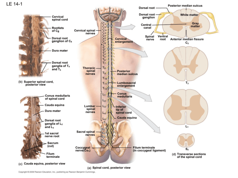
LE 14-1 Posterior median sulcus Dorsal root Dorsal root ganglion Cervical spinal cord Rootlets of C8 White matter Gray matter Central canal Cervical spinal nerves Dorsal root ganglion of C8 Spinal nerve Cervical enlargement Ventral Anterior median fissure root C3 Dura mater Dorsal root ganglia of T4 and T5 Thoracic spinal nerves Superior spinal cord, posterior view Lumbosacral enlargement Dura mater Lumbar spinal nerves Sacrum (cut) Inferior tip of spinal cord Cauda equina Dorsal root ganglia of L2 and L3 1st sacral nerve root T3 Conus medullaris Conus medullaris of spinal cord Cauda equina Posterior median sulcus L1 Sacral spinal nerves Coccygeal nerve(Co1) Filum terminale (in coccygeal ligament) Filum terminale S2 Transverse sections of the spinal cord Cauda equina, posterior view Spinal cord, posterior view LE 14-1a,d_1 Dorsal root Dorsal root ganglion Central canal Posterior median sulcus White matter Gray matter Cervical spinal nerves Cervical enlargement Spinal nerve Ventral root Anterior median fissure C3 Thoracic spinal nerves Posterior median sulcus Spinal cord, posterior view T3 Transverse sections of the spinal cord LE 14-1a,d_2 Thoracic spinal nerves Lumbar spinal nerves Lumbosacral enlargement Conus medullaris Inferior tip of spinal cord Cauda equina L1 Sacral spinal nerves Filum terminale (in coccygeal ligament) Coccygeal nerve(Co1) Spinal cord, posterior view S2 Transverse sections of the spinal cord LE 14-1bc Cervical spinal cord Conus medullaris of spinal cord Rootlets of C8 Cauda equina Dura mater Dorsal root ganglion of C8 Dorsal root ganglia of L2 and L3 1st sacral nerve root Dura mater Sacrum (cut) Dorsal root ganglia of T4 and T5 Superior spinal cord, posterior view Filum terminale Cauda equina, posterior view LE 14-1d Posterior median sulcus Dorsal root Dorsal root ganglion White matter Gray matter Central canal Spinal nerve Ventral root Anterior median fissure C3 T3 L1 S2 Transverse sections of the spinal cord LE 14-2 Spinal cord White matter Anterior median fissure Gray matter Ventral root Spinal nerve Pia mater Dorsal root Dorsal root ganglion Pia mater Denticulate ligaments Arachnoid mater Dura mater Arachnoid mater (reflected) Dura mater (reflected) Spinal blood vessel Posterior view Dura mater Dorsal root of sixth cervical nerve Anterior view Vertebral body Ventral root of sixth cervical nerve ANTERIOR Arachnoid mater Subarachniod space Autonomic (sympathetic) ganglion Pia mater Ventral root of spinal nerve Rami communicantes Spinal cord Ventral ramus Filum terminale Subarachnoid space containing cerebrospinal fluid and spinal nerve roots L5 vertebra Terminal portion of filum terminale S2 vertebra MRI, sectional view Spinal cord Adipose tissue in epidural space Dorsal ramus Denticulate ligament POSTERIOR Sectional view Dorsal root ganglion LE 14-2a Spinal cord Anterior median fissure Pia mater Denticulate ligaments Arachnoid mater (reflected) Dura mater (reflected) Spinal blood vessel Dorsal root of sixth cervical nerve Anterior view Ventral root of sixth cervical nerve LE 14-2b Spinal cord Filum terminale Subarachnoid space containing cerebrospinal fluid and spinal nerve roots L5 vertebra Terminal portion of filum terminale S2 vertebra MRI, sectional view LE 14-2c White matter Gray matter Ventral root Spinal nerve Dorsal root Dorsal root ganglion Pia mater Arachnoid mater Dura mater Posterior view LE 14-2d Dura mater ANTERIOR Arachnoid mater Subarachniod space Vertebral body Autonomic (sympathetic) ganglion Pia mater Ventral root of spinal nerve Rami communicantes Ventral ramus Dorsal ramus Spinal cord Denticulate ligament Adipose tissue in epidural space POSTERIOR Sectional view Dorsal root ganglion LE 14-3 Occipital bone Spinal cord emerging from foramen magnum Cervical plexus (C1–C5) Cervical spinal nerves (C1–C8) Brachial plexus (C5–T1) Thoracic spinal nerves (T1–T12) Lumbar spinal nerves (L1–L5) Sacral plexus (L4–S4) Coccygeal nerves (Co1) Lumbar plexus (T12–L4) Sciatic nerve Sacral spinal nerves (S1–S5) emerging from sacral foramina LE 14-3_1 Occipital bone Spinal cord emerging from foramen magnum Cervical plexus (C1–C5) Cervical spinal nerves (C1–C8) Brachial plexus (C5–T1) Thoracic spinal nerves (T1–T12) LE 14-3_2 Thoracic spinal nerves (T1–T12) Lumbar spinal nerves (L1–L5) Lumbar plexus (T12–L4) Sciatic nerve Sacral plexus (L4–S4) Coccygeal nerves (Co1) Sacral spinal nerves (S1–S5) emerging from sacral foramina LE 14-4 Dura mater Epidural space Body of third lumbar vertebra Interspinous ligament Lumbar puncture needle Cauda equina in subarachnoid space Filum terminale Cauda equina LE 14-4a Dura mater Epidural space Body of third lumbar vertebra Interspinous ligament Lumbar puncture needle Cauda equina in subarachnoid space Filum terminale LE 14-4b Cauda equina Organization of Spinal Cord Gray Matter • Recall, it is divided into horns – Dorsal, lateral (only in thoracic region), and ventral • Dorsal half – sensory roots and ganglia • Ventral half – motor roots • Based on the type of neurons/cell bodies located in each horn, it is specialized further into 4 regions – – – – Somatic sensory (SS) - axons of somatic sensory neurons Visceral sensory (VS) - neurons of visceral sensory neur. Visceral motor (VM) - cell bodies of visceral motor neurons Somatic motor (SM) - cell bodies of somatic motor neurons Gray Matter: Organization Figure 12.31 White Matter in the Spinal Cord • Divided into three funiculi (columns) – posterior, lateral, and anterior – Columns contain 3 different types of fibers (Ascend., Descend., Trans.) • Fibers run in three directions – Ascending fibers - compose the sensory tracts – Descending fibers - compose the motor tracts – Commissural (transverse) fibers - connect opposite sides of cord White Matter Fiber Tract Generalizations • Pathways decussate (most) • Most consist of a chain of two or three neurons • Most exhibit somatotopy (precise spatial relationships) • All pathways are paired – one on each side of the spinal cord White Matter: Pathway Generalizations Descending (Motor) Pathways • Descending tracts deliver motor instructions from the brain to the spinal cord • Divided into two groups – Pyramidal, or corticospinal, tracts – Indirect pathways, essentially all others • Motor pathways involve two neurons – Upper motor neuron (UMN) – Lower motor neuron (LMN) • aka ‘anterior horn motor neuron” (also, final common pathway) Pyramidal (Corticospinal) Tracts • Originate in the precentral gyrus of brain (aka, primary motor area) – I.e., cell body of the UMN located in precentral gyrus • Pyramidal neuron is the UMN – Its axon forms the corticospinal tract • UMN synapses in the anterior horn with LMN – Some UMN decussate in pyramids = Lateral corticospinal tracts – Others decussate at other levels of s.c. = Anterior corticospinal tracts • LMN (anterior horn motor neurons) – Exits spinal cord via anterior root – Activates skeletal muscles • Regulates fast and fine (skilled) movements Corticospinal tracts 1. 2. 3. 4. Location of UMN cell body in cerebral cortex Decussation of UMN axon in pyramids or at level of exit of LMN Synapse of UMN and LMN occurs in anterior horn of s.c. LMN axon exits via anterior root Extrapyramidal Motor Tracts • Includes all motor pathways not part of the pyramidal system • Upper motor neuron (UMN) originates in nuclei deep in cerebrum (not in cerebral cortex) • UMN does not pass through the pyramids! • LMN is an anterior horn motor neuron • This system includes – – – – Rubrospinal Vestibulospinal Reticulospinal Tectospinal tracts • Regulate: – Axial muscles that maintain balance and posture – Muscles controlling coarse movements of the proximal portions of limbs – Head, neck, and eye movement Extrapyramidal Tract Note: 1. UMN cell body location 2. UMN axon decussates in pons 3. Synapse between UMN and LMN occurs in anterior horn of sc 3. LMN exits via ventral root 4. LMN axon stimulates skeletal muscle Extrapyramidal (Multineuronal) Pathways • Reticulospinal tracts – originates at reticular formation of brain; maintain balance • Rubrospinal tracts – originate in ‘red nucleus’ of midbrain; control flexor muscles • Tectospinal tracts - originate in superior colliculi and mediate head and eye movements towards visual targets (flash of light) Main Ascending Pathways • The central processes of first-order neurons branch diffusely as they enter the spinal cord and medulla • Some branches take part in spinal cord reflexes • Others synapse with second-order neurons in the cord and medullary nuclei Three Ascending Pathways • The nonspecific and specific ascending pathways send impulses to the sensory cortex – These pathways are responsible for discriminative touch (2 pt. discrimination) and conscious proprioception (body position sense). • The spinocerebellar tracts send impulses to the cerebellum and do not contribute to sensory perception Nonspecific Ascending Pathway • • • • • • Include the lateral and anterior spinothalamic tracts Lateral: transmits impulses concerned with pain and temp. to opposite side of brain Anterior: transmits impulses concerned with crude touch and pressure to opposite side of brain 1st order neuron: sensory neuron 2nd order neuron: interneurons of dorsal horn; synapse with 3rd order neuron in thalamus 3rd order neuron: carry impulse from thalamus to postcentral gyrus Specific and Posterior Spinocerebellar Tracts • Dorsal Column Tract 1. AKA Medial lemniscal pathway 2. Fibers run only in dorsal column 3. Transmit impulses from receptors in skin and joints 4. Detect discriminative touch and body position sense =proprioception • 1st O.N.- a sensory neuron • synapses with 2nd O.N. in nucleus gracilis and nucleus cuneatus of medulla • 2nd O.N.- an interneuron • decussate and ascend to thalamus where it synapses with 3rd O.N. • 3rd-order (thalamic neurons) •transmits impulse to somatosensory cortex (postcentral gyrus) Spinocerebellar Tract • Transmit info. about trunk and lower limb muscles and tendons to cerebellum • No conscious sensation LE 14-5a POSTERIOR Posterior median sulcus Posterior gray commissure Posterior gray horn Dura mater Arachnoid mater (broken) Lateral gray horn Central canal Dorsal root Anterior gray horn Anterior gray commissure Anterior median fissure Pia mater ANTERIOR Dorsal root ganglion Ventral root LE 14-5b Posterior gray horn Posterior median sulcus From dorsal root Posterior gray commissure Lateral gray horn Somatic Visceral Anterior gray horn Anterior gray commissure Anterior median fissure To ventral root Visceral Somatic Sensory nuclei Motor nuclei LE 14-5c Leg Posterior white column (funiculus) Lateral white column (funiculus) Hip Trunk Arm Flexors Extensors Hand Forearm Arm Shoulder Trunk Anterior white column (funiculus) Anterior white commissure LE 14-6 Epineurium covering peripheral nerve Blood vessels Perineurium (around one fascicle) Endoneurium Schwann cell Myelinated axon Fascicle Blood vessels Perineurium (around one fascicle) Endoneurium LE 14-6a Epineurium covering peripheral nerve Blood vessels Perineurium (around one fascicle) Endoneurium Schwann cell Myelinated axon Fascicle LE 14-6b Blood vessels Perineurium (around one fascicle) Endoneurium LE 14-7a To skeletal muscles of back Postganglionic fibers to smooth muscles, glands, etc., of back Dorsal root ganglion Dorsal root Visceral motor Somatic motor Dorsal ramus Ventral ramus Spinal nerve To skeletal muscles of body wall, limbs Ventral root Postganglionic fibers to smooth muscles, glands, etc., of body wall, limbs Gray remus (postganglionic) Rami communicantes White remus (preganglionic) Sympathetic ganglion Sympathetic nerve Somatic motor commands Visceral motor commands Motor fibers Postganglionic fibers to smooth muscles, glands, visceral organs in thoracic cavity Preganglionic fibers to sympathetic ganglia innervating abdominopelvic viscera LE 14-7b From exteroceptors, proprioceptors of back From interoceptors of back Dorsal root Somatic sensory Visceral sensory Dorsal ramus Ventral ramus From exteroceptors, proprioceptors of body wall, limbs Dorsal root ganglion From interoceptors of body wall, limbs Rami communicantes Ventral root Somatic sensations Visceral sensations Sensory fibers From interoceptors of visceral organs LE 14-8 ANTERIOR POSTERIOR LE 14-9 Lesser occipital nerve Great auricular nerve Cervical plexus Brachial plexus Transverse cervical nerve Supraclavicular nerve Phrenic nerve Axillary nerve Musculocutaneous nerves Thoracicl nerves Radial nerve Lumbar plexus Ulnar nerve Median nerve Sacral plexus Iliohypogastric nerve Ilioinguinal nerve Genitofemoral nerve Femoral nerve Obturator nerve Superior Inferior Gluteal nerves Pudendal nerve Sciatic nerve Lateral femoral cutaneous nerve Saphenous nerve Common fibular nerve Tibial nerve Medial sural cutaneous nerve LE 14-9_1 Lesser occipital nerve Great auricular nerve Cervical plexus Brachial plexus Transverse cervical nerve Supraclavicular nerve Phrenic nerve Axillary nerve Musculocutaneous nerves Thoracicl nerves LE 14-9_2 Radial nerve Lumbar plexus Ulnar nerve Median nerve Iliohypogastric nerve Sacral plexus Ilioinguinal nerve Genitofemoral nerve Femoral nerve Obturator nerve Superior Inferior Gluteal nerves Pudendal nerve Sciatic nerve Lateral femoral cutaneous nerve Saphenous nerve Common fibular nerve Tibial nerve Medial sural cutaneous nerve LE 14-10 Accessory nerve (N XI) Cranial nerves Hypoglossal nerve (N XII) Great auricular nerve Lesser occipital nerve C1 C2 Nerve roots of cervical plexus C3 C4 C5 Geniohyoid muscle Transverse cervical nerve Thyrohyoid muscle Ansa cervicalis Omohyoid muscle Supraclavicular nerves Clavicle Phrenic nerve Sternohyoid muscle Sternothyroid muscle LE 14-11a Roots (ventral rami) Trunks Divisions Cords Peripheral nerves Nerve to subclavius muscle Dorsal scapular nerve C5 SUPERIOR TRUNK C6 Suprascapular nerve Lateral cord MIDDLE TRUNK C7 Posterior cord Lateral pectoral nerve C8 Medial pectoral nerve Subscapular nerves T1 Axillary nerve INFERIOR TRUNK Medial cord Musculocutaneous nerve Medial antebrachial cutaneous nerve Median nerve Posterior brachial cutaneous nerve Long thoracic nerve Thoracodorsal nerve Ulnar nerve Radial nerve Anterior view BRACHIAL PLEXUS LE 14-11b Dorsal scapular nerve C4 C5 C6 C7 C8 T1 Suprascapular nerve Superior trunk BRACHIAL PLEXUS Middle trunk Inferior trunk Musculocutaneous nerve Median nerve Ulnar nerve Radial nerve Lateral antebrachial cutaneous nerve Deep radial nerve Superficial branch of radial nerve Ulnar nerve Median nerve Anterior interosseous nerve Deep branch of ulnar nerve Superficial branch of ulnar nerve Palmar digital nerves Anterior view LE 14-11b_1 Dorsal scapular nerve C4 Suprascapular nerve BRACHIAL PLEXUS C5 C6 Superior trunk Middle trunk Inferior trunk C7 C8 T1 Musculocutaneous nerve Median nerve Ulnar nerve Radial nerve Anterior view LE 14-11b_2 Lateral antebrachial cutaneous nerve Deep radial nerve Ulnar nerve Superficial branch of radial nerve Median nerve Anterior interosseous nerve Radial nerve Deep branch of ulnar nerve Ulnar nerve Superficial branch of ulnar nerve Median nerve Palmar digital nerves Anterior Anterior view Posterior Distribution of cutaneous nerves LE 14-11c Musculocutaneous nerve Axillary nerve Branches of axillary nerve Radial nerve Ulnar nerve Median nerve Posterior antebrachial cutaneous nerve Deep branch of radial nerve Superficial branch of radial nerve Dorsal digital nerves Posterior view LE 14-11c_1 Musculocutaneous nerve Axillary nerve Branches of axillary nerve Radial nerve Ulnar nerve Median nerve Posterior antebrachial cutaneous nerve Posterior view LE 14-11c_2 Deep branch of radial nerve Superficial branch of radial nerve Dorsal digital nerves Radial nerve Ulnar nerve Median nerve Anterior Posterior Distribution of cutaneous nerves Posterior view LE 14-12 Clavicle, cut and removed Cervical plexus Right common carotid artery Deltoid muscle Musculocutaneous nerve Right axillary artery over axillary nerve Median nerve Radial nerve Biceps brachii, long and short heads Brachial plexus (C5-T1) Sternocleidomastoid muscle, sternal head Sternocleidomastoid muscle, clavicular head Right subclavian artery Ulnar nerve Coracobrachialis muscle Skin Right brachial artery Median nerve Retractor holding pectoralis major muscle (cut and reflected) LE 14-12_1 Right common carotid artery Clavicle, cut and removed Musculocutaneous nerve Cervical plexus Brachial plexus (C5-T1) Deltoid muscle Right axillary artery over axillary nerve Median nerve Radial nerve Biceps brachii, long and short heads Sternocleidomastoid muscle, sternal head Sternocleidomastoid muscle, clavicular head Right subclavian artery LE 14-12_2 Clavicle, cut and removed Brachial plexus (C5-T1) Deltoid muscle Musculocutaneous nerve Sternocleidomastoid muscle, sternal head Right axillary artery over axillary nerve Sternocleidomastoid muscle, clavicular head Median nerve Radial nerve Right subclavian artery Biceps brachii, long and short heads Coracobrachialis muscle Skin Right brachial artery Ulnar nerve Median nerve Retractor holding pectoralis major muscle (cut and reflected) LE 14-13ab T12 subcostal nerve Lumbosacral trunk Iliohypogastric nerve Superior gluteal nerve LUMBAR PLEXUS Ilioinguinal nerve Inferior gluteal nerve Genitofemoral nerve Lateral femoral cutaneous nerve SACRAL PLEXUS Sciatic nerve Branches of Femoral branch genitofemoral nerve Genital branch Femoral nerve Posterior femoral cutaneous nerve Lumbosacral trunk Obturator nerve Lumbar plexus, anterior view Co1 Pudendal nerve Sacral plexus, anterior view LE 14-13a T12 subcostal nerve Iliohypogastric nerve Ilioinguinal nerve LUMBAR PLEXUS Genitofemoral nerve Lateral femoral cutaneous nerve Branches of genitofemoral nerve Femoral branch Genital branch Femoral nerve Obturator nerve Lumbosacral trunk Lumbar plexus, anterior view LE 14-13b Lumbosacral trunk Superior gluteal nerve Inferior gluteal nerve SACRAL PLEXUS Sciatic nerve Posterior femoral cutaneous nerve Co1 Pudendal nerve Sacral plexus, anterior view LE 14-13c-d Subcostal nerve Superior gluteal nerve Iliohypogastric nerve Ilioinguinal nerve Inferior gluteal nerve Genitofemoral nerve Pudendal nerve Lateral femoral cutaneous nerve Posterior femoral cutaneous nerve Femoral nerve Sciatic nerve Superior gluteal nerve Inferior gluteal nerve Obturator nerve Pudendal nerve Sciatic nerve Posterior femoral cutaneous nerve (cut) Saphenous nerve Sural nerve Saphenous nerve Fibular nerve Tibial nerve Medial sural cutaneous nerve Common fibular nerve Superficial fibular nerve Deep fibular nerve Common fibular nerve Sural nerve Lateral sural cutaneous nerve Tibial nerve Saphenous nerve Sural nerve Saphenous nerve The lumbar and sacral plexuses, anterior view Tibial nerve Sural nerve Fibular nerve Medial plantar nerve Lateral plantar nerve The sacral plexus, posterior view LE 14-13c Subcostal nerve Iliohypogastric nerve Ilioinguinal nerve Genitofemoral nerve Lateral femoral cutaneous nerve Femoral nerve Superior gluteal nerve Inferior gluteal nerve Obturator nerve Pudendal nerve Saphenous nerve Sciatic nerve Posterior femoral cutaneous nerve (cut) Sural nerve Fibular nerve Saphenous nerve Common fibular nerve Tibial nerve Superficial fibular nerve Sural nerve Saphenous nerve Deep fibular nerve Saphenous nerve The lumbar and sacral plexuses, anterior view Tibial nerve Sural nerve Fibular nerve LE 14-13d Superior gluteal nerve Inferior gluteal nerve Pudendal nerve Posterior femoral cutaneous nerve Sciatic nerve Saphenous nerve Sural nerve Fibular nerve Tibial nerve Sural nerve Saphenous nerve Tibial nerve Common fibular nerve Tibial nerve Medial sural cutaneous nerve Saphenous nerve Lateral sural cutaneous nerve Sural nerve Fibular nerve Medial plantar nerve Sural nerve Lateral plantar nerve The sacral plexus, posterior view LE 14-14a Gluteus maximus Superior gluteal nerve Inferior gluteal nerve Gluteus medius Gluteus minimus Internal pudendal artery Tibial branch Common fibular branch Pudendal nerve Components of sciatic nerve Greater trochanter of femur Posterior femoral cutaneous nerve Nerve to gemellus and obturator internus Gluteus maximus Posterior gluteal region LE 14-14b Biceps femoris Sartorius Tibial nerve Lateral sural cutaneous nerve Gracilis Semimembranosus Common fibular nerve Popliteal artery Plantaris Semitendinosus Nerve to lateral head of gastrocnemius Nerve to medial head of gastrocnemius Gastrocnemius, lateral head Gastrocnemius, medial head Medial sural cutaneous nerve Popliteal region LE 14-14c_1 Gluteus maximus (cut) Gluteus medius (cut) Gluteus minimus Inferior gluteal nerve Pudendal nerve Superior gluteal nerve Piriformis Perineal branch Hemorrhoidal branch Posterior femoral cutaneous nerve Perineal branches Sciatic nerve Descending cutaneous branch Semitendinosus Biceps femoris (cut) Posterior view LE 14-14c_2 Tibial nerve Common fibular nerve Popliteal artery and vein Medial sural cutaneous nerve Lateral sural cutaneous nerve Gastrocnemius Small saphenous vein Sural nerve Calcaneal tendon Tibial nerve (medial calcaneal branch) Posterior view LE 14-15 Dorsal root Activation of a sensory neuron Arrival of stimulus and activation of receptor Receptor Sensation relayed to the brain by collateral REFLEX ARC Stimulus Effector Ventral root Information processing in CNS Response by effector Activation of a motor neuron Sensory neuron (stimulated) Excitatory interneuron Motor neuron (stimulated) LE 14-16 • Genetically determined by development can be classified • Learned • Control skeletal muscle contractions • Include superficial and stretch reflexes by response by processing site by complexity of circuit • One synapse • Control actions of smooth and cardiac muscles, glands • Processing in the spinal cord • Processing in the brain • Multiple synapses (two to several hundred) LE 14-17 Sensory receptor Ganglion Sensory neuron Ganglion Sensory neuron Motor neuron Interneurons Circuit 1 Circuit 2 Sensory receptor (muscle spindle) Motor neurons Skeletal muscle Monosynaptic reflex Skeletal muscle 1 Skeletal muscle 2 Polysynaptic reflex LE 14-18a Stimulus. Stretching of muscle stimulates muscle spindles Response. Contraction of muscle Activation of a sensory neuron Activation of a motor neuron Stretch reflex Information processing at motor neuron LE 14-18b Stretch Receptor (muscle spindle) Spinal cord REFLEX ARC Stimulus Effector Contraction Sensory neuron (stimulated) Motor neuron (stimulated) Response Patellar reflex


