Test yourself on lesions in section pictures (includes explanation)
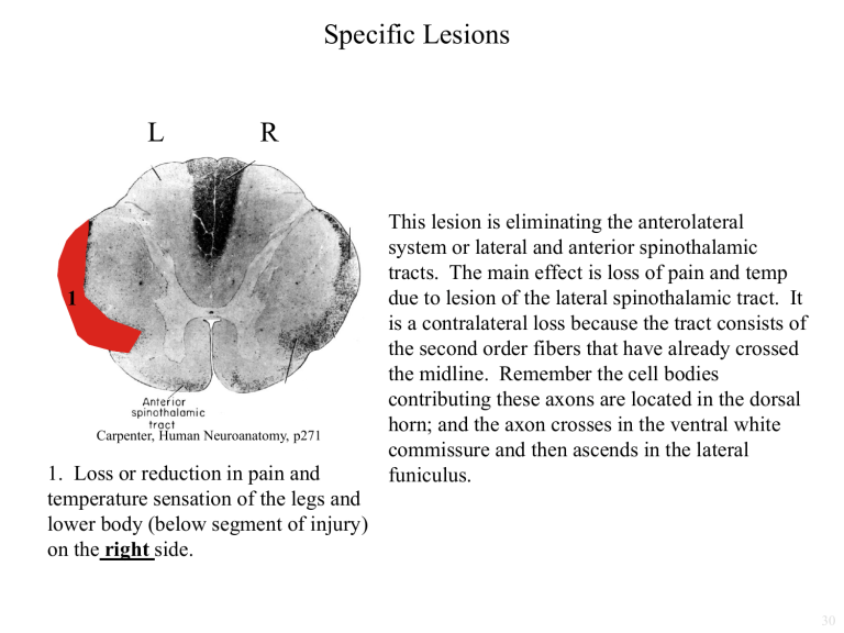
Specific Lesions
L R
1
Carpenter, Human Neuroanatomy, p271
1. Loss or reduction in pain and temperature sensation of the legs and lower body (below segment of injury) on the right side.
This lesion is eliminating the anterolateral system or lateral and anterior spinothalamic tracts. The main effect is loss of pain and temp due to lesion of the lateral spinothalamic tract. It is a contralateral loss because the tract consists of the second order fibers that have already crossed the midline. Remember the cell bodies contributing these axons are located in the dorsal horn; and the axon crosses in the ventral white commissure and then ascends in the lateral funiculus.
30
Specific Lesions
L
2
R
Carpenter, Human Neuroanatomy, p271
2. Bilateral loss in pain and temperature sensitivity of the body, specifically the trunk (head and face not affected)
Here you have bilateral loss of pain and temperature due to lesion of the second order fibers as they are crossing the midline prior to ascending as the lateral spinothalamic tract.
This lesion will affect primarily dermatomes at the level of the lesion and 2 segments below. It will affect those 2 segments below, because the primary afferent in this pathway ascends several spinal segments within Lissauer’s tract
(the cap of the dorsal horn) before synapsing on the second order neurons crossing here.
Dermatomes more than 2 segments below the level of the lesion will be little affected because the fibers subserving those regions have already crossed in segments below the lesion and are now traveling out laterally in the funiculus as the lateral spinothalamic tract.
31
Specific Lesions
L
3
R
Carpenter, Human Neuroanatomy, p271
3. Loss of pain and temperature sensation in right leg and trunk (begins two dermatomes below)
This lesion induces an ipsilateral loss because the cell bodies of the second order neurons in the
Anterolateral System (ALS) pathway are being eliminated. It is not contralateral because the axons have not yet crossed. The main region affected will be 2 dermatomes below the level of the lesion since the primary afferents synapsing on these second order neurons entered the spinal cord ~2 segments below and then ascended to this level in Lissauer’s tract. This lesion would affect all of the right leg if it extended in caudal spinal cord segments. If the lesion was more focal it may only affect the upper part of the leg, since neurons receiving input from the lower leg would be located in the dorsal horn of more caudal segments and the axons of those neurons would be unaffected by a focal lesion, since they are traveling out laterally in the lateral spinothalamic tract.
32
Specific Lesions
Loss of pain and temperature in the ipsilateral face is due to elimination of the spinal trigeminal tract and nucleus. This pathway has not yet crossed, since these are the primary afferents and cell bodies of the second order neurons. Loss of pain and temperature in the contralateral body occurs due to elimination of the lateral spinothalamic tract. This tract crossed back in the spinal cord, so effects are contralateral . Loss of fine discrimination touch in the ipsilateral arm is due to lesion of the cuneate nucleus. These are the cell bodies of the second order neurons, so this information has not yet crossed the midline and the cuneate is specifically for fine discrimination touch from the upper body. The ataxia would be due to lesion of the spinocerebellar tracts which are located in the lateral part of the section.
4
L R
4. Loss of pain and temperature sensation on the left side of the face, loss of pain and temperature sensation in the right leg, arm and trunk. ALSO
– dorsal column effects – some loss of fine discrimination touch in the left arm.
Also ataxia could be present.
32
Specific Spinal Cord Lesions
5.
L
5
R
Carpenter, Human Neuroanatomy, p271
5. Loss or reduction in discriminative, positional, and vibratory tactile sensations of the legs and lower body
(below segment of injury) on the right side.
The lesioned area is the Gracile
Fasciculus. This contains the central processes of the primary afferents subserving fine discrimination
(conscious proprioception) from the legs and lower part of the body. Because these are the first order neurons, the pathway has NOT yet crossed. If the pathway hasn’t crossed, the lesion is ipsilateral. This will affect the dermatomes subserving segments below the level of the lesion. This is true because the information from these lower dermatomes has already entered the dorsal horn and is now traveling within this medial location. Information from the upper part of the body is traveling more laterally.
Specific Spinal Cord Lesions
6.
L
This is an ipsilateral effect because this region contains the central processes of the primary afferent neurons. The upper body is affected because this region is the cuneate fasciculus and lower body information is carried more medially in the gracile fasciculus.
R
6. Loss or reduction in discriminative, positional, and vibratory tactile sensations of the arms and upper body
(below segment of injury) on the left side.
L
Specific Brain Stem Lesions
R L R
These lesions will result in loss of conscious proprioception (fine discrimination touch, pressure, vibratory sense) in the right leg and lower trunk. What is lesioned = Gracile fasciculus. This contains the central processes of the primary afferents ascending to ultimately synapse in the nearby Gracile nucleus. The pathway has not crossed! So the effect is ipsilateral. This region carries information from the lower part of the body. This is the dorsal column system so it mediates conscious proprioception.
L
Specific Brain Stem Lesions
R L R
Result is loss of conscious proprioception in the left leg and lower body. What is lesioned = Gracile nucleus. These are the cell bodies of the second order neurons. The pathway has not yet crossed so the effect is ipsilateral. It is lower body because it is
Gracile nucleus (not Cuneate which carries upper body info). It is conscious proprioception because these are components of the dorsal column system.
L
Specific Brain Stem Lesions
R L R
The result is a loss in conscious proprioception in the right arm and upper body. What is lesioned is the cuneate nucleus. It is ipsilateral because these are the cell bodies of the second order neurons and the pathway has not yet crossed. It is the upper body because it is the cuneate nucleus (lower body = gracile nucleus). It is conscious proprioception because this is part of the dorsal column system.
L
Specific Brain Stem Lesions
R L R
The effect is loss of conscious proprioception on the right side of the body. What is lesioned = internal arcuate fibers. These are the axons of the second order neurons just
BEFORE they have crossed the midline. The effect is ipsilateral because they have not YET crossed. It will affect both upper and lower parts of the body because axons from both gracile and cuneate nuclei are running here. Modality is conscious proprioception (fine discrimination touch) because these are part of the dorsal column system.
L
Specific Brain Stem Lesions
R L
Dorsal
R
Ventral
Result is loss of conscious proprioception (fine discrimination touch) on the right side of the body. What is lesioned = medial lemniscus. The effect is contralateral because the axons of this tract have crossed the midline (as the internal arcuate fibers). Both upper and lower parts of body will be affected, because the axons of both the gracile and cuneate nuclei contribute to this tract. The gracile neurons send their axons to the ventral portion of the tract and the cuneate neurons send their axons to the dorsal portion of the tract.
Practice Question 1
Which statement is true regarding the shaded area below?
A.
Pathway terminates on the left side of the spinal cord
B.
Pathway arises from cells in the dorsal horn on the left side of the spinal cord
C.
Pathway terminates in the left VPL
D.
Pathway arises from cells in the dorsal root ganglion on the right
E.
Pathway arises from cells in the right motor cortex
Level of section = pyramidal decussation. Lesioned = lateral spinothalamic tract. Answer = C
Answer: C
A is wrong because this pathway is ascending it will terminate above the level of this lesion, not below in the spinal cord. B is wrong because these are the second order fibers in the pathway, so they crossed the midline near the level of their cell bodies – crossed back in the ventral white commissure of the spinal cord. So the pathway actually arises from the right spinal cord. C is correct the lateral spinothalamic tract ascends to terminate in the ipsilateral
VPL of the thalamus.
D is wrong because these fibers are the second order neurons, not the first order neurons. This pathway WILL receive input from the cells in the right dorsal root ganglion. E is wrong because this is ascending sensory information, not a descending motor tract.
33
Practice Question 2
Which statement is true regarding the shaded area below?
A.
Cells project to the left VPM
B.
Pathway arises from cells in the right trigeminal ganglion
C.
Pathway arises from cells in the dorsal horn on the left side of the spinal cord
D.
Cells project to the right VPL
E.
Cells project to the right VPM
Answer: E
Area lesioned = Spinal trigeminal Nucleus. Answer = E
A is wrong because this nucleus contains the cell bodies of the second order neurons whose axons have not yet crossed the midline, so they will project to the contralateral hemisphere. B is wrong because the axons arising from the trigeminal ganglion neurons are located in the spinal trigeminal tract, just lateral to this nucleus, but also that afferent input comes from the ipsilateral side. C is wrong because the dorsal horn cells carry information about the body and do not project into the trigeminal system. D is wrong because this information is about the face and head – all somatosensory info from the face head goes to VPM. E is correct, these second order neurons will project to the contralateral thalamus.
34
Practice Question 3
Which statement is true regarding the shaded area below?
A.
Lesion results in loss of pain and temperature from the right side of the face
B.
Axons end in the left VPM
C.
Axons arise from the right trigeminal ganglion
D.
Axons arise from the right ganglion of IX
E.
Lesion results in a loss of pain and temperature from the left side of the face
Area lesioned = Spinal Trigeminal Nucleus
Left
Right
Midline
Answer: E
A is incorrect because these are the cell bodies of the second order neurons, so the pathway has not yet crossed, thus sensory deficits would be ipsilateral. B is incorrect because the axons of these neurons cross the midline in the reticular formation to terminate in the contralateral (right)
VPM. C is incorrect because the axons terminating on these second order neurons arise from the left trigeminal ganglion and left geniculate ganglion and superior ganglia of IX and X. D is incorrect for the same reason. E is correct because these second order neurons receive information from the ipsilateral head and face via the trigeminal nerves and tract.
35
Clinical Problems
Where is the lesion??
Q1. Loss of muscle joint sense of the left leg. Right trunk and leg
- total analgesia, thermoanesthesia
A1. Left hemisection at thoracic level
With a hemisection, the dorsal column system and the anterolateral system will be eliminated. The dorsal column system carries muscle joint sense
(as well as tactile discrimination, vibration, and pressure sense). This information travels ipsilateral within the dorsal funiculus of the spinal cord
(to synapse in the brainstem where the second order fibers cross the midline). The anterolateral system carries pain and temperature senses.
Analgesia = absence of pain, thermoanesthesia = absence of temperature sense. A lesion in the left spinal cord will affect the right side of the body because the fibers in the lateral spinothalamic tract have already crossed the midline. Lower body lesion, including the trunk area suggests thoracic level since dermatomes at and below the level of the lesion will be affected. Upper body is not affected, so it cannot be cervical cord.
Clinical Problems
What is the clinical problem and where is the lesion?
Q2. Analgesia and thermoanesthesia on the left hand, had previously burned the little finger of left hand but was unaware until smelled the burning skin. On Exam: Reduced pain and temperature sense involving the 8 th cervical and first thoracic dermatomes of the left hand, but tactile discrimination normal. Right arm similar but much less severe.
A2. Early signs of syringomyelia. Gliosis and cavitation interrupts ALS at the level of the 8 th cervical and 1 st thoracic segments of cord. Uneven growth of cavitation – causes one side to be worse – not that large since does not involve fasciculus cuneatus.
Syringomyelia is expansion of the CSF into the spinal cord (starting from the central canal). This causes a midline lesion and interruption of the fibers crossing from both sides to ascend as the lateral spinothalamic tract. This tract carries pain and temperature information.
Clinical Problems
Q3. 60 yr old man – raises his feet unnecessarily high and brings them to the ground in a stamping manner, stands with feet wide apart. Finding it difficult to walk, especially in dark. Asked to stand with toes and heels together and then close eyes – patient begins to sway. Loss of muscle joint sense, vibration sense of both legs.
A3. Lesion involving fasciculus gracilus in both posterior white columns – learn that patient has previously had syphilis. Diagnosis = Tabes Dorsalis.
Both legs are affected, so the lesion is bilateral . It is the lower part of the body, therefore gracilus. As part of the dorsal column system, this region conveys conscious proprioception, which includes a sense of the location of your body in space (muscle joint sense, as well as vibration, pressure, and fine tactile discrimination).


