The Nervous System
advertisement
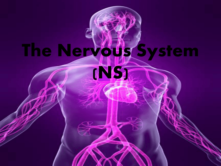
The Nervous System (NS) Introduction • 1. Distinguishing between the two major types of cells that make up the NS i. Neuron ii. Neurolgia • 2. Name and describe the two major groups of NS organs. i. Central Nervous System (CNS) ii. Peripheral Nervous System (PNS) Introduction Continued: • The nervous system is the major controlling, regulatory, and communicating system of the body. It is the center for all mental activity. It works closely with the endocrine system, and aids in maintaining homeostasis. Functions of the NS • Sensory function- Sensory receptors detect changes called stimuli that occur inside and outside the body. They monitor outside things, such as temperature, light, and sound. Inside the body they monitor pressure, pH, carbon dioxide concentration, and levels of electrolytes. All of this gathered information is called sensory input • Integration-sensory input is converted into electrical signals called nervous impulses, which are sent to the brain for processing. They are brought together to create sensations, produce thoughts, or add to memory. • • Motor function- The NS can respond by signals to the muscles once sensory and integration has taken place. The signals will cause the muscles to contract, or to the glands, causing them to produce secretions. Muscles and glands are called effectors because they cause an effect in response to directions from the NS. Types of Cells I. The Neuron (nerve cell) The Neuron (nerve cell) • The neuron conducts nerve impulses. • They are highly specialized and amitotic. • Amitotic =If they are destroyed they can not be replaced. They do not go through mitotic division. Types of Neurons Structure of the Neuron • Each neuron has a cell body (soma) • One or more dendrites, and a single axon • Dendrites and axons are cytoplasmic extensions, or processes, that project from the cell body. • They are sometimes referred to as fibers. Dendrites and Axon • The number of dendrites vary; however, they are referred to as afferent because they transmit impulses to the nerve cell. The axon transmits impulses away from the cell body so it is referred to as an efferent process. Axons • Axons are covered by a myelin sheath giving it a white appearance (white matter) found in the brain and spinal cord CNS. • The gray matter is composed of cell bodies and appears gray (no myelin sheath). • Narrow gaps between schwann cells called nodes of Ranvier • Another axon covering is the neurilemma, it is a membranous sheath on nerves of the PNS. Cell Body • The area within the neuron that contains all the components of a typical eukaryotic cell. • Genetic material (DNA) is stored here. Ganglia-(sing. Ganglion) sm. Collection of nerve cell bodies outside the CNS. The Neuroglia Cells • These cells do not conduct nerve impulses. • They support, protect, and nourish the neuron. • They are more numerous than the neuron. • They can go through mitotic division. Neuroglia Types of Neuroglia CellsMicroglial cells 1. Microglial cells- scattered throughout the CNS Function: Support neurons Phagocytize bacterial cells Removes cellular debris Forms scars in damaged areas Types of Neuroglia CellsOligodendrocytes 2. Oligodendrocytes- align along the nerve fibers Function: Provide insulating layers of myelin (Myelin sheath) in CNS Types of Neuroglia CellsAstrocytes 3. Astrocytes- Most commonly found between nerves and blood vessels. Functions: Provide structural support Join parts by their cellular processes Regulate concentrations of nutrients & ions within the tissues Form scar tissue in CNS Creates “Blood-Brain Barrier.” Types of Neuroglia CellsEpendymal cells 4. Ependymal cells- epithelial-like membrane that covers specialized brain parts (choroid plexuses) Function: Forms inner linings that enclose spaces in the brain and central canal of the spinal cord. Ependymal cells • Nerve impulse: Nerve Impulse • Functional characteristics of neurons are excitability and contractility. Excitability is the ability to react to stimuli. Conductivity is the ability to transmit a nerve impulse from one point to another. • The resting membrane is the cell membrane of a non-conducting, or resting neuron. The membrane is impermeable to the passive diffusion of Na+ (sodium)and K+ (potassium). Events Leading to a Nerve Impulse • • • • • • Neuron membrane maintains a resting potential Threshold stimulus is received Na+ channel in trigger zone of neuron open Na+ diffuses inward, depolarizes membrane K+ channel in membrane opens K+ ions diffuse outward, re-polarizing the membrane • The action potential causes local bioelectric current • A wave of action potential travels the length of the axon. Cell Membrane Potential There are 2 major components the PNS and CNS CNS & PNS • Central NS- brain and spinal cord • • • Peripheral NS- Cranial nerves (12 pairs, Spinal nerves (31 pairs) and the Autonomic nerves (parasympathetic and sympathetic) • • Sympathetic nerves stimulate the body in times of stress and crisis. They increase heart rate and forcefulness, dilate (relax airways so more oxygen can enter. Increase bp, stimulate adrenal glands to secrete epinephrine (adrenaline) , and inhibit intestinal contractions slowing digestion. • • Parasympathetic nerves normally act to as a balance to the sympathetic nerves. They slow down heart rate, contract pupils of the eye, lower bp, stimulate peristalsis to clear rectum, and increase quantity of saliva. • Called fight or flight responses. • Plexus • A plexus is a network of nerves in the PNS. Chemical Messengers • Neurotransmitters: chemical messenger released at the end of nerve cells. It either stimulates or inhibits other cells. It can be another nerve cell, gland or muscle cell. Neurotransmitters The Meninges • Meninges: 3 layers of connective tissue membranes that surround the brain and spinal cord. Spinal Cord Function of the Spinal Nerves The Brain This three-pound organ is the seat of intelligence, interpreter of the senses, initiator of body movement, and controller of behavior Cerebellum • The hindbrain includes the upper part of the spinal cord, the brain stem, and a wrinkled ball of tissue called the cerebellum Function: The hindbrain controls the body’s vital functions such as respiration and heart rate. The cerebellum coordinates movement and is involved in learned rote movements. Cerebrum • The forebrain is the largest and most highly developed part of the human brain: it consists primarily of the cerebrum two halves (hemispheres) mirror images of each other Midbrain • Uppermost part of the brainstem –midbrain Function: Controls some reflex actions and is part of the circuit involved in the control of eye movements and other voluntary movements Neurological Disorders • Neurological Disorders When the brain is healthy it functions quickly and automatically. But when problems occur, the results can be devastating. Some 50 million people in this country—one in five—suffer from damage to the nervous system. The National Institute of Neurological Disorders and Stroke (NINDS) supports research on more than 600 neurological diseases. Some of the major types of disorders include: neurogenetic diseases (such as Huntington’s disease and muscular dystrophy), developmental disorders (such as cerebral palsy), degenerative diseases of adult life (such as Parkinson’s disease and Alzheimer’s disease), metabolic diseases (such as Gaucher’s disease), cerebrovascular diseases (such as stroke and vascular dementia), trauma (such as spinal cord and head injury), convulsive disorders (such as epilepsy), infectious diseases (such as AIDS dementia), and brain tumors. References: • NINDShttp://www.ninds.nih.gov/disorders/brain_ba sics/know_your_brain.htm • Hole’s Essentials of Anatomy and Physiology 11th ed. • http://www.youtube.com/watch?v=0p_HHHO umRo • http://www.youtube.com/watch?v=4M82Ww FACLg


