Limulus molt observation study sheets.
advertisement
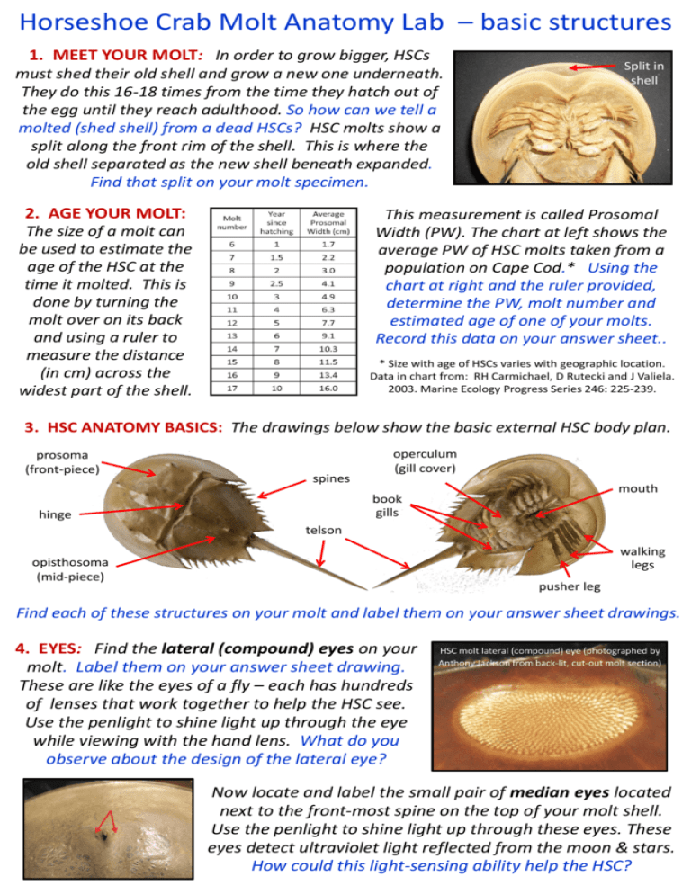
Horseshoe Crab Molt Anatomy Lab – basic structures 1. MEET YOUR MOLT: In order to grow bigger, HSCs must shed their old shell and grow a new one underneath. They do this 16-18 times from the time they hatch out of the egg until they reach adulthood. So how can we tell a molted (shed shell) from a dead HSCs? HSC molts show a split along the front rim of the shell. This is where the old shell separated as the new shell beneath expanded. Find that split on your molt specimen. 2. AGE YOUR MOLT: The size of a molt can be used to estimate the age of the HSC at the time it molted. This is done by turning the molt over on its back and using a ruler to measure the distance (in cm) across the widest part of the shell. Split in shell This measurement is called Prosomal Width (PW). The chart at left shows the average PW of HSC molts taken from a population on Cape Cod.* Using the chart at right and the ruler provided, determine the PW, molt number and estimated age of one of your molts. Record this data on your answer sheet.. * Size with age of HSCs varies with geographic location. Data in chart from: RH Carmichael, D Rutecki and J Valiela. 2003. Marine Ecology Progress Series 246: 225-239. 3. HSC ANATOMY BASICS: The drawings below show the basic external HSC body plan. prosoma (front-piece) spines operculum (gill cover) mouth book gills hinge telson walking legs opisthosoma (mid-piece) pusher leg Find each of these structures on your molt and label them on your answer sheet drawings. 4. EYES: Find the lateral (compound) eyes on your molt. Label them on your answer sheet drawing. These are like the eyes of a fly – each has hundreds of lenses that work together to help the HSC see. Use the penlight to shine light up through the eye while viewing with the hand lens. What do you observe about the design of the lateral eye? Now locate and label the small pair of median eyes located next to the front-most spine on the top of your molt shell. Use the penlight to shine light up through these eyes. These eyes detect ultraviolet light reflected from the moon & stars. How could this light-sensing ability help the HSC? Horseshoe Crab Molt Anatomy Lab – finer details Now locate the small, claw-tipped chelicerae. just above the mouth, and the two paddle-like chilaria located at the lower end of the mouth. Both structures serve to direct food to the mouth. Label these parts on your answer sheet drawing. 5. MOUTH: One of the HSCs most unique features is its between-the-legs-located mouth. Observe the many bristles on the leg bases that surround your molt’s mouth. These are the gnathobases. Considering their location and structure, what purpose do you think they serve? 6. PUSHER LEGS: Observe one of the hind legs of your molt. This is called the pusher leg. What do you see that is different about the pusher leg (compared to the other legs)? Follow the pusher leg down to where it meets the mouth, and look for a strong spine there (in area at right arrow). These spines are used to open up clams for eating. How do you think the HSC does that? Find the part on your molt that’s labeled F in the photo at left. This little organ is called the flabellum. It holds hundreds of thousands of special sensory receptors. Considering its location, what do you think the function of these sensors might be? 7. SEXING YOUR MOLT: The underside of the HSC operculum (gill cover) holds the pores through which eggs (in adult females) and sperm (in males) come out in spawning. Observing the shape and feel of these pores can tell us which sex of HSC your molt was. This is the only way we have of telling the boys from the girls in young horseshoe crabs. Find the operculum (plate that covers the gills) on your molt. Gently lift it up and run your fingers along its underside (in the area at the pencil tip in the photo at right) Can you tell which kind of HSC you have? The circled area above shows you where to look on the underside of the molt operculum. In male HSCs, the pores will stick out like little bumps and feel hard to the touch. When you run your hand along a female’s operculum, it will feel soft and smooth. Horseshoe Crab Molt Anatomy Lab – Bonus challenge MORE on MOLTING: Replacing a shell isn’t easy. Using its old shell as a mold, the HSC grows a larger one underneath. This new shell is soft and wrinkled. As it takes in water, the front of the old shell splits. The crab crawls out leaving its old shell behind. The photo shows a series of fine zig-zag lines that can be found on the side of an HSC shell. Using the magnifying glass and light, see if you can find such lines on the front side of your molt. What do you think caused them? Zig-zag lines MOLTING THEIR GUTS OUT: When HSCs molt, along with forming all new external shell parts, they also re-form certain chitinous parts of their digestive system, including the lining of the esophagus, crop-gizzard and rectum. Observe the molt specimen in your kit that has the top shell separated from the bottom so you can see what its underside looks like (as in photo at right). Use that photo to help you find the structure pointed to on your molt specimen. Thinking about where this structure is located (including what sits above it), what do you think it might be used for? Now look at the bottom part of the underside of the top of your dissected molt (as shown in the photo at left). Notice the deeply vaulted space inside the bottom of the shell. If you look carefully inside that space, you’ll see two lines (one on each side, running from the hinge area down) each with 6 thin protruding ridges of chitin (see arrows at left). Using the underside of one of your whole molts as reference, figure out what key HSC body parts lie above these ridges, and describe how they could help those body parts work.

