Introduction Dermatoglyphics is the scientific study of epidermal
advertisement

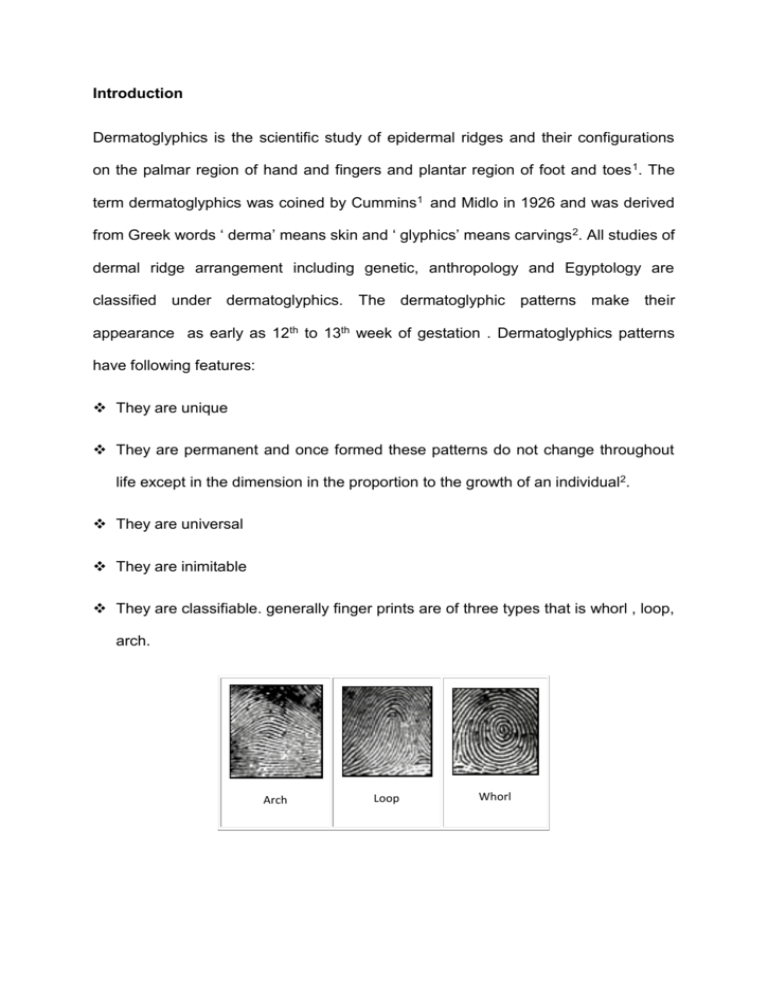
Introduction Dermatoglyphics is the scientific study of epidermal ridges and their configurations on the palmar region of hand and fingers and plantar region of foot and toes 1. The term dermatoglyphics was coined by Cummins1 and Midlo in 1926 and was derived from Greek words ‘ derma’ means skin and ‘ glyphics’ means carvings 2. All studies of dermal ridge arrangement including genetic, anthropology and Egyptology are classified under dermatoglyphics. The dermatoglyphic patterns make their appearance as early as 12th to 13th week of gestation . Dermatoglyphics patterns have following features: They are unique They are permanent and once formed these patterns do not change throughout life except in the dimension in the proportion to the growth of an individual2. They are universal They are inimitable They are classifiable. generally finger prints are of three types that is whorl , loop, arch. Arch Loop Whorl Development of dermatoglyphic pattern is under genetic control .this is evident from the clear resemblance of dermatoglyphics among related person. Dermatoglyphics as a diagnostic aid is now well established in a number of diseases , which have a strong hereditary basis , and is employed as a method of screening abnormal anaomalies3,4 . Myocardial Infarction is almost always caused by coronary artery disease (CAD). The most common cause of myocardial ischemia is atherosclerotic disease of an epicardial coronary artery or arteries sufficient to cause a regional reaction in myocardial blood flow and inadequate reperfusion of the myocardium supplied by the involved coronary arteries. Coronary artery is the leading cause of mortality and morbidity worldwide , with >4.5 million deaths occurring in the developing world. Despite a recent decline in developed countries both CAD mortality and the prevalence of CAD risk factors continue to rise in rapidly in developing world.5 In the United States, diseases of the heart are the leading cause of death, causing a higher mortality than cancer. At least 250000 people die of a heart attack before they reach the hospital.6 IHD is likely to become the most common cause of death worldwide by 2020. 6,7 The etiology of CAD is multifactorial with genetics playing an important role. The knowledge of dermatoglyphics in patients with CAD can be utilized to find out the genetic correlation. Thus, with regard to high incidence of CAD in the world, the existence of such relation might be important in screening program for prevention of myocardial infarction. Therefore, there is a scope of further research in this field. The present study is a step towards the same. AIMS : To do a comparative study of the dermatoglyphics (finger tip pattern) in patients with myocardial infarction and control group. To assess the usefulness of finger tip pattern in serving as a predictor for myocardial infarction MATERIAL AND METHODS The present study was carried out in the Department Of Anatomy , MGM Medical College Indore Madhya-pradesh The study was conducted on 200 subjects out of these 100 were confirmed cases of myocardial infarction having coronary heart disease.The total number of controls were 100 and having no clinically proven CAD. The age group selected for cases and control were between 40-75 years.The confirmed cases of MI were taken from the Division of Intensive Coronary Care Unit of the Department of Medicine, M.Y. Hospital and M.G.M. Medical College, Indore. The controls were taken from attendant of patients. The individuals with history/ family history of hypertension , diabetes , or any cardiac problem , mental illness , congenital abnormality were excluded from control and baseline ECG , serum TGS and serum cholesterol was done for the controls. The patient and control group were explained about the purpose of study and written consent was taken from them. The ink method as described by Cummins and Midlo was used for the study for taking the fingerprints.8 It required ink pad, kores duplicating ink, roller, proforma for taking fingerprints, magnifying lens, soap, cardboard. The hands were thoroughly washed with soap before taking prints and well dried. Then requisite amount of ink was placed on ink pad and evenly distributed with the help of roller. Then each finger of both hands was rolled with medial side to laterally on the pad or cardboard and then placed on paper with one lateral edge and then rolled over in opposite direction. Fingerprints were recorded carefully and analyzed for pattern type. Percentage of whorl, loop and arches were compared into two groups. STATISTICS For each subject 10 fingerprints were taken. Overall 1000 fingerprints for cases and 1000 fingerprints for control group was obtained for analysis. In statistical analysis SPSS software was used and large sample test Z test was used. For the comparison of percentage of different finger prints taken Z test was used for difference of proportion. For calculation of mean age Z test for difference of means was calculated. RESULTS In this study 200 persons were selected according to criteria given in the material and methods. Out of 200 persons, 100 cases who were known cases of CAD were treated as Study Group and 100 were Healthy Controls. The age group selected for both cases and controls was between 40 to 75 years. Out of 100 cases, 84 were males and 16 were females. The mean age of cases and controls for male and female is as follows: Total male cases : Mean age Total number of females cases 52.33 ± 9.08. : 16 Mean age Mean age of total no. of cases 84 : : 59.25±7.16 53.44±9.10 Total number of controls were : 100 74 were males and 26 were females Total number of male controls : 74 Mean age of male Total number of female controls : : Mean age of female controls Mean age of total no. of controls 26 : : 57.70±7.95 50.32±8.33 51.57 ± 7.99. Table No. 1 shows comparison of total number of different types of fingerprints in cases (MI patients) and controls. It is observed that total number of whorls in patients with myocardial infarction were 39.6% and total number of whorls in control is 25.4. The P value < 0.001, it means that total number of whorls was significantly higher in patients with myocardial infarction as compared to control group. While there was less number of loops (51.8%) in patients with myocardial infarction as compared to control group (68.0%). The P value < 0.001, means that total number of loops is significantly lower in the patients with myocardial infarction. Table No. 1 Comparison of the total number of finger prints between patients and control group Type of finger prints Cases Controls P value Significance (%) (%) Whorls 39.6 25.4 < 0.001 Significant Loops 51.8 68.0 < 0.001 Significant Arches 8.6 6.6 > 0.001 Not significant Since there was significant difference in total number of loops and whorls in the two groups, they were further compared in individual digit. Table No. 2(a) and 2(b) shows comparison of percentage of whorls in individual digit in two groups. Table No. 2(a) Comparison of Percentage of Whorls in Each Digit in Two Groups of Right Hand Group Right hand Index Middle Thumb Little Ring finger finger finger finger Whorls Cases 48 42 28 64 42 Controls 22 26 26 38 20 P value < 0.001 > 0.001 > 0.001 < 0.001 < 0.001 Significant Not Not Significant Significant significant significant Significance Table No. 2(b) Comparison of Percentage of Whorls in Each Digit in Two Groups of Left Hand Group Left Hand Index Middle Thumb Little Ring finger finger finger finger Whorls Cases 38 32 22 50 38 Controls 20 34 18 32 18 P value < 0.001 > 0.001 > 0.001 > 0.001 < 0.001 Significant Not Not Not Significant significant significant significant Significance WHORLS All the digits in patients shows higher percentage of whorls. For the right hand, the difference being significant statistically in right thumb, right ring finger and right little finger, there value in cases were 48%, 64% and 42% respectively for cases and 22%, 38% and 20% respectively for the control group, which is statistically highly significant (P<0.001). For the left hand, the difference being significant in left thumb and little finger, there values for cases were 38% and 38% respectively and for control group, they were 20% and 18% respectively. It was statistically highly significant (P<0.001). P value is < 0.001 which shows that whorls were significantly higher in patients of myocardial infarction as compared to controls. Table No. 3(a) and 3(b) shows comparison of percentage of loops in individual digit in two groups. Table No. 3(a) Comparison of Percentage of Loops in Each Digit in Two Groups of Right Hand Group Right hand Index Middle Thumb Little Ring finger finger finger finger Loops Cases 42 40 72 34 48 Controls 78 62 72 46 72 P value < 0.001 < 0.001 > 0.001 > 0.001 < 0.001 Significant Significant Not Not Significant significant significant Significance Table No. 3(b) Comparison of Percentage of Loops in Each Digit in Two Groups of Left Hand Group Left Hand Index Middle Thumb Little Ring finger finger finger finger Loops Cases 60 56 68 54 44 Controls 72 54 78 62 84 P value > 0.001 > 0.001 > 0.001 > 0.001 < 0.001 Not Not Not Not Significant significant significant significant significant Significance LOOPS The percentage of loops were significantly lower in right thumb, right index finger and right little finger, they were for the cases were 42%, 40% and 48% respectively and for control group, they were 78%, 62% and 72% respectively. It was statistically highly significant (P<0.001). For the left hand, loops were significantly lower in the left little finger, their values for the cases were 44% and for control group it was 84% respectively. It was statistically highly significant (P<0.001). P value is < 0.001 which shows that loops were significantly lower in patients of myocardial infarction as compared to controls. Table No. 4 shows comparison of different types of finger tips in controls and CAD patients. Table No. 4 Percentage of Persons Showing Different Finger Tip Patterns Finger tip pattern Cases Controls P value Significance (%) (%) (n=100) (n=100) Whorls 90 60 < 0.001 Significant Loops 94 98 > 0.05 Not significant Arches 22 20 > 0.05 Not significant It shows that significantly more number of CAD patients shows presence of whorl in one or more finger as compared to control. It was 90% in cases and 60% in control (p value < 0.001). The difference in incidence of loops and arches was not statistically significant. DISCUSSION The present study recorded the finger print pattern of all 10 fingers of 200 persons of age group 40-75 years. Out of these 200 persons, 100 persons were known cases of CAD and 100 were the healthy controls. The total number of fingerprints pattern recorded was 2000, out of these 1000 were for cases and 1000 were for controls. Fingerprints patterns of both cases and controls were analyzed by using Z test. The percentage of total number of different types of finger prints, comparison of fingerprint patterns between case and control in individual digit and comparison of different types of fingertip patterns in control and MI patients. It was found that the total number of whorls is significantly higher in patients with MI and total number of loops are significantly lower in patients with CAD. Such difference were significant only in right thumb, left thumb, right ring finger and left little finger. Similarly, loops were significantly less in right thumb, right index finger, right and left little finger. It is observed that 90% of CAD patients shows whorls on one or more finger as compared to 60% controls. The observations in this study is supported by observation found in earlier studies. Rashad & Mi9 in 1975 reported significantly higher frequency of whorl in myocardial infarction patients. In 1978, Rashad et al10 also reported significantly higher frequency of whorls and less number of loops in his patients and they also found higher absolute ridge count in patients with myocardial infarction. In 1981 Anderson et al11 also noted decrease in loop pattern and increase in whorl pattern in myocardial infarction but not statistically significant difference when compared with controls. Rao12 in 1995 reported that person who have predominant whorls in fingerprints have higher incidence of widespread arterial and venous thrombosis. Bhatt13 in 1996 presented data showing significant higher incidence of whorl and lower incidence of loops in patients with MI. In 1994 Kaseem & Mokhtar14 reported significant association in finger tip pattern, ridge count, and palm pattern in patient with CAD and control group. In 1998 Mishra et al 15 reported the significant increase in whorls in patient with coronary heart disease. In 2000, Dhall et al16 reported increased incidence of whorl and less number of loops in patients with MS as compared to control group. In 2002, Jalali & Hajian- Tilaki17 reported that increase number of arch type of finger tip pattern in patient with MI. In 2012 Chimne & Ksheersagar18 reported decreased loops and increased whorls in CAD patients. So observations of present study add support to these earlier observations. It suggests that an increase risk of myocardial infarction. All these studies done regarding fingertip pattern in patients with MI indicate relation between the dermatoglyphics pattern as indicator of genetic susceptibility in incidence of myocardial infarction. Such persons with these fingerprint patterns may be identified with this simple and economical technique for having fingerprint. These persons may be screened regularly for CAD as a preventive measure. ACKNOWLEDGEMENT: It is with a deep sense of gratitude and reverence that I express my sincere thanks to my guide Dr. Mrs. Sudha Shrivastava, Professor and Head, Department of Anatomy, and Dr. Anil Bharani, Professor, Department of Medicine, for allowing me to undertake this study in the institution and I am also thankful to Pawan goyal (Statistician) & K.K. Dwivedi, Finger Print Expert, Crime Branch, S.P. Office, Indore, who helped me in analysing the finger tip patterns taken in the study. . REFERENCES: 1. Harold Cummins and Charles Midloo. Palmar and plantar epidermal configurations (Dermatoglyphics) in European Americans. Am J Phys Anthropol 1926; 9 :471-502. 2. Penros LS. Finger prints,palms and chromosomes. Nature 1963; 197: 933938. 3. Sir Francis Galton. Fingerprints. London: MacMillan & Co. 4. Schaumann B , Alter M. Dermatoglyphics in Medical Disorders. SpringerVerlag New YorK 1976, 187-189. 5. Okrainec K, Banerjee DK, Eiseberg MJ. Coronary Artery Disease in developing world. Amer.Heart J.;2004;148:7-15. 6. Andrew PS,Braunwald E. Harrison's Textbook of Internal Medicine. 16thed. United States of America : Mc Graw Hill Publication; 2005. 7. Kumar V,Abbas AK,Fausto N, Robbins and Cotran Pathologic Basis Of Disease.2007, 7THed, Saunders Elsevier Publication, Philadelphia, Indian Reprint, pp:571-87. 8. Cummins H, Midloo C, Finger Prints, Palms and Soles. An Introduction To Dermatoglyphics. New York ;Dovar pub.INC;1961. 9. Rashad MN, Mi MP. Dermatoglyphics traits in patients with cardiovascular disorders. Amer J Physl Anthropol 1975;42(2):281-83. 10. Rashad MN, Mi MP, Rhoads G. Dermatoglyphic studies of myocardial infarction patients .Hum Hered 1978; 28:16. 11. Anderson MW, Haug PJ, Critchfield G. Dermatoglyphic features of myocardial infarction patients. Amer J Physl Anthropol 1981; 55(4):523-27. 12. Rao UR. Anticardiolipin antibodies. Medicine Update APICON 1995;17-19. 13. Bhatt SH. New signs of myocardial infarction. Medicine Update 1996; Sept : 411-416 14. Kaseem NS, Mokhtar MM, Elbel- Bessy MF. Genetic markers in coronary heart diseas. J Egypt Public health Asso. 1994; (5-6):359-78. 15. Misra S, Dhiman SR , Kaur K . Finger Dermatoglyphics and Coronary Heart Disease. Man in India 1998; 78(1-2): 127-33. 16. Dhall U, Rathee S,Dhall A. Utility of finger prints in myocardial infarction patients. J Anat Soc India 2000; 49(2):153-54. 17. Jalili F, Hajian-Tilaki KO. A comparative study of dermatoglyphic pattern in patients with myocardial infarction and control group. Acta Medica Iranica 2002; 40(3):187-91. 18. Chimne DH,Ksheersagar DD. Dermatoglyphic patterns in angiographically proven coronary artery disease. J Anat Soc India 2012; 61(2) :262-68.


