Juvenile Rheumatoid Arthritis (JRA)
advertisement
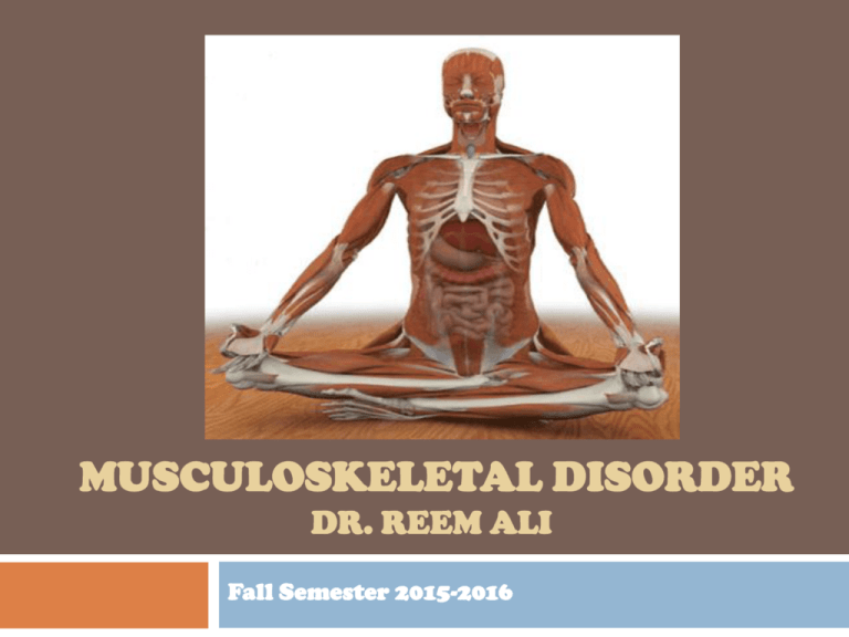
MUSCULOSKELETAL DISORDER DR. REEM ALI Fall Semester 2015-2016 Musculoskeletal The main organs and tissues that are part of the musculoskeletal system in humans are the cartilages the bones the muscles Musculoskeletal Main functions of MS are: To support & protect vital organs (the brain, heart and lungs) To keep structure and maintenance of the body spatial conformation. Allows the body to move (walking, standing, bending). Because soft tissues are resilient in children, dislocations and sprains are less common than in adults Nutrient storage (glycogen in muscles, calcium and phosphorus in bones) Musculoskeletal: Physical Assessment Inspect child undressed Observe child walking Spinal alignment ROM Muscle strength Reflexes Assessment of concerns Musculoskeletal: Physical Assessment Assessment of concerns: Pain or tenderness & Muscle spasm Masses Soft tissue swelling assess injury site at the last Musculoskeletal: Diagnostic procedures X-ray & Bone scan Alkaline phosphatase (ALP): ALP is an enzyme found mainly in bone, liver, placenta, and kidney; levels may be elevated in bone disease, fractures, trauma, or liver disease and during periods of rapid growth. Electromyography (EMG): studies the electrical activity of skeletal muscle and nerve conduction Muscle or bone biopsy Arthoscopy: direct visualization of a joint with a fiberoptic instrument Kyphosis Is an abnormally increased convex angulation in the curvature of the thoracic spine It can occur secondary to disease process such as tuberculosis, chronic arthritis Treatment Postural exercises Bracing (Milwaukee) for more marked deformity Lordosis Is an accentuation of the lumbar curvature beyond physiologic limits Lordosis is a normal observation in toddlers It may be a complication of a disease process, the result of trauma or idiopathic In older children is often seen in association with flexion contractures of the hip, obesity, congenital dislocated hip Scoliosis Is a spinal deformity which may involve lateral curvature, spinal rotation causing rib asymmetry, and thoracic hypokyphosis. It is the most common spinal deformity. It can be congenital, or it can develop during infancy or childhood, but it is most common during adolescence (peaks between 8-15 years) It may be genetic and transmitted as an autosomal dominant trait It may be multifactorial Scoliosis: Types Functional scoliosis caused by a secondary problem such as unequal leg length. The curve tends to be a C-shaped curve treated by treating the primary cause first Structural scoliosis the cause is idiopathic with a positive family history in some cases It involves a permanent curvature of spine accompanied by damage to the vertebra. The curve tends to be S-shaped curve. Scoliosis: Clinical Manifestation From standing position ( feet together and arms at sided) Unequal shoulders level Curved spinal column Uneven level of the elbows From bending position ( child bends and touch his toes): Rotation of the spine becomes more prominent. Hump in the back One shoulder blade is more prominent than other is In some cases there are back pain, fatigue and dyspnea 12 Scoliosis: Management Non-surgical management aimed to : promoting self-esteem and positive body image maintain spinal stability prevent further progression of deformity until bone growth is complete and surgical repair can be performed mild cases (less than 20%), observation and exercises- swimming is advised & Long-term monitoring. Moderate (20-40%), exercises, traction, bracing. Bracing (Milwaukee brace) is successful in halting or slowing the progression of curvatures Severe (more than 40%), bracing until the skeletal system mature and then surgical intervention Surgery includes realignment and straightening of the spine with internal fixation Internal fixation 14 External fixation: Milwaukee Brace 15 Congenital clubfoot (Talipes) Deformity of the ankle and foot Categories of Talipes Positional clubfoot (transitional, mild or postural), Syndromic (tetralogic ) clubfoot may occur from intrauterine crowding responds to simple stretching and casting. associated with other congenital abnormalities such as myelomeningocele, more severe form of clubfoot that is often resistant to treatment. Congenital clubfoot (idiopathic ) has a wide range of rigidity and prognosis Congenital clubfoot: Management goal of management: is Correction of the deformity & Maintenance of the correction until normal muscle is gained Management Casts begin immediately or shortly after birth and continue until marked overcorrection is reached. Weekly manipulation and cast changes proceed for the first 6 to 12 weeks of life. Surgery If casting and manipulation are not successful Followed by brace and cast Congenital clubfoot: Management Nursing Care Observation of skin and circulation (particularly important in young infants because of their normally rapid growth rate): swelling in the toes, foot temperature Parents need to understand the diagnosis, the overall treatment program, the importance of regular cast changes Developmental/congenital hip dysplasia/dislocation (DDH/CHD) Improper formation and function of the hip socket. Cause of DDH is unknown, but there are predisposing factors such as: Genetic factors & birth order Physiologic factors: maternal hormone Mechanical factors: intrauterine position (breech), oligohydraminos, twining and fetus size, delivery type, postnatal positioning DDH occurs more commonly in females. DDH/CHD: Degrees Acetabular dysplasia (or preluxation) Subluxation Dislocation DDH/CHD: Degrees Acetabular dysplasia (or preluxation) The mildest form The femoral head remains in the acetabulum DDH/CHD: Degrees Subluxation Accounts for the largest percentage of DDH. It implies incomplete dislocation The femoral head remains in contact with the acetabulum, but a stretched capsule and ligamentum tears cause the head of the femur to be partially displaced. Dislocation Femoral head loses contact with the acetabulum and is displaced posteriorly and superiorly over the fibrocartilaginous rim DDH/CHD: diagnosis Ortolani test Abduction of the thighs with external rotation. If the femoral head can be felt to slip forward into the acetabulum on pressure from behind, it is dislocated (positive Ortolani sign) Sometimes an audible “clunk” can be heard. Barlow test Pressure from the front If the femoral head is felt to slip out over the posterior lip of the acetabulum and immediately slips back in place when pressure is released, there is dislocation or “unstable” (positive Barlow sign) DDH/CHD: diagnosis Other signs of DDH are: Shortening of the limb on the affected side (limping and toe walking Positive Trendelenburg sign or gait) Asymmetric thigh and gluteal folds Broadening of the perineum(in bilateral dislocation) Ultrasounds DDH/CHD: Management Newborn to six months Pavlik harness The harness is used to maintain the infant’s hips in flexion and abduction and external rotation Pavlik harness device is to be worn continuously. The child in a Pavlik harness needs special attention to skin care because the infant’s skin is sensitive and the harness may cause irritation. 6-8 months: Gradual reduction by traction is used for approximately 3 weeks. If the hip is not reducible, an open operative reduction is performed. Following reduction the child is placed in a hip spica cast for 2-4 months DDH/CHD: Management Older child: Operative reduction After cast removal and before weight bearing is permitted, range-of-motion exercises & rehabilitative measures The former practice of doubleor triple-diaper for DDH is not recommended because it promotes hip extension, thus worsening proper hip development DDH/CHD: Nursing Diagnosis & Management Knowledge deficit regarding care of harness or cast Impaired physical mobility Risk for impaired skin integrity related to pressure from casts or braces Risk for altered skin perfusion due to casts or braces Risk for altered growth and development due to limited mobility Nursing Management Compliance with corrective devices by parents Not removed for bathing Prevent skin irritation Cast care & Diaper area Because DDH may reoccur it is important to follow-up until the child reaches skeletal maturity Juvenile Rheumatoid Arthritis (JRA) (self-reading) Is an inflammatory disease of the body joints and sometimes affects blood vessels and connective tissue Unknown cause but a slight tendency to occur in families Peak ages : 2 – 5 years and between 9 - 12 years of age JRA is similar to the adult disease with some distinguishing features onset before puberty a negative rheumatoid factor (RF). Juvenile Rheumatoid Arthritis (JRA): Courses Pauciarticular Polyarticular Systemic Juvenile Rheumatoid Arthritis (JRA): Pauciarticular 4 or fewer joints are affected The most common form of JRA; about half of all children with JRA have this type Affects large joints, such as the knees Girls under age 8 are most likely to develop this type of JRA. Some children have special kinds of antibodies in the blood. Antinuclear antibody (ANA) Rheumatoid factor Juvenile Rheumatoid Arthritis (JRA): Pauciarticular Eye disease affects about 20 to 30% of children with pauciarticular JRA Up to 80% of those with eye disease also test positive for ANA The disease tends to develop at a particularly early age in these children Regular examinations by an ophthalmologist are necessary to prevent serious eye problems such as iritis or uveitis Some children with pauciarticular disease outgrow arthritis by adulthood, although eye problems can continue and joint symptoms may recur in some people Juvenile Rheumatoid Arthritis (JRA): Polyarticular 30% of all children with JRA have polyarticular disease 5 or more joints are affected. The small joints (hands and feet) are most commonly involved, though large joints may be affected symmetrical; that is, it affects the same joint on both sides of the body Some children have an antibody in their blood called IgM rheumatoid factor (RF) These children often have a more severe form of the disease Juvenile Rheumatoid Arthritis (JRA): Systemic (Still’s disease) Joint swelling, & fever and a light skin rash May also affect internal organs such as the heart, liver, spleen, and lymph nodes Almost all children with this type of JRA test negative for both RF and ANA Affects 20% of all children with JRA. A small percentage of these children develop arthritis in many joints and can have severe arthritis that continues into adulthood. Juvenile Rheumatoid Arthritis (JRA): Clinical Manifestations Joint swelling Stiffness that typically is worse in the morning or after a nap Pain may limit/loss movement of the affected joint Commonly affects the knees and joints in the hands and feet One of the earliest signs of JRA may be limping in the morning because of an affected knee. Besides joint symptoms, children with systemic JRA have A high fever and a light skin rash. The rash and fever may appear and disappear very quickly. Swelling in the lymph nodes located in the neck and other parts of the body In some cases (< 50%), internal organs (heart and, very rarely, the lungs) may be involved. Juvenile Rheumatoid Arthritis (JRA): Clinical Manifestations Eye inflammation Sometimes occurs in children with pauciarticular JRA. Not present until some time after a child first develops JRA Typically, there are periods when the symptoms of JRA are better or disappear (remissions) and times when symptoms are worse (flare-ups). Growth problems Depending on the severity of the disease and the joints involved it may cause joints to grow unevenly or to one side. causing one leg or arm to be longer than the other. Overall growth may also be slowed. Juvenile Rheumatoid Arthritis (JRA): Diagnosis Diagnosis of JRA is based on: Age of onset Arthritis in one or more joints for 6 weeks or longer Exclusion of other etiologies. Laboratory tests-- Blood may be taken to test for RF and ANA, and to determine the Erythrocyte Sedimentation Rate (ESR). positive RF is detected in just 10% of the cases. The RF test helps the doctor tell the difference among the three types of JRA. ANA is found in the blood more often than RF, and both (ANA & RF) are found in only a small portion of JRA patients. ESR indicates inflammation in the body. Not all children with active joint inflammation have an elevated ESR. Lab. tests may include elevated WBCs Juvenile Rheumatoid Arthritis (JRA): Therapeutic Management • Preserve joint function, • Prevent physical deformities, and • Relieve symptoms Goals • Nonsteroidal antiinflammatory drugs • Diseasemodifying antirheumatic drugs • Corticosteroids • Biologic agent • Physical therapy Treatment • Reye Syndrome Free of treatment related harm Juvenile Rheumatoid Arthritis (JRA): Treatment NSAIDs: Aspirin, ibuprofen; may cause Reye’s Syndrome Disease-modifying anti-rheumatic drugs (DMARDs): most given in combination with NSAIDs to slow the progression of JRA Corticosteroids: to control severe symptoms; can interfere with a child's normal growth, a round face, weakened bones, and increased susceptibility to infections. Biologic agents: Etanercept (Enbrel) blocks the actions of tumor necrosis factor, a naturally occurring protein in the body that helps cause inflammation Physical therapy: Exercise to maintain muscle tone and preserve and recover the range of motion of the joints; rest of affect body part and heat application Juvenile Rheumatoid Arthritis (JRA): Side effect of Aspirin Bleeding Stomach upset Liver problems Reye’s Syndrome: Is sudden (acute) brain damage (encephalopathy) & liver function problems Abnormal accumulations of fat begin to develop in the liver and other organs of the body, along with a severe increase of pressure in the brain Without proper treatment death is common within a few days Juvenile Rheumatoid Arthritis During painful episodes of the disease Proper positioning is important to support and protect affected joints. Isometric exercises and passive range-ofmotion exercises will prevent contractures and deformities. Swimming in warm water provides strengthening and range-of-motion exercises while protecting the joints. After discharge routine ophthalmologic examinations Swimming Cerebral palsy (CP)* It is a group of non-progressive disorders (meaning the brain damage does not worsen, but secondary orthopedic difficulties are common) of motor neuron impairment that result in motor dysfunction may be accompanied by perceptual problems, language deficits, and intellectual involvement. The disabilities usually result from injury to the cerebellum, basal ganglia, or motor cortex. The exact cause is unknown; it may result from injury to the brain before, during, or shortly after birth Cerebral palsy (CP) Risk factors include: Prematurity LBW Asphyxia Infections (meningitis, encephalitis) Head injuries Metabolic problems such as hyperbilirubinemia and hypoglycemia Sever dehydration Cerebral palsy (CP): Types Spastic Dyskinetic/ athetoid Ataxic Mixed type/dystonic Cerebral palsy (CP): Types Spastic : (most common type) May involve one or both sides Hypertonicity with poor control of posture, balance, and coordinated motion (rigid & jerky movement). Impairment of fine and gross motor skills Dyskinetic/ athetoid: Abnormal involuntary movement Slow, wormlike, writhing (rolling & twisting) movements that usually involve the extremities, trunk, neck, facial muscles, and tongue. Involvement of the pharyngeal, laryngeal, and oral muscles causes drooling and dysarthria (imperfect speech articulation) Involuntary irregular jerking movements Cerebral palsy (CP): Types Ataxic Rapid, repetitive movements performed poorly. Disintegration of movements of the upper extremities when the child reaches for objects Mixed type/dystonic: Combination of spasticity and athetosis. Cerebral palsy (CP): Clinical Manifestations Reflex abnormalities: persistence of primitive infantile reflexes is one of the earliest clues to CP Delayed gross motor development. Alteration in muscle tone: increased or decreased resistance to passive movements. Abnormal posture :opisthotonic postures (exaggerated arching of the back) and may feel stiff on handling or dressing Possible Associated disabilities and problems: Convulsion/seizure Visual and hearing impairments Communication and speech difficulties Some may have varying levels of mental retardation. Cerebral palsy (CP): Treatment Goals of treatment: Establish locomotion, communication and self-help. Gain optimum appearance and integration of motor functions. Correct associated defects. Adaptation. Management: Provide safe environment to prevent injury. Prevent physical deformities by using braces and provide ROM exercises. Appropriate motor activities. Medications such as sedatives, muscle relaxants, anticonvulsants. Encourage self-care Occupational therapy to improve small muscles development Speech therapy Cerebral palsy (CP): Treatment Fracture: Clinical Manifestations (self-reading) Swelling, pain or tenderness Diminished functional use of the affected part; inability to bear weight Bruising, severe muscular rigidity Sometimes crepitus Less frequent neurological and vascular damage (ischemia), which can be assessed using 5 Ps ( Pain, Pallor, Pulselessness ,Paresthesia تنمل, Paralysis) Fractures in children younger than 1 year are unusual because a large amount of force is necessary to fracture their bones. A child younger than 1 year with a fracture should be evaluated for possible physical abuse or an underlying musculoskeletal disorder that would cause spontaneous bone injury Fracture: Therapeutic Management Cast: fiberglass or plaster application to immobilize affected body part Tractions :Is the direct application of force to produce equilibrium at the fracture site Distraction: involves the use of an external device to separate opposing bones to encourage regeneration of new bone & used to immobilize fractures or correct defects when the bone is rotated or angled Fracture: Cast Risk for altered tissue perfusion R/F pressure from cast Keep extremity elevated to decrease edema. assess circulation Q 15 minutes after applying the cast then hourly assess skin warmth and 5 Ps Risk for impaired skin integrity R/F pressure from cast Cast edges must smoothed/covered Cast remains in place for 4-8 weeks Discourage itching under the cast Fracture: Cast Possible concerns Unusual odor under the cast Drainage from cast Tingling وخز, numbness and swelling in the casted body part Loose or cracked cast Unexplained fever Unusual fussiness or irritability and pain Discoloration of finger or toes Musculoskeletal: Neurovascular Assessment Neurovascular checks should be done at least every 1 to 2 hours during the first 48 hours, and usually for as long as the child is in traction Pain: (location, alleviated and aggravating factors; Does the pain become worse when fingers or toes are flexed) Sensation: Can the child feel/ touch on the affected extremity Motion: Can the child move fingers or toes below area of injury / nerve injury Temperature: Is the extremity warm or cool to touch Capillary refill; Color; Pulses (distal to injury or cast) Fracture: Complications Circulatory impairment: Careful assessment of the pulses, skin color, and temperature is crucial Nerve compression syndromes (e.g., carpal tunnel syndrome, tarsal tunnel syndrome) Sensory testing with touch and pinprick evaluating motor strength by asking the child to move the unaffected joint distal to the injury Fracture: Complications Compartment syndromes is a tissue ischemia due to a compression of nerves, blood vessels, and muscle inside a closed space (compartment) within the body due to increased pressure in that part The most frequent causes are: tight dressings or casts, hemorrhage, trauma, burns, and surgery Signs & symptoms include: motor weakness and pain that does not decrease with medication the muscle may feel tight or full Burning sensation Fracture: Complications Epiphyseal damage: leads to unequal growth Infection: osteomyelitis (potential problem in open fractures, from pressure ulcers, or when bone surgery) Pulmonary emboli: blood, air, or fat emboli may produce a lifethreatening vascular obstruction and ischemia. Primary symptom is shortness of breath and chest pain. Interventions should include: Place patient in high fowlers Administer O2 and check chest X-ray

