Protein synthesis
advertisement
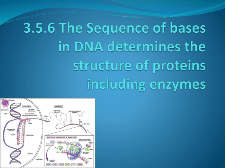
Quick recap! What can you remember from before you exam? The Central Dogma All organisms, from the simplest bacteria to ourselves, use the same basic mechanisms of reading and expressing genes – so fundamental to life it is known as the “Central Dogma”. rRNA – Ribosomal RNA Is the type of RNA found in ribosomes, during protein synthesis it provides the site where polypeptides are assembles. mRNA- messenger RNA mRNA – long single strand, is a mirror copy of one of the two strands, it is small enough to leave the nucleus, via the nuclear pores, it acts as a template for proteins synthesis, it is adapted for its function by being specific for each protein and can easily be broken down so is only manufactured when the protein is needed. tRNA- transfer RNA tRNA – small’ish ~ 80 bases, it is single stranded folded into a clover leaf shape, with one end extended further creating the point of attachment for amino acids. There are several different types of tRNA each able to carry a single amino acid. At the opposite end of the tRNA is the anticodon, this is used to line up the amino acids during protein synthesis. Comparing DNA, mRNA and tRNA DNA mRNA tRNA Double polynucleotide chain Single polynucleotide chain Single polynucleotide chain Large Medium Small Double helix Single helix Clover leaf shaped Deoxyribose Ribose Ribose A, T, C & G A, C, G & U A, C, G & U Found in nucleus Manufactured in the nucleus, but found through out the cell. Manufactured in the nucleus, but found through out the cell. Quantity is constant in a cells for a species (except....) Varies from cell to cell and with metabolic activity Varies from cell to cell and with metabolic activity Stable Unstable In- between stability! Protein synthesis - overview Transcription – production of pre-mRNA Splicing – removal of introns from the premRNAto make mRNA Translocation – mRNA moving out of the nucleus Translation – where mRNA is used as a template to which complementary tRNA molecules attached to amino acids, start to form a polypeptide chain with the help of a ribosome. Transcription in detail Transcription the steps! Task – Using page 224 and you imagination draw a cartoon strip to show how mRNA is transcribed. Posttranscriptional Modifications 5’cap – Transcription usually begins with an A or a G, in eukaryotes the terminal 5’ A or G is removed then a 5’ – 5’ linkage forms with GTP – this is called a 5’cap. It protects the 5’ end from nucleases and phoshatases on its journey out of the nucleus. 3’ poly-A tail – At the 3’ end of the mRNA a special enzyme known as poly-A polymerase adds about 250 A ribonucleiotides on, again this protects the mRNA from nucleases. Posttranscriptional Modifications DNA is made up of sections of exons which code for proteins an introns which do not. If left the introns could disrupt protein synthesis. In eukaryotic cells the introns are removed and the exons are joined together in a process known as splicing. NOTE: - After the introns have been removed the exons can be joined together in a variety of combinations – this means that a single gene can code for up to a dozen different proteins. Important point Mutations can affect the splicing of premRNA Certain disorders such as Alzheimer’s are a result of an error in splicing – why could this be a problem? Task Add splicing on to your cartoon strip. Amino acid activation Before amino acids can be used for protein synthesis, each amino acid has to be linked to a specific tRNA. Specific enzymes will only link a particular amino acid to a tRNA with a particular anticodon. This process takes place in the cytoplasm. Translation in detail http://www- class.unl.edu/biochem/gp2/m_biology/animation/gen e/gene_a3.html http://vcell.ndsu.edu/animations/translation/movieflash.htm http://highered.mcgrawhill.com/olc/dl/120077/micro06.swf Task Using page 226 add translation on to your cartoon strip. Final stage – Assembling a protein The steps following translation depend on the protein being made but usually consist of: Coiling or folding into α – helix or β – pleated sheets, into its secondary structure. Folding of the secondary structure into a 3-D shape of the tertiary structure. Sometimes the addition of different polypeptide chains and non-proteins groups to make its quaternary structure. Application stuff! You have to be able to interpret data from experimental work investigating the role of nucleic acids. Answer all of the HSW questions from page 228 – 229. Homework Work through the Exam style questions on page 234 - 235
