ANP_Course Review_Sem_1
advertisement
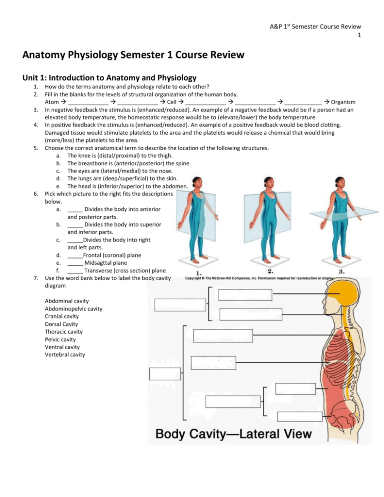
A&P 1st Semester Course Review 1 Anatomy Physiology Semester 1 Course Review Unit 1: Introduction to Anatomy and Physiology 1. 2. 3. 4. 5. 6. 7. How do the terms anatomy and physiology relate to each other? Fill in the blanks for the levels of structural organization of the human body. Atom _____________ _____________ Cell _____________ _____________ ____________ Organism In negative feedback the stimulus is (enhanced/reduced). An example of a negative feedback would be if a person had an elevated body temperature, the homeostatic response would be to (elevate/lower) the body temperature. In positive feedback the stimulus is (enhanced/reduced). An example of a positive feedback would be blood clotting. Damaged tissue would stimulate platelets to the area and the platelets would release a chemical that would bring (more/less) the platelets to the area. Choose the correct anatomical term to describe the location of the following structures. a. The knee is (distal/proximal) to the thigh. b. The breastbone is (anterior/posterior) the spine. c. The eyes are (lateral/medial) to the nose. d. The lungs are (deep/superficial) to the skin. e. The head is (inferior/superior) to the abdomen. Pick which picture to the right fits the descriptions below. a. _____ Divides the body into anterior and posterior parts. b. _____ Divides the body into superior and inferior parts. c. _____Divides the body into right and left parts. d. _____Frontal (coronal) plane e. _____ Midsagttal plane f. _____ Transverse (cross section) plane Use the word bank below to label the body cavity diagram Abdominal cavity Abdominopelvic cavity Cranial cavity Dorsal Cavity Thoracic cavity Pelvic cavity Ventral cavity Vertebral cavity A&P 1st Semester Course Review 2 Unit 2: Basic Chemistry Match the category of biological macromolecule with the descriptions below. 1. 2. 3. 4. A. Carbohydrates B. Lipids _____ Energy storage (more than one answer). _____ Monosaccharides _____ Genetic information _____ Composed of 3 fatty acids and a glycerol C. Nucleic acids D. Proteins 5. _____ Composed of amino acid chains 6. _____ Composed of nucleotides 7. _____ Enzymes 8. _____ Cushions and protects organs Match the type of carbohydrate with the structural descriptions below (HONORS). 9. 10. 11. 12. 13. 14. A. Disacchardies B. Monosaccharides C. Polysaccharides _____ Polymers of simple sugars linked together _____ Single chain or ring _____ Two monosaccharides joined together What is the main source of fuel for cellular respiration (HONORS)? _______________________________________ What is the structure of a fatty acid (HONORS)? ______________________________________________________ Types of Lipids Chart (HONORS) Type of Lipid Triglyceride Structure Function Phospholipid Steroid 15. Types of Triglyceride Chart (HONORS) Structure Name State of matter 16. The diagram to the right is an amino acid. Label the R group, amine group and carboxyl group (HONORS). 17. Which structure on the diagram in question 16 distinguishes this amino acid from another amino acids (HONORS)?______________________________________________________________________ 18. What type of bonding occurs between amino acids to form a protein (HONORS)? ________________________________________________________________________ Animal or Plant A&P 1st Semester Course Review 3 19. Types of Proteins Chart (HONORS) Type of Protein Structure Fibrous Function Globular Match the terms regarding cellular respiration to the definitions below (HONORS) A. Aerobic Respiration B. Anaerobic Respiration C. ATP 22. ____ The chemical reaction is C6H12O6 → 2C3H6O3 20. ____ Energy rich molecule that powers all cellular (lactic acid)+ 2 ATP. activities. 23. ____ Occurs when cells require large amounts of 21. ____ The chemical reaction is C6H12O6 + 6O2 ATP quickly. 6H2O + 6CO2 + 32 ATP +heat. 24. ____ Provides a high yield of ATP slowly and steadily. 25. How do enzymes increase the rate of chemical reactions? _____________________________________________________ _____________________________________________________________________________________________________ 26. How does fever affect enzyme activity?_____________________________________________________________________ ____________________________________________________________________________________________________ 27. What are two factors in the human body that influence the activity of enzymes? a. _______________________________ b. _________________________________ 28. Label the picture that shows a chemical reaction with an enzyme and which shows a chemical reaction without an enzyme. Unit 3: Tissues Matching: Use the terms below to identify the characteristics of each of the four types of tissue. A. Connective 1. 2. 3. 4. 5. 6. B. Epithelial ___ Supports, protects, binds, insulates and transports ___ Avascular but innervated ___ Creates electrochemical impulses ___ Specialized to contract and shorten ___ Apical surface ___ Irritability and conductivity C. Muscular 7. 8. 9. 10. D. Nervous ___ Cells fit closely together in sheets ___ Can be striated or non-striated ___ Consists of cells and extracellular matrix ___ Covering/lining that provides protection, absorption, excretion, filtration, and secretion A&P 1st Semester Course Review 4 11. Fill in the following chart regarding the types of epithelial tissue. Name Simple squamous Number of layers Cell Shape Function Location in body Simple cuboidal Simple columnar Stratified squamous Pseudostratified ciliated columnar Matching: Match the following types of connective tissue to their characteristics below. A. Adipose B. Blood C. Bone 13. ___ Extracellular matrix consists of calcium and collagen. 14. ___ Pulls bones. 15. ___ Transport vehicle for the cardiovascular system. 16. ___ Osteocytes, osteoblasts, osteoclasts. 17. ___ Very flexible and found at the ends of bones and in between vertebrae. 23. Fill in the following chart on the types of muscle tissue. Muscle Type Structure Function Skeletal D. Cartilage 18. 19. 20. 21. 22. E. Ligaments/Tendons ___ Insulates and protects organs. ___ Consists of dense rows of collagen fibers. ___ Chondrocytes. ___ Adipocytes store fat for cellular fuel. ___ Only connective tissue that has a fluid matrix Location Cardiac Smooth 24. The cell cycle under normal circumstances will (loosely control/tightly control) cell division. When (carcinogens/mutations) occur in protein-encoding genes that regulate (cell growth/immunity) an abnormal cell mass will develop. If the abnormal cell mass is local, slow growing and surrounded by a capsule it is (benign/malignant). If the abnormal cell mass is nonencapsulated, fast growing and aggressively invading their surroundings then it is (benign/malignant). 25. What role do carcinogens play in cancer?___________________________________________________________________ A&P 1st Semester Course Review 5 Unit 4: Integumentary System 1. Label the diagram below using the word bank: Blood Vessels, Dermis, Epidermis, Hair, Hypodermis, Nervous structure, Sebaceous Glands, Sudoriferous Glands 2. What is the histology of the following layers of the skin? A. Epidermis-____________________________________________________ B. Dermis- ______________________________________________________ C. Hypodermis- __________________________________________________ 3. Identify which layers in question 2 are vascular and avascular. Fill in the blank: Structures and functions of the skin 4. 5. 6. 7. ____________ Cell that produces a protein that gives the epidermis its protective properties. ____________ Cell that synthesizes a pigement that protects underlying cells from ultraviolet radiation. ____________ An oily substance that contains bactericidal enzymes to provide a chemical barrier to bacteria. ____________ Secretes sebum typically into a hair follicle. 8. ____________ Body temperature regulation through secretions that evaporate heat off the skin. 9. ____________ Cutaneous sensation. 10. ____________ Aids in excretion of urea and uric acid. 11. ____________ Ingest foreign substances and pathogens preventing them from penetrating into deeper body tissues. 12. ____________ Controls blood flow in relation to heat loss and heat retention. 13. Why is it important for skin to synthesize vitamin D by sunlight?_________________________________________________ A&P 1st Semester Course Review 6 Unit 5: Skeletal System 1. Label the bones on the skeleton to the right using the following words: carpals, clavicle, coccyx, cranium, fibula, femur, humerus mandible, metacarpals, metatarsals, patella, phalanges, phalanges, pelvis, radius, ribs, sacrum, scapula, sternum, tarsals, tibia, ulna, vertebral column a. Color code the bones that belong to the appendicular skeleton and those that belong to the axial. A&P 1st Semester Course Review 7 2. 3. 4. Classify the bones labeled in question 1,2 and 3 according to their shape and list them in the chart below. Short Irregular Flat 5. Which shape was predominant in the appendicular skeleton? Axial skeleton? ______________________________________ _____________________________________________________________________________________________________ What is the difference in function between the axial and appendicular skeleton? _________________________________ _____________________________________________________________________________________________________ _____________________________________________________________________________________________________ What is an articulation? _________________________________________________________________________________ Classify the bones labeled in questions 1, 2, and 3 according to their joint classification and list them in the chart below. Cartilaginous Synovial Long 6. 7. 8. Fibrous Label the bones of the cranium using the following words: foramen magnum, frontal, mandible, occipital, parietal, temporal, zygomatic Label the sections of the vertebral column using the following words: cervical, coccyx, lumbar, sacrum, thoracic, A&P 1st Semester Course Review 8 9. List three reasons why bone markings are important anatomical landmarks (HONORS) a. _____________________________________________________________________________________________ b. _____________________________________________________________________________________________ c. _____________________________________________________________________________________________ Bone Marking Matching: Match the bone marking with its description below (HONORS) A. Head B. Foramen C. Fossa D. Process 10. ___ Shallow depression, often for articular surfaces 11. ___ Bony expansion carried on a narrow neck to help form joint 12. ___ Small rounded projection for muscle attachment E. Sinus F. Tubercle 13. ___ Any bony prominence 14. ___ Round/oval openings for nerves or blood vessels 15. ___ Air filled cavity lined with mucous membrane 16. Which bone markings create the following types of synovial joints (HONORS)? a. Ball and socket:________________________________________________________________________________ b. Hinge:________________________________________________________________________________________ c. Gliding:______________________________________________________________________________________ d. Saddle:_______________________________________________________________________________________ 17. Identify the following structures on the long bone below: articular cartilage, compact bone, diaphysis, distal epiphysis, epiphyseal plate/line, foramen, medullary cavity, periosteum, proximal epiphysis, spongy bone, yellow marrow 18. List the function for the following parts of a long bone a. Red marrow:___________________________________________________________________________________ A&P 1st Semester Course Review 9 b. Yellow marrow:_________________________________________________________________________________ c. Articular cartilage:______________________________________________________________________________ d. Periosteum:____________________________________________________________________________________ e. Ligaments:_____________________________________________________________________________________ 19. Use the following terms to describe the diagram of the microscopic anatomy of compact bone: blood vessel, central (Haversian) canal (used twice), lacuna, lamellae (used twice), osteocyte, osteon, perforating (Volkmann’s) canal, periosteum 20. Next to each of the structures labeled on the diagram of the histology of the bone, briefly describe its function. 21. What is the difference in structure and function of compact and spongy bone? ____________________________________ ____________________________________________________________________________________________________ ____________________________________________________________________________________________________ 22. List the organic and inorganic components of the bony matrix. Describe how these components contribute to the function. _____________________________________________________________________________________________________ _____________________________________________________________________________________________________ A&P 1st Semester Course Review 10 Unit 6: Muscular System 1. Use the following terms to label the diagram of the anatomy of the belly of a skeletal muscle: actin myofilaments, bone, cross bridge, endomysium, epimysium, fascicle, muscle fiber, myosin myofilaments, perimysium, sarcolemma, sarcomere, sarcomere, sarcomplasmic reticulum, tendon, Z disc, Z disc 2. What is the function of the three layers of connective tissue (epimysium, perimysium, endomysium) surrounding the belly of the muscle and how do they work with tendons to create movement? _________________________________________ _____________________________________________________________________________________________________ _____________________________________________________________________________________________________ A&P 1st Semester Course Review 11 3. 4. 5. 6. 7. Label the diagram below with the following words: belly, insertion, origin When observing skeletal muscle underneath a microscope it appears (non-striated/striated). The cells of a muscle are (cylindrical, spindle) shaped, (involuntary/voluntary), and (multinucleate/uninucleate). Fill in the flow chart of muscle organization from largest to smallest. Muscle Belly _____________ _____________ Myofibril ______________ Label the diagram below of the neuromuscular junction with the following terms: axon, axon terminal, muscle fiber, myofibril, neuromuscular unit, neurotransmitter, sarcolemma, synaptic cleft The following statements are steps in the stimulation and contraction of a skeletal muscle. Organize the letters into their correct order in the blanks below. A. Myosin forms a cross bridge with actin. Q. Acetylcholine attaches to receptors on the B. Sarcoplasmic reticulum releases calcium. sarcolemma. C. Action potential is propagated down the R. Actin is covered and myosin can no longer axon of a motor neuron. attach. D. Acetylcholine diffuses into synaptic cleft. S. Acetylcholine is removed from the E. Action potential stimulates the release of receptors of the sarcolemma acetylcholine. F. Myosin requires ATP to release from actin. G. Action potential across sarcolemma stimulates sarcoplasmic reticulum. H. Action potential reaches the axon terminal. I. Calcium exposes actin. J. Myosin pulls actin towards the center of the sarcomere. K. Muscle relaxes. L. Exchange of sodium and potassium propagates an action potential across the sarcolemma. M. Muscle contracts. N. Action potential ceases across sarcolemma and sodium and potassium move back. O. Calcium returns to sarcoplasmic reticulum. P. Acetylcholine causes sodium to rush into the muscle cell and potassium to rush out. C____ E ____ ____ P____ G____ ____--> ____ J___ F S___ O____ K 8. Use the table below to explain the role of the following molecules in myoneural signaling and muscle contraction (HONORS). Role in myoneural signaling and muscle contraction Molecule ATP Toponin Tropomyosin Calcium Potassium Sodium 9. Fill in the following terms on the diagram to the right: Aerobic respiration, anaerobic respiration, and glycolysis (HONORS) 10. Label which of the processes labeled in previous question occurs in the cytoplasm on the cell and which occurs in the mitochondria (HONORS). 11. How does aerobic and anaerobic respiration work together to generate ATP during muscle contraction (Honors)?_____________________________________ ___________________________________________________________________ __________________________________________________________________ 12. Use the following terms to label the muscle man below: Masseter, Platysma, Sternocleidomastoid, Pectoralis major, Rectus abdominis, External and internal obliques, Trapezius, Latissimus dorsi, Deltoid, Biceps brachii, Triceps brachii, Gluteus maximus, Adductor group, Hamstring group, Quadriceps group, Tibialis anterior, Gastrocnemius 13. Match the antagonistic pairs below with the following movements. A. Flexion/Extension B. Medial/Lateral Rotation ____ Biceps brachii/Triceps brachii ____Deltoid/Pectoralis major ____ Right/Left Sternocleidomastoids ____Hamstring group/Quadriceps group C. Abduction/Adduction Unit 7: Fundamentals of Nervous System 1. 2. The diagram to the right demonstrates the functional classification of the nervous system. Label each classification and provide an explanation for each part below. A. ________________________________________________ ________________________________________________ ________________________________________________ B. ________________________________________________ ________________________________________________ ________________________________________________ C. ________________________________________________ ________________________________________________ ________________________________________________ Fill in chart below on the structural organization of the nervous system. 3. For the three functional classifications of the nervous system in question 1, indicate if the functional classification is part of the central or peripheral nervous system. 4. The (autonomic/somatic) nervous system innervates skeletal muscles. The (autonomic/somatic) nervous system innervates smooth and cardiac muscles. The (parasympathetic/sympathetic) nervous system stimulates an organ while the parasympathetic/sympathetic) nervous system inhibits an organ. 5. What is a neuron? _______________________________________________________________________________________ _____________________________________________________________________________________________________ Match the parts of the neuron with their function below: A. Axon B. Cell body C. Dendrites D. Myelin sheath E. Axon terminal 6. 7. 8. 9. 10. 11. _____ increase the transmission rate of nerve impulses _____conductive region; generates an action potential _____contains neurotransmitters _____ input area; receives signals from other neurons _____ input area; main nutritional and metabolic area Label the following terms on the diagram below: Cell body, Myelin sheath, Dendrites, Axon, Axon terminal. 12. In the diagram in question 11, label where the sensory portion and motor portion of the neuron. 13. How is the transmission of nerve impulse created across an axon?_______________________________________________ ____________________________________________________________________________________________________ 14. Label the diagram below of synapse using the following words: Axon terminal, neurotransmitters, receiving neuron, synaptic cleft, transmitting neuron. 15. Use the terms in question 14 to explain the process of communication between neurons. __________________ _________________________________________________ _________________________________________________ __________________________________________________ ___________________________________________________ _________________________________________________ ___________________________________________________ ____________________________________________________ _____________________________________________________ _______________________________________________________ _______________________________________________________ ______________________________________________________ ______________________________________________________ 16. A/an (action/graded) potential is short-lived, localized membrane potential that is found in (motor/sensory) receptors (HONORS). 17. A/an (action/graded) potential causes (complete/partial) depolarization of the membrane that is then propagated down the (axon/dendrites) of a (motor/sensory) neuron (HONORS). 18. How do graded potentials, action potentials and synapses work together to create communication between neurons and organs (HONORS)?____________________________________________________________________________________ _____________________________________________________________________________________________________ _____________________________________________________________________________________________________ 19. Membrane Potentials (HONORS) Type of Role in nerve impulse conduction Membrane Potential Resting Membrane Na concentration inside neuron (High/Low) K concentration inside neuron (High/Low) Depolarization (High/Low) (High/Low) Hyperpolarization (High/Low) (High/Low) Charge inside neuron Unit 9: Central Nervous System 1. Label the following structures on the diagram below: Cerebrum, Medulla oblongata, Pons, Midbrain, Hypothalamus, Thalamus, Epithalamus, Cerebellum, and Pituitary Gland. Use the structures in question 1 to identify their functions below. 10. _________________Houses the pineal gland that regulates sleep wake cycles. 11. _________________Control of breathing. 12. _________________Relay center for sensory impulses 13. _________________Regulation of body temperature, water balance and metabolism. 14. _________________Provides precise timing for skeletal muscle activity and controls our balance and equilibrium. 15. _________________Endocrine gland that releases hormones that regulate various bodily functions. 16. _________________Speech, memory logical and emotional response as well as consciousness, interpretation of sensation and voluntary movement. 17. _________________ Reflex center involved with vision and hearing. 18. _________________Control heart rate, blood pressure, breathing, swallowing, and vomiting. 19. Fill in the functions of the lobes of the cerebrum and use directional terms to describe where they are located. Lobe of Cerebrum Function Location (using directional terms) Frontal Parietal Occipital Temporal 20. Compare and contrast the function and structure of meninges with cerebrospinal fluid. _____________________________ _____________________________________________________________________________________________________ _____________________________________________________________________________________________________ 21. What are the two major functions of the spinal cord? A. _____________________________________________________________________________________________ B. _____________________________________________________________________________________________ 22. What are the structural differences between gray and white matter?_____________________________________________ _____________________________________________________________________________________________________ _____________________________________________________________________________________________________ 23. Label the steps of the reflex arc below. Unit 9: Special Senses 1. Identify the following structures of the eye: aqueous humor, cornea, lens, optic nerve, pupil, retina (photoreceptors), sclera, vitreous humor Fill in the blank: Use the words from the diagram in question one to fill in the blanks for the definitions below. 2. ______________ Firm, white fibrous layer that protects and maintains eyeball shape. 3. ______________ An opening in the center of the iris that regulates the amount of light entering the eye. 4. ______________ and _______________ Prevents the eyeball from collapsing inward. 5. 6. 7. 8. 9. ______________ Transparent, protective layer that allows light to enter the eye. ______________ Light sensitive layer of the eye that contains rods and cones. ______________ Convex structure that focuses the light entering the eye on the retina. ______________ Transmits nerve impulses to the optic cortex which results in vision. Label the diagram of the ear using the following words: Tympanum, Incus, Malleus, Stapes, Cochlea, Semicircular Canal, Pharyngotympanic (auditory) tube, Vestibule, Vestibulocochlear nerve. Write the structure from question 9 that corresponds with its function below. 10. _________________In many animals it collects and directs soundwaves, in humans it’s something to pierce. 11. _________________Connects ear to throat and when we swallow or yawn it equalizes the pressure in the middle ear cavity with the external environment. 12. _________________Where sound waves enter and is lined with ear wax which traps foreign bodies and repels insects. 13. _________________, ______________ and ____________ transmit the vibratory motion of the eardrum to the fluids in the inner ear. 14. ________________Sound waves hit this and cause it to vibrate. 15. ________________houses the hearing receptors. 16. ________________ and _____________________help with balance. 17. What is a chemoreceptor? _______________________________________________________________________________ _____________________________________________________________________________________________________ 18. What is the function and location of olfactory receptors and taste buds in the body?________________________________ _____________________________________________________________________________________________________ _____________________________________________________________________________________________________
