Day 1 – External Anatomy & the Integumentary System
advertisement
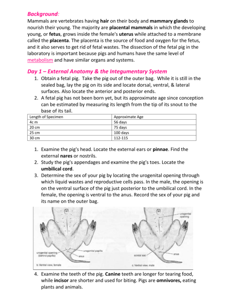
Background: Mammals are vertebrates having hair on their body and mammary glands to nourish their young. The majority are placental mammals in which the developing young, or fetus, grows inside the female's uterus while attached to a membrane called the placenta. The placenta is the source of food and oxygen for the fetus, and it also serves to get rid of fetal wastes. The dissection of the fetal pig in the laboratory is important because pigs and humans have the same level of metabolism and have similar organs and systems. Day 1 – External Anatomy & the Integumentary System 1. Obtain a fetal pig. Take the pig out of the outer bag. While it is still in the sealed bag, lay the pig on its side and locate dorsal, ventral, & lateral surfaces. Also locate the anterior and posterior ends. 2. A fetal pig has not been born yet, but its approximate age since conception can be estimated by measuring its length from the tip of its snout to the base of its tail. Length of Specimen 4c m 20 cm 25 cm 30 cm Approximate Age 56 days 75 days 100 days 112-115 1. Examine the pig's head. Locate the external ears or pinnae. Find the external nares or nostrils. 2. Study the pig's appendages and examine the pig's toes. Locate the umbilical cord. 3. Determine the sex of your pig by locating the urogenital opening through which liquid wastes and reproductive cells pass. In the male, the opening is on the ventral surface of the pig just posterior to the umbilical cord. In the female, the opening is ventral to the anus. Record the sex of your pig and its name on the outer bag. 4. Examine the teeth of the pig. Canine teeth are longer for tearing food, while incisor are shorter and used for biting. Pigs are omnivores, eating plants and animals.
