Macrovesicular or Large Droplet Fat
advertisement
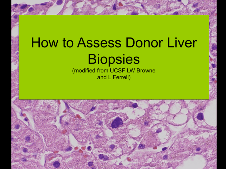
How to Assess Donor Liver Biopsies (modified from UCSF LW Browne and L Ferrell) Learn the Fat Terminology • Macrovesicular fat: TWO types – Large droplet (old term macrovesicular) – Small droplet (old term microvesicular) • Microvesicular: VERY small droplets filling hepatocytes, very rare and not easily seen on H&E (EM) Macrovesicular or Large Droplet Fat is defined as a fat droplet(s) occupying greater than one-half of the hepatocyte. Small Droplet Fat is defined as a fat droplet(s) occupying less than one-half of the hepatocyte and not displacing nuclei (This is a form of macrovesicular fat but is not as bad as large droplet fat for graft outcome.) “True” microvesicular steatosis is reserved for very small, uniform fat globules packed within hepatocytes, visible at least as patches at 10X, as is seen, for example, in fatty liver of pregnancy. Trichrome Large Droplet Fat Volume • The overall volume of large droplet fat occupying the total liver biopsy should be estimated (For example, at 10x, just look to see about what % of the liver parenchyma has large droplet fat, do not include portal zones/scar.) • This is the key factor for estimating usability of the graft >50% large droplet 10-15% large droplet Less than 5% large droplet; gylcogenated nuclei, don’t confuse with fat Small Droplet Fat • The percentage of the total cells affected by small droplet fatty change should be estimated. – The total volume of large droplet volume and of small droplet fatty change % cells together should not exceed 100%. Small and large droplet is frequently seen together in the same cells. This total may be used to determine graft usage in borderline cases. Note: On the old form, microvesicular fat was used as the term for this small droplet fat 20-30% large droplet steatosis; >60% small droplet steatosis Determining the Presence of “True” Microvesicular Fat • “True” microvesicular steatosis can be very difficult to impossible to appreciate on frozen sections. – However, it is very uncommon in donor liver biopsies, and it is highly unlikely that you will really see it so think again if you’re tempted to make this diagnosis. – One can see “tiny bubble artifact” in hepatocyte cytoplasm, not to be confused with true microvesicular fat. Important Fat Amounts Large droplet fat: <30%: Team will probably use graft 30-50%: May or may not use graft >50%: Would discard graft Small droplet fat may become important in the borderline cases Fibrosis and Inflammation • Can be estimated for form • Make sure bridging fibrosis or cirrhosis is noted when present- may preclude transplant 30-40% large droplet steatosis; portal and periportal fibrosis, possible bridging 30-40% large droplet steatosis Portal inflammationmoderate Other • Significant necrosis • Presence of tumor • Granulomata
