Microscope Skills / Microscope Lab Study Guide for Quiz
advertisement

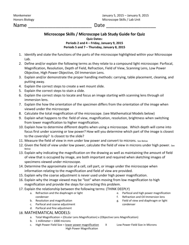
Monkemeier Honors Biology January 5, 2015 – January 9, 2015 Microscope Skills / Lab Unit Name _______________________ Date __________________ Microscope Skills / Microscope Lab Study Guide for Quiz Quiz Dates: Periods 2 and 4 – Friday, January 9, 2015 Periods 5 and 7 – Thursday, January 8, 2015 1. Identify and state the functions of the parts of the microscope highlighted within your Microscope Lab. 2. Define and/or explain the following terms as they relate to a compound light microscope: Parfocal, Magnification, Resolution, Depth of Field, Refraction, Field of View, Scanning Lens, Low Power Objective, High Power Objective, Oil Immersion Lens. 3. Explain and/or demonstrate the proper handling methods: carrying, table placement, cleaning, and putting away. 4. Explain the correct steps to create a wet mount slide. 5. Explain the correct steps to stain a slide. 6. Explain the correct steps to locate and focus an image starting with scanning lens through oil immersion lens. 7. Explain the how the orientation of the specimen differs from the orientation of the image when viewed under the microscope 8. Calculate the total magnification of the microscope. (see Mathematical Models below) 9. Explain what happens to the: field of view, magnification, resolution, brightness when switching from lower magnification to higher magnification. 10. Explain how to determine different depths when using a microscope. Which depth will come into focus first under scanning or low power? How will you determine which part of the image is closest to the coverslip? Is closest to the slide? 11. Measure the field of view in mm under low power and convert to microns. See Below 12. Given the field of view under low power, calculate the field of view in microns under high power. See Below 13. Explain why indicating the magnification on the drawing as well as maintaining the amount of field of view that is occupied by image, are both important and required when sketching images of specimens viewed under microscope. 14. Determine the approximate size of a cell, cell part, or image under the microscope when information relating to the magnification and field of view are provided. 15. Explain why the coarse adjustment is never used under high power magnification. 16. Explain why the image viewed may be “lost” when moving from low magnification to high magnification and provide the steps for correcting this problem. 17. Explain the relationship between the following terms: (THINK DEEPLY) a. b. c. d. Refraction and the diaphragm or light condenser Resolution and magnification Parfocal and coarse adjustment Parfocal and fine adjustment e. f. g. Parfocal and high power magnification Refraction and Oil Immersion lens Field of view and diaphragm or light condenser 18. MATHEMATICAL MODELS: a. b. c. Total Magnification = (Ocular Lens Magnification) x (Objective Lens Magnification) 1 millimeter = 1000 microns High Power Field Size = lower power magnification X Low Power Field Size in Microns High Power Magnification


