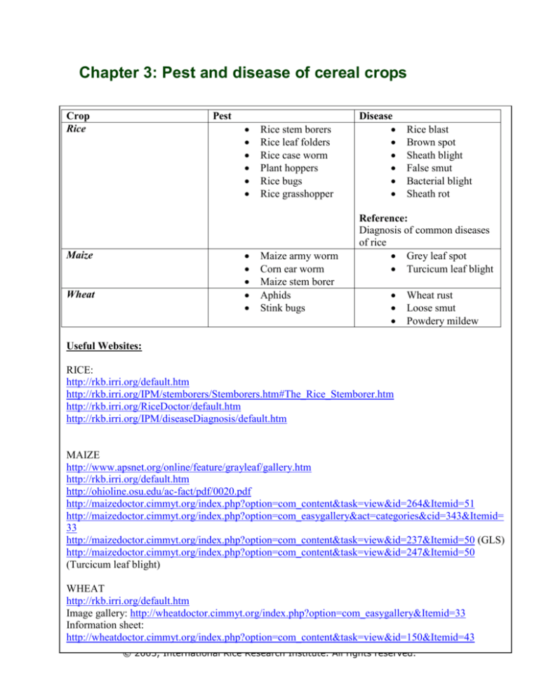
Chapter 3: Pest and disease of cereal crops
Crop
Rice
Pest
Maize
Wheat
Rice stem borers
Rice leaf folders
Rice case worm
Plant hoppers
Rice bugs
Rice grasshopper
Maize army worm
Corn ear worm
Maize stem borer
Aphids
Stink bugs
Disease
Rice blast
Brown spot
Sheath blight
False smut
Bacterial blight
Sheath rot
Reference:
Diagnosis of common diseases
of rice
Grey leaf spot
Turcicum leaf blight
Wheat rust
Loose smut
Powdery mildew
Useful Websites:
RICE:
http://rkb.irri.org/default.htm
http://rkb.irri.org/IPM/stemborers/Stemborers.htm#The_Rice_Stemborer.htm
http://rkb.irri.org/RiceDoctor/default.htm
http://rkb.irri.org/IPM/diseaseDiagnosis/default.htm
MAIZE
http://www.apsnet.org/online/feature/grayleaf/gallery.htm
http://rkb.irri.org/default.htm
http://ohioline.osu.edu/ac-fact/pdf/0020.pdf
http://maizedoctor.cimmyt.org/index.php?option=com_content&task=view&id=264&Itemid=51
http://maizedoctor.cimmyt.org/index.php?option=com_easygallery&act=categories&cid=343&Itemid=
33
http://maizedoctor.cimmyt.org/index.php?option=com_content&task=view&id=237&Itemid=50 (GLS)
http://maizedoctor.cimmyt.org/index.php?option=com_content&task=view&id=247&Itemid=50
(Turcicum leaf blight)
WHEAT
http://rkb.irri.org/default.htm
Image gallery: http://wheatdoctor.cimmyt.org/index.php?option=com_easygallery&Itemid=33
Information sheet:
http://wheatdoctor.cimmyt.org/index.php?option=com_content&task=view&id=150&Itemid=43
© 2003, International Rice Research Institute. All rights reserved.
Diagnosis of Common
Diseases of Rice
Francisco Elazegui
EPPD, IRRI
Zahirul Islam
Training Center, IRRI
Table Of Contents
Introduction .................................................................................................... 1
1. Introduction ..................................................... Error! Bookmark not defined.
2.1. Diagnosis ............................................................................................... 3
2.2. Symptoms ............................................................................................. 3
2.3. Dwarfing or stunting ............................................................................... 3
2.4. Yellowing or Chlorosis .............................................................................. 3
2.6. Spot ...................................................................................................... 4
2.7. Lesion ................................................................................................... 4
2.8. Signs ..................................................................................................... 5
2.9. Spores ................................................................................................... 5
2.10. Conidia ................................................................................................ 5
2.11. Hypha.................................................................................................. 5
2.12. Mycelium ............................................................................................. 5
2.13. Sclerotia .............................................................................................. 6
2.14. Bacterial ooze ....................................................................................... 6
2.Terms and Definitions .................................................................................... 7
2.1. Diagnosis ............................................................................................... 7
2.2. Symptoms ............................................................................................. 7
2.3. Dwarfing or stunting ............................................................................... 7
2.4. Yellowing or Chlorosis .............................................................................. 7
2.5. Rotting .................................................................................................. 8
2.6. Spot ...................................................................................................... 8
2.7. Lesion ................................................................................................... 9
2.8. Signs ..................................................................................................... 9
2.9. Spores ................................................................................................... 9
2.10. Conidia .............................................................................................. 10
2.11. Hypha................................................................................................ 10
2.12. Mycelium ........................................................................................... 10
2.13. Sclerotia ............................................................................................ 11
i
Table Of Contents
2.14. Bacterial ooze ..................................................................................... 11
3. Diagnosis of Rice Diseases ........................................................................... 13
3.1. Fungal Diseases .......................................... Error! Bookmark not defined.
3.1. Fungal Diseases ................................................................................. 13
3.1.1. Rice Blast [Pyricularia grisea (Cooke) Sacc.] ....................................... 13
3.1.2. Sheath Blight [Rhizoctonia solani Kuhn] ............................................. 14
3.1.3. Brown Spot [Bipolaris oryzae (Breda de Haan) Shoemaker] .................. 15
3.1.4. Leaf Scald [Microdochium oryzae (Hashioka &Yokogi) Samuels & I.C.
Hallett].................................................................................................... 15
3.1.5. Narrow Brown Spot [Cercospora janseana (Racib.) O. Const.] ............... 16
3.1.6. Stem Rot [Sclerotium oryzae Cattaneo] ............................................. 18
3.1.7. Sheath Rot [Sarocladium oryzae (Sawada) W. Gams & D. Hawksworth] . 19
3.1.8. Bakanae [Fusarium fujikuroi Nirenberg] ............................................. 19
3.1.9. False Smut [Ustilaginoidea virens (Cooke) Takahashi] ......................... 20
3.2. Bacterial Diseases ................................................................................. 22
3.2. Bacterial Diseases .................................... Error! Bookmark not defined.
3.2.1. Bacterial Blight [Xanthomonas oryzae pv. oryzae (Ishiyama) Swing et al.]
.............................................................................................................. 22
3.2.2. Bacterial Leaf Streak [Xanthomonas oryzae pv. oryzicola (Fang et al.)
Swing et al.] ............................................................................................ 22
3.3. Virus Diseases ...................................................................................... 23
3.3. Virus Diseases ......................................... Error! Bookmark not defined.
3.3.1. Tungro [Rice tungro bacillifor virus and spherical virus]........................ 23
3.3.2. Grassy Stunt [Rice grassy stunt virus] ............................................... 23
3.3.3. Ragged Stunt [Rice ragged stunt virus] .............................................. 24
3.4. Nematode Diseases ............................................................................... 24
3.4.1. Ufra or Stem Nematode [Ditylenchus angustus Butler] ......................... 24
3.4.2. White Tip [Aphelenchoides besseyi Christie] ....................................... 25
3.4.3. Root Knot [Meloidogyne graminicola Golden & Birchfield] ..................... 25
Contributors .................................................................................................. 26
ii
Introduction
A disease is an abnormal condition that injures the plant or causes it to function
improperly. Diseases are readily recognized by their symptoms - associated visible
changes in the plant. The organisms that cause diseases are known as pathogens. Many
species of bacteria, fungus, nematode, virus and mycoplasma-like organisms cause
diseases in rice. Disorders or abnormalities may also cause by abiotic factors such as low
or high temperature beyond the limits for normal growth of rice, deficiency or excess of
nutrients in the soil and water, pH and other soil conditions which affect the availability
and uptake of nutrients, toxic substances such as H2S produced in soil, water stress and
reduced light. In broad sense such disorders and abnormalities refer as physiological
diseases. However, here we will cover only the common diseases of rice those cause by
pathogen. Before attempting diagnosis of rice diseases it is important to understand some
frequently used terms.
1
2.1. Diagnosis
Diagnosis is the investigation or analysis of the cause or nature of a condition, situation,
or problem. Diagnosis of plant disease is the identification of specific plant disease
through the symptoms, signs, or other factors.
2.2. Symptoms
Symptoms are external manifestations of diseases or visible abnormalities arising from
disease. Symptoms may vary according to time, environment, host variety, and race of
the pathogen.
2.3. Dwarfing or stunting
Size of the entire plant or of some of its organs becomes smaller than the normal size
(Photo 1).
Photo 1: Dwarfed or stunted rice plant.
2.4. Yellowing or Chlorosis
Yellowing of normally green tissue due to chlorophyll destruction or failure of
chlorophyll formation.
Photo 2: Chlorosis in rice leaf.
3
1. Introduction
Photo 3: Rotting.
Rotting is disintegration and decomposition of host tissue (Photo 3). The rot may be dry
or soft. Dry rot is firm or dry decay, while soft rot is soft, watery decomposition.
2.6. Spot
A spot is a localized necrotic or dead area (Photo 4). It may be circular, angular, or
irregular in shape. Several spots may run together or coalesce forming larger necrotic
areas. Based on color and location, spots may be characterized viz. brown spot, black
spot, leaf spot, fruit spot etc.
Photo 4: Spots on rice leaf.
2.7. Lesion
A localized area of discolored, diseased tissue.
4
1. Introduction
2.8. Signs
Sign is the structure of the pathogen that is found associated with an infected plant. Most
of the signs are best seen and distinguished under the microscope. Examples of signs are
spores, hypha, mycelium, sclerotia, bacterial ooze, etc.
2.9. Spores
Spores are reproductive structure of the fungus (Photo 5). A spore is analogous to the
seed of plants. They can be sexual or asexual.
Photo 5: Spores.
2.10. Conidia
Conidia are asexual spores of fungus (Photo 6).
Photo 6: Conidia of fungus.
2.11. Hypha
Hypha is the single thread or filament of the fungus (Photo 7).
Photo 7: Hypha of fungus.
2.12. Mycelium
Mycelium is the body of the fungus consisting of individual filaments or hyphae (Photo
8).
5
1. Introduction
Photo 8: Fungal mycelium.
2.13. Sclerotia
Sclerotia are compact or hard masses of mycelium (Photo 9).
Photo 9: Fungal sclerotia.
2.14. Bacterial ooze
Bacterial ooze exudates bacterial cells on the surface of plant parts infected with bacteria
(Photo 10).
Photo 10: Bacterial ooze.
6
2.Terms and Definitions
2.1. Diagnosis
Diagnosis is the investigation or analysis of the cause or nature of a condition, situation,
or problem. Diagnosis of plant disease is the identification of specific plant disease
through the symptoms, signs, or other factors.
2.2. Symptoms
Symptoms are external manifestations of diseases or visible abnormalities arising from
disease. Symptoms may vary according to time, environment, host variety, and race of
the pathogen.
2.3. Dwarfing or stunting
Size of the entire plant or of some of its organs becomes smaller than the normal size
(Photo 1).
Photo 1: Dwarfed or stunted rice plant.
2.4. Yellowing or Chlorosis
Yellowing of normally green tissue due to chlorophyll destruction or failure of
chlorophyll formation.
Photo 2: Chlorosis in rice leaf.
7
2.Terms and Definitions
Photo 3: Rotting.
Rotting is disintegration and decomposition of host tissue (Photo 3). The rot may be dry
or soft. Dry rot is firm or dry decay, while soft rot is soft, watery decomposition.
2.5. Rotting
Rotting is disintegration and decomposition of host tissue (Photo 3). The rot may be dry
or soft. Dry rot is firm or dry decay, while soft rot is soft, watery decomposition.
Photo 3: Rotting.
2.6. Spot
A spot is a localized necrotic or dead area (Photo 4). It may be circular, angular, or
irregular in shape. Several spots may run together or coalesce forming larger necrotic
areas. Based on color and location, spots may be characterized viz. brown spot, black
spot, leaf spot, fruit spot etc.
8
2.Terms and Definitions
Photo 4: Spots on rice leaf.
2.7. Lesion
A localized area of discolored, diseased tissue.
2.8. Signs
Sign is the structure of the pathogen that is found associated with an infected plant. Most
of the signs are best seen and distinguished under the microscope. Examples of signs are
spores, hypha, mycelium, sclerotia, bacterial ooze, etc.
2.9. Spores
Spores are reproductive structure of the fungus (Photo 5). A spore is analogous to the
seed of plants. They can be sexual or asexual.
Photo 5: Spores.
9
2.Terms and Definitions
2.10. Conidia
Conidia are asexual spores of fungus (Photo 6).
Photo 6: Conidia of fungus.
2.11. Hypha
Hypha is the single thread or filament of the fungus (Photo 7).
Photo 7: Hypha of fungus.
2.12. Mycelium
Mycelium is the body of the fungus consisting of individual filaments or hyphae (Photo
8).
Photo 8: Fungal mycelium.
10
2.Terms and Definitions
2.13. Sclerotia
Sclerotia are compact or hard masses of mycelium (Photo 9).
Photo 9: Fungal sclerotia.
2.14. Bacterial ooze
Bacterial ooze exudates bacterial cells on the surface of plant parts infected with bacteria
(Photo 10).
Photo 10: Bacterial ooze.
11
3. Diagnosis of Rice Diseases
3.1. Fungal Diseases
3.1.1. Rice Blast [Pyricularia grisea (Cooke) Sacc.]
3.1.1.1. Symptoms
The fungus attacks all aboveground parts of the rice plant. Depending on the site of
symptom rice blast is referred as leaf blast, collar blast, node blast and neck blast. In leaf
blast, the lesions on leaf blade are elliptical or spindle shaped with brown borders and
gray centers (Photo 11). Under favorable conditions, lesions enlarge and coalesce
eventually killing the leaves. Collar blast occurs when the pathogen infects the collar that
can kill the entire leaf blade. The pathogen also infects the node of the stem that turns
blackish and breaks easily; this condition is called node blast. Neck of the panicle can
also be infected. Infected neck is girdled by a grayish brown lesion that makes panicle
fall over when infection is severe. The pathogen also causes brown lesions on the
branches of panicles and on the spikelets.
Photo 11: Different kinds of rice blasts.
3.1.1.2. Causal Organism
Mature conidia are usually three-celled or 2 septate (Photo 12) (rarely 1 or 3), pyriform
(pear-shaped), hyaline or colorless to pale olive, 19-27 x 8-10
basal appendage at the point of attachment to the conidiophore. Conidia usually
germinate from the apical or basal cells. The conidiophores are pale brown, smooth and
straight, or bending. The perfect stage is rarely found in the field.
13
3. Diagnosis of Rice Diseases
Photo 12: Rice blast conidia.
3.1.2. Sheath Blight [Rhizoctonia solani Kuhn]
3.1.2.1. Symptoms
The lesions are usually observed on the leaf sheaths although leaf blades may also be
affected. The initial lesions are small, ellipsoid or ovoid, and greenish-gray (Photo 13)
and usually develop near the water line in lowland fields. Under favorable conditions,
they enlarge and may coalesce forming bigger lesions with irregular outline and grayishwhite center with dark brown borders. The presence of several large spots on a leaf
sheath usually causes the death of the whole leaf.
Photo 13: Sheath blight disease of rice plant.
3.1.2.2. Causal Organism
Instead of spores, the rice sheath blight fungus produces sclerotia measuring usually 1 to
3 mm in diameter and relatively spherical (Photo 14). Sclerotia are formed on or near the
spots and can be easily detached from the plant. Under natural conditions, sclerotia
usually occur singly but may sometimes coalesce to form larger masses. They are whitish
when young and turn brown or dark brown when old.
Photo 14: Sclerotia of rice sheath blight disease.
14
3. Diagnosis of Rice Diseases
3.1.3. Brown Spot [Bipolaris oryzae (Breda de Haan) Shoemaker]
3.1.3.1. Symptoms
Brown spot may be manifested as seedling blight or as a foliar and glume disease of
mature plants. On seedlings, the fungus produces small, circular, brown lesions, which
may girdle the coleoptile and cause distortion of the primary and secondary leaves. In
some cases, the fungus may also infect and cause a black discoloration of the roots.
Infected seedlings are stunted or killed. On the leaves of older plants, the fungus produces
circular to oval lesions that have a light brown to gray center surrounded by a reddish
brown margin (Photo 15). On moderately susceptible cultivars, the fungus produces tiny,
dark specks. When infection is severe, the lesions may coalesce, killing large areas of
affected leaves. The fungus may also infect the glumes, causing dark brown to black oval
spots, and may also infect the grain, causing a black discoloration.
Photo 15: Brown spot symptoms on rice leaf and grains.
3.1.3.2. Causal Organism
The brown spot fungus produces multiseptate (three or more septae) conidiophore, singly
a are generally
curved, boat, or club-shaped, with 6 to 14 transverse septa or cross walls (Photo 16), 63153 x 14attachment to a conidiophore).
Photo 16: Conidia of rice brown spot disease pathogen.
3.1.4. Leaf Scald [Microdochium oryzae (Hashioka &Yokogi) Samuels &
I.C. Hallett]
3.1.4.1. Symptoms
Leaf scald disease exhibits a variety of symptoms. The characteristic symptoms are
zonate lesions of alternating light tan and dark brown starting from the leaf edges or tips
(Photo 17). The lesions usually occur on mature leaves, and are more or less oblong with
light brown halos. Individual lesions are 1-5 cm long, 0.5-1 cm broad and may enlarge to
15
3. Diagnosis of Rice Diseases
as long as 25 cm. The continuous enlargement and coalescing of lesions may result in the
blight of a large part of the leaf blade. The zonation on the lesions fades as they become
old and affected areas dry out, giving the leaf a scalded appearance.
Photo 17: Lessons of rice leaf scold.
3.1.4.2. Causal Organism
The conidia are borne on superficial stromata (compact masses of specialized vegetative
hyphae) arising on lesions. They are bow to new-moon shaped (Photo 18), single-celled
when young and 2-celled when mature, occasionally 2-3 septate, pink in mass and
hyaline under the microscope. The teleomorph produces brown, globose perithecia that
are embedded in the leaf tissue, except for the opening called ostiole. Asci are
cylindrical/club-shaped and unitunicate (an ascus in which both the inner and outer walls
are more or less rigid and do not separate during spore ejection); ascospore are fusoid,
straight or somewhat curved, 3-5 septate.
Photo 18: Conidia of rice leaf scald pathogen.
3.1.5. Narrow Brown Spot [Cercospora janseana (Racib.) O. Const.]
3.1.5.1. Symptoms
The characteristic symptoms of the disease are usually observed during the late growth
stages and are characterized by the presence of short, linear, brown lesions mainly on the
leaves, although it may also occur on leaf sheaths, pedicels, and glumes (Photo 19). The
lesions are 2 to 10 mm long and 1 mm wide. The lesions tend to be narrower, shorter and
darker brown on tolerant varieties and wider and lighter brown with gray necrotic centers
on more susceptible ones. Leaf necrosis may also occur on susceptible cultivars. A net
blotch-like pattern often forms on leaf sheaths, where the cell walls turn dark brown and
the intercellular areas are tan to yellow. The disease usually appears at mature crop
stages.
16
3. Diagnosis of Rice Diseases
Photo 19: Narrow brown spots on rice leaves .
3.1.5.2. Causal Organism
The conidiophores are emerging from stromata, solitary or in group of two or three, dark,
paler at apex, three or more septate, 88-140 x 4.5
clavate (Photo 19), 3-10 septate, 20-
Photo 20: Conidia of narrow brown spot disease pathogen.
17
3. Diagnosis of Rice Diseases
3.1.6. Stem Rot [Sclerotium oryzae Cattaneo]
3.1.6.1. Symptoms
The first symptoms are generally observed in the field after the mid tillering stage.
Initially, the disease appears as small, blackish, irregular lesion on the outer leaf sheath
near the water line. The lesion enlarges as the disease progresses with the fungus
penetrating into the inner leaf sheaths. Eventually, the fungus penetrates and rots the
culm while the leaf sheath is partially or entirely rotted (Photo 21). Infection of the culm
may result in lodging, unfilled panicles, chalky grains, and in severe cases, death of the
tiller. Brownish-black lesions appear and finally one or two internodes of the stem rot
and collapse. Upon opening infected stem, dark grayish mycelium may be found within
the hollow stem and numerous tiny, black sclerotia are embedded all over the diseased
leaf sheath tissues. Sclerotia and mycelium of the fungus are generally present inside
infected culms (Photo 22). The presence of sclerotia is usually a positive and easy way of
diagnosing the disease.
Photo 21: Lesion of stem rot disease on rice leaf sheath.
Photo 22: Rice stem rot symptom and sclerotia of pathogen.
3.1.6.2. Causal Organism
Perithecia of the fungus are found embedded in the leaf sheaths, and are dark, globose,
and 250tissue. Asci are cylindrical, short-stalked, and 104-165 x 8.7(dissolve or liquefy) at maturity and contain eight ascospores. Ascospores are fusiform,
somewhat curved, 3-septate, and 35globose, smooth, and usually 180and septate. Conidia are fusiform, three-septate, curved, 29-49 x 10produced on pointed sterigmata.
18
3. Diagnosis of Rice Diseases
3.1.7. Sheath Rot [Sarocladium oryzae (Sawada) W. Gams & D.
Hawksworth]
3.1.7.1. Symptoms
Rotting occurs on the leaf sheath enclosing the young panicles (Photo 23). The lesions
start as oblong or somewhat irregular spots, 0.5-1.5 cm long, with gray to light brown
centers surrounded by distinct dark reddish brown margins. As the disease progresses,
lesions enlarge and coalesce and may cover most of the leaf sheath. Lesions may also
consist of diffuse reddish brown discoloration in the sheath. An abundant whitish
powdery growth may be found inside affected sheaths; the leaf sheath may look normal
from the outside. With early or severe infection, the panicle may fail to emerge
completely or not at all; the young panicles remain within the sheath or only partially
emerge. Panicles that have not emerged tend to rot, and florets turn red-brown to dark
brown. Most grains are sterile, shriveled, partially or unfilled and discolored.
Photo 23: Symptoms of rice sheath rot disease.
3.1.7.2. Causal Organism
The fungus produces white mycelium, sparsely branched, septate, and measures 1.5in diameter. Conidiophores arising from the mycelium are slightly thicker than the
vegetative hyphae, branched once or twice, each time with 3-4 branches in a whorl.
Conidia are borne simply on the tip, produced consecutively, hyaline, smooth, singlecelled, cylindrical, and are 4-9 x 1-
3.1.8. Bakanae [Fusarium fujikuroi Nirenberg]
3.1.8.1. Symptoms
The classic and most conspicuous symptom of the disease is the hypertrophic effect or
abnormal elongation of plant (Photo 24). These symptoms can even be observed from a
distance. The affected plants may be several inches taller than normal plants, thin,
yellowish green and may produce adventitious roots at the lower nodes of the culm.
Diseased plants bear few tillers and leaves dry up quickly. The affected tillers usually die
before reaching maturity; when infected plants survive, they bear empty panicles.
19
3. Diagnosis of Rice Diseases
Photo 24: Bakanae infected rice plant.
3.1.8.2. Causal Organism
The fungus has micro and macroconidiophores bearing micro and macroconidia;
respectively (Photo 25). Microconidiophores are single, lateral and formed from hyphae,
while macroconidiophores consist of a basal cell bearing 2-3 apical phialides which
produce macroconidia. Macroconidia are multi-celled (3 to 7 septate), slightly curved or
bent at pointed ends, typically canoe-shaped and measure 25-60 x 2.5-4 µm.
Microconidia are one-celled, ovoid or oblong, borne singly in chains or false head on
laterally borne conidiophores and measure 5-12 x 1.5-2.5 µm. Some conidia are
intermediate, with two or three cells, oblong or slightly curved.
Photo 25: Microconidia and macroconidia of rice bakanae disease pathogen.
3.1.9. False Smut [Ustilaginoidea virens (Cooke) Takahashi]
3.1.9.1. Symptoms
The disease occurs in the field at hard dough to mature stage of the crop. The fungus
transforms individual grains of the panicle into greenish spore balls that have velvety
appearance. The spore balls are small at first and visible in between glumes, grow
gradually to reach 1 cm or more in diameter and encloses the floral parts. They are
covered with a membrane that bursts as a result of further growth. The color of the ball
becomes orange and later yellowish green, or greenish black (Photo 26). At this stage, the
surface of the ball cracks. The outermost layer of the ball is green and consists of mature
spores together with the remaining fragments of mycelium. The outer soporiferous region
is three-layered. The outermost layer is greenish black with powdery spores; the middle
layer, orange; the innermost, yellowish.
20
3. Diagnosis of Rice Diseases
Photo 26: False smut on rice grains.
3.1.9.2. Causal Organism
Chlamydospore (a thick- or double-walled asexual spore formed directly from a
vegetative hyphal cell that functions as a resistant or overwintering stage) formed on the
spore balls are born laterally on minute sterigmata (a small, arclike, usually pointed
hyphal branch or structure supporting a spore) on radial hyphae, and are spherical to
elliptical, warty, olivaceous, 3-5 x 4-6 µm; younger spores are smaller, paler, almost
smooth. Chlamydospores germinate in culture by germ tubes, which become septate and
form conidiophores bearing conidia at the tapering apex. These conidia are ovoid and
very minute (Photo 27).
Photo 27: Conidia of rice false smut pathogen.
21
3. Diagnosis of Rice Diseases
3.2. Bacterial Diseases
3.2.1. Bacterial Blight [Xanthomonas oryzae pv. oryzae (Ishiyama) Swing
et al.]
Water-soaked lesson usually starting at leaf margins, a few cm from the tip, and
spreading towards the leaf base; affected areas increase in length and width, and become
yellowish to light brown due to drying; with yellowish border between dead and green
areas of the leaf (Photo 28). It is usually observed at maximum tillering stage and
onwards. In severely diseased fields grains may also be infected. In the tropics infection
may also cause withering of leaves or entire young plants (refers as kresek) and
production of pale yellow leaves at a later stage of the growth.
Photo 28: Bacterial blight infected rice leaf and fields.
3.2.2. Bacterial Leaf Streak [Xanthomonas oryzae pv. oryzicola (Fang et
al.) Swing et al.]
Bacterial Leaf Streak first appears as short, water-soaked streaks between the veins,
which become longer and translucent and turn to light brown or yellowish brown. Thus,
large areas of the leaf may become dry due to numerous streaks. At the late stage the
disease is indistinguishable from the bacterial leaf blight.
Photo 29: Rice fields with bacterial leaf streak infested plants
22
3. Diagnosis of Rice Diseases
3.3. Virus Diseases
3.3.1. Tungro [Rice tungro bacillifor virus and spherical virus]
Plants affected by tungro exhibit stunting and reduced tillering. Their leaves become
yellow or orange-yellow, may also have rust-colored spots. The leaf discoloration starts
from the tip and may or may not extend to the lower part of the leaf blade; often only the
upper portion is discolored. Young leaves may have a mottled appearance and old leaves
show rusty-colored specks of various sizes (Photo 30). Infected plants have delayed
flowering. The panicles are small and not completely exerted, and bear mostly sterile or
partially-filled grains often covered with dark brown specks. Tungro are transmitted by
the green leafhoppers.
Photo 30: Tungro disease infected rice leaf and field.
3.3.2. Grassy Stunt [Rice grassy stunt virus]
Plants affected by this disease, show severe stunting; excessive tillering, with short leaves
that are narrow and, pale green to pale yellow in color (Photo 31). They may have newlyexpanded leaves that maybe mottled or striped and may also have numerous small,
irregular, dark brown or rust-colored spots. Brown plant hopper transmit this disease.
Photo 31: Grassy stunt infected rice hills.
23
3. Diagnosis of Rice Diseases
3.3.3. Ragged Stunt [Rice ragged stunt virus]
Affected plants show stunting that may have reduced tillering. The leaves are short, dark
green, and serrated along one or both edges giving a ragged appearance (Photo 32). The
leaf blades are often twisted form a spiral. The vein swellings appear on leaf sheaths, leaf
blade and culms and nodal branches are developed at later growth stages. Brown plant
hopper transmits the disease.
Photo 32: Ragged stunt infected rice plants
3.4. Nematode Diseases
3.4.1. Ufra or Stem Nematode [Ditylenchus angustus Butler]
Affected seedlings or plants show whitish discoloration (chlorosis) at the early stage of
infection (Photo 33). Plants than are stunted and have deformed and twisted leaves. These
plants have panicles, which are exserted, or panicles that inside the leaf sheath of the flag
leaf. However, panicles that are exserted, become twisted and deformed, with unfilled
grains.
Photo 33: Ufra disease damage symptoms at different growth stages.
24
3. Diagnosis of Rice Diseases
3.4.2. White Tip [Aphelenchoides besseyi Christie]
Affected plants shows a characteristic symptoms on leaf tips; which become chlorotic or
whitened for a distance of up to 5 cm (Photo 34). Eventually the infected leaf becomes
dry and shreds while flag leaf becomes twisted and panicles may not emerge or if
emerged may have high sterility, distorted glumes, and small and distorted kernels.
Photo 34: White tip diseased rice plants
3.4.3. Root Knot [Meloidogyne graminicola Golden & Birchfield]
Affected plants are stunted and become yellow in color. They have reduced tillering and
their most diagnostic symptom is the presence of root galls. The disease is more serious
in upland than in lowland rice.
Photo 35: Rice plant with knot or gall on roots.
25
3. Diagnosis of Rice Diseases
Contributors
Authors
Francisco Elazegui
EPPD, IRRI
Zahirul Islam
Training Center, IRRI
RoboHelp Development
Albert Dean Atkinson
Training Center, IRRI
26


