JIA
Arthritis
Juvenile idiopathic Arthritis (JIA):
•
One of the more common chronic childhood diseases
– almost as common as childhood-onset diabetes mellitus
– at least 4 times more common than sickle cell disease or cystic fibrosis
– at least 10 times more common than leukemia, hemophilia, chronic renal failure, or muscular dystrophy
•
Objective symptoms lasting minimum of 3 days in same joints
•
Pain is usually insidious onset, but can be significant enough to affect daily activities
•
Morning stiffness – due to gelling phenomenon very common
•
Night pain not common
• Can have accompanying constitutional symptoms
•
Affected joints can grow faster causing leg length discrepancies
•
Local and general growth delay seen
– Jaw
–
MCP/MTP
CRITERIA JIA
• Age of onset < 16 years
•
Arthritis (swelling or effusion, limitation of motion, tenderness or pain on motion, and/or increased heat) in one or more joints
• Duration of disease 6 weeks or longer
•
Onset type defined by type of disease in the first 6 months:
Polyarticular: 5 or more inflamed joints
Oligoarticular: < 5 inflamed joints
Systemic arthritis: Characteristic fever and rash, very inflammatory
•
Exclusion of other forms of arthritis
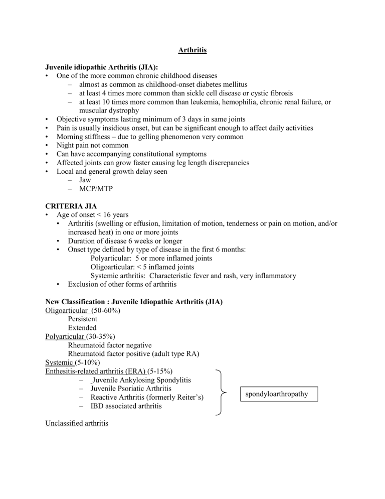
New Classification : Juvenile Idiopathic Arthritis (JIA)
Oligoarticular (50-60%)
Persistent
Extended
Polyarticular (30-35%)
Rheumatoid factor negative
Rheumatoid factor positive (adult type RA)
Systemic (5-10%)
Enthesitis-related arthritis (ERA) (5-15%)
–
Juvenile Ankylosing Spondylitis
– Juvenile Psoriatic Arthritis
–
Reactive Arthritis (formerly Reiter’s)
–
IBD associated arthritis
Unclassified arthritis spondyloarthropathy
OLIGO JIA
•
Fewer than 5 joints: larger joints and lower extremities
– Ankle or wrist arthritis and symmetric arthritis predict worse disease
•
Peak onset between 2-4 years
•
One knee most common presentation (50%)
•
Girls:Boys - 3:1
•
Labs may be normal or mild elevation of acute phase reactants
•
ANA positive (<1:640) in 65-85%
• RF usually negative
•
Associated with uveitis
•
Prognosis: Overall better outcome with less damage
– many will go into remission but subset of children will develop a polyarticular course
–
20-30% will get uveitis with the sequelae of blindness if unrecognized and untreated
UVEITIS
• Up to 21% of oligoarticular and 10% of polyarticular patients
•
ANA+ young girls with oligoarticular disease have highest risk
•
Usually asymptomatic
• Can occur anytime during the course of disease (joint disease does not correlate)
•
Complications: cataracts, glaucoma, visual impairment up to blindness, posterior synechiae, band keratopathy
POLYARTICULAR JIA
•
> 5 joints involved: Usually symmetric joints involving the large and small joints
–
DIP involvement
– Can see boutonnière and swan neck deformities
•
Age of onset: biphasic
•
Girls:Boys – up to 5:1
•
RF positive patients onset in late childhood or adolescence, similar to adult onset rheumatoid arthritis
–
Associated with rheumatoid nodules (5-10%)
– early erosive disease
– chronic course persisting into adulthood, poorer prognosis than if RF negative
•
Can see systemic symptoms, such as elevated acute phase reactants
•
Anti Cyclic Citrullinated Peptide antibodies (anti-CCP antibodies)
– up to 75% of children with RF+ poly JIA have positive anti CCP antibodies
•
Differential Diagnosis: poly JIA
Acute (within 72 hours)
–
Infection: Septic arthritis, acute rheumatic fever
– Malignancy: Leukemia, Neuroblastoma
–
Toxic Synovitis
–
Blood disorders: Hemophilia, Sickle Crisis (dactylitis)
– Trauma
Chronic (more than 4 weeks)
–
Infection: TB, Lyme disease, Parvovirus, Rubella
–
Pigmented villonodular synovitis (PVNS)
–
Other rheumatic diseases (SLE, Sarcoid, vasculitis)
• Prognosis: Guarded outcome with potential damage
– prognosis worse if RF+, small joint or hip involvement, or erosive disease
– patients who had not gone into remission by age 16 are likely to have chronic course
SYSTEMIC JIA
• Aka Still’s disease
•
Girls:Boys - 1:1
•
Peak age of onset is variable
•
Characteristic features: fever 39°C or higher for >2 weeks and salmon colored rash
•
Systemic symptoms predominate early in the disease
• Other features include anemia and serositis
•
Child looks ill during fever, but well when afebrile
•
Arthritis joint involvement variable or maybe just arthralgias
• Characteristic fever pattern: twice daily peaks with dips into below normal temps
SJIA LABS
•
Significant inflammatory markers
–
Elevated ESR, CRP, WBC, Platelets, Ferritin, d-dimers, AST, ALT
– Decreased Hgb, Albumin
•
Concern for Macrophage Activation Syndrome (MAS) (consumptive process)
–
See drop in ESR, Platelets, WBC
–
See coagulopathy (increased PT/PTT)
– Mental status changes and seizures
–
Can be life threatening: an emergency, needs immediate treatment
Differential Diagnosis SJIA
•
Periodic Fever Syndromes: PFAPA (periodic fever, aphthous ulcers, pharyngitis, and adenopathy), Familial Mediterranean Fever (FMF), Hyper IgD syndrome
• And…
•
Prognosis: Systemic JIA – Guarded outcome
–
50% will recover without problems
–
Significant morbidity and mortality can occur with macrophage activation syndrome
Characteristic Polyarticular Oligoarticular Systemic
60
≤4
10
Variable
Percent of cases 30
Number of joints
≥5 involved 1
Age at onset
Sex ratio (F:M)
Throughout childhood; peak @1-3 y.o.
3:1
Systemic involvement
Occurrence of uveitis
Systemic dz generally mild; possibility of unremitting articular dz
5%
Frequency of seropositivity
RF
ANA
10% (increase w/ age)
40-50%
Prognosis
1 In the first 6 months after diagnosis
2 In girls w/ uveitis
Guarded to moderately good
Early childhood; peak @ 1-2 y.o.
5:1
Systemic disease absent; major morbidity is uveitis
5-15%
Throughout childhood; no peak
1:1
Systemic disease often self-limited; arthritis chronic and destructive in half
Rare
Rare
75-85% 2
Rare
10%
Excellent except for eyesight Moderate to poor
Enthesitis Related Arthritis (ERA)/Seronegative Enthesitis Arthritis (SEA)/
Spondyloarthropathies
• Refers to a group of rheumatic diseases that includes:
–
Joints of axial skeleton and enthesitis as well as peripheral joint disease
•
Associated with HLA B27
– Positive in 60-75% of children with spondyloarthropathy.
–
Positive in 90% of children with juvenile ankylosing spondylitis.
–
Positive in 15% of children with psoriatic arthritis.
–
Prognosis depends on presence/absence of HLA-B27
•
Spondyloarthropathies account for approximately 13% of children with a rheum condition.
•
Frequently have enthesitis, iritis, and are RF negative (seronegative)
•
Often symptoms evolve gradually
Physical Exam
•
Examination of the entheses – pain on palpation at 2, 6, 10 o’clock positions on the patella, the tibial tuberosity, the attachment of the Achilles tendon or plantar fascia.
•
Examination of the peripheral joints – often asymmetric and involves the LE, including small joints of the toes.
•
Examination of the axial skeleton
- Abnormalities in contour of the spine in the standing and fully flexed position
- Schober test measurement <6 cm
- Pain with direct pressure over the SI joints or with compression of the pelvis.
- Pain in the costosternal/costovertebral joints
- Decreased thoracic excursion (<5 cm).
Psoriatic arthritis
Joint involvement shows radial pattern
Nail changes/ pits and psoriatic patches on the face, scalp, and extensor surfaces of the extremities.
3 to 12% of JIA patients
Age of Onset – preschool years and middle to late childhood
Slightly more common in girls; M:F of 1:2.5.
Scattered, asymmetric oligoarthritis of large and small joints, most commonly knee, finger, toes.
Dactylitis , concomitant inflammation of the flexor sheath (sausage-like swelling), seen in approximately 50% of children.
Pitting of the nails, seen in 75% of children with disease, especially if they have IP joint disease.
Chronic anterior uveitis, in approximately 17% of children. o Asymptomatic uveitis -15-20% and assoc with +ANA o Symptomatic uveitis - rare in kids and assoc with +HLA-B27
Preceded, accompanied, or followed by psoriasis o 10% of children the onset of skin and joint disease is concurrent or within a few weeks of each other. o 40% the psoriasis precedes arthritis, up to 9 years later.
o 50% the arthritis precedes the psoriasis, by up to 14 years.
There is little correlation between skin and joint disease severity.
Psoriasis affects 1-3% of general population and 20-30% of those patients have associated arthritis
Can be aggressive and damaging
Radiology findings include: Juxta-articular osteoporosis, fluffy periostitis, and occasional asymmetric erosions accompanied by periosteal new bone
IBD associated arthropathy
A noninfectious arthritis occurring before or during the course of Crohn’s disease or ulcerative colitis.
Occurs in 7 to 21% of children with IBD.
8 to 15% of children have a first degree relative with IBD. More common in the Jewish population
Two types: o 1) SI arthritis – least common; pain and stiffness in the lower back, buttocks, or thighs. Frequently accompanied by enthesitis. Does NOT correlate with gut disease activity. o 2) Peripheral polyarthritis – frequently affects LE joints. Most children have 2 or more exacerbations lasting 4 to 6 weeks. DOES correlate with GI inflammation.
Erythema nodosum – nodular paniculitis seen in the pretibial subcutaneous tissue. Often accompanied by articular pain/synovitis.
Pyoderma gangrenosum – painful, chronic ulcer with a red, raised border following minor trauma. Rare in children.
•
Labs include: Anemia, high ESR!, negative RF and ANA, Hemoccult positive.
•
X-rays show soft tissue thickening, joint effusions, periostitis, spur formation with enthesitis.
Reactive Arthritis
•
Follows salmonella, shigella, campylobacter, yersinia and chlamydial infection
• A post-infectious/reactive arthritis following diarrheal illness, urethritis or cervicitis.
•
Asymmetric arthritis (usually oligo pattern, affecting knees and ankles)
•
May see TRIAD of acute iritis, urethritis (Reiter’s syndrome- may become chronic)
• Antibiotics NOT indicated with diarrheal associated reactive arthritis
•
Antibiotic treatment may improve rate of recovery with Chlamydial reactive arthritis
•
Associated with keratoderma blennorrhagica: o May look like psoriasis or syphilis o Can occur in patches or as sterile pustules
•
Also often associated with:
•
Inflammatory eye disease
• Balanitis, oral ulceration
•
Enthesopathy especially around patella and calcaneous
•
Sacroiliitis
•
M:F ratio of 4:1.
•
90% of patients have HLA-B27.
•
Features may occur simultaneously or over 3 to 4 weeks.
•
Urethritis – inflammation of the meatus, wbc on urinalysis, or sterile, purulent discharge.
•
Conjunctivitis – erythema of bulbar conjunctiva, photophobia. Present at the onset of disease in 2/3 of patients.
•
LABs: elevated ESR (40 to 130), increased wbc with left shift, UA with 5 to 1000 wbc
•
X-rays show soft tissue swelling, juxta-articular osteoporosis, erosions/spur formation at tendon insertion sites.
•
Treatment:
•
NSAIDS, especially sulindac, are beneficial
•
Sulfasalazine in chronic cases
Juvenile Ankylosing Spondylitis
•
Onset late childhood or adolescence.
• M:F of 7:1
•
HLA-B27 is present in approximately 91% of children with JAS. o Risk to develop AS with positive HLA-B27 about 1-3%
• Arthritis – 82% of children have peripheral joint symptoms (distal>proximal, lower>upper extremities) at onset, vs. 24% who have pain, stiffness, limited ROM of the LS spine or SI joints.
•
Enthesitis – characteristic early finding
• Extra-articular manifestations: o Iritis – red, painful, photophobic eye, usually unilateral. May occur in 6 to 27% of patients. Typically lasts 6 weeks. Eye and joint disease do not correlate. o Cardiac – Mild inflammatory aortic regurgitation can occur o Pulmonary - may have decreased vital capacity on PFTs due to diminished chest expansion.
•
Labs: anemia of chronic inflammation, ESR and platelet count may be elevated, RF and
ANA negative and HLA-B27 positive.
•
X-rays - SI joint – (30 degree/oblique view pelvis) pseudowidening, sclerosis, and fusion of the SI joint. (May be seen w/in 1 year of symptom onset)
•
X-rays - LS spine – later finding; periostitis w/ flattening of the anterior margin of the vertebra and straightening of the lumbar spine.
•
X-rays: Entheses – soft tissue density changes, erosion (Achilles insertion) or spur formation (plantar fascia insertion).
•
TREATMENT: o NSAIDs, for symptomatic improvement o Oral/IV pulse steroids for severe arthritis o Intra-articular corticosteroids o PT/OT to improve range of motion o Sulfasalazine o TNF inhibitor: etanercept (Enbrel) and infliximab (Remicade), adalimumab
(Humira).
GENERAL PRINCIPLES IN THE TREATMENT OF JIA
•
Objectives: control pain and inflammation and prevent damage and disability
•
Most of the damage in polyarticular and systemic course occurs within 2 years and in oligoarticular course within 5 years
•
Can now detect cartilage damage via MRI earlier
•
Start with nonsteroidal antiinflammatory drugs (NSAIDS)
•
If significant synovitis involving multiple joints persists for 3-6 months, or radiologic evidence of destructive disease is present consider initiation of DMARD (disease modifying antirheumatic drug) o Methotrexate, Sulfasalazine, Leflunomide (Arava), Azathioprine (Imuran),
Hydroxychloroquine, cyclosporine, thalidomide, tacrolimus (FK-506), IVIG
•
If significant synovitis persists despite DMARD consider adding biologic agent o TNF alpha inhibitors – Etanercept (receptor blocker), Infliximab (monoclonal antibody), Adalimumab (humanized monoclonal antibody) o IL-1 inhibitors – anakinra o CTLA4-Ig – abatacept (Orencia) o Anti-CD 20 ab - Rituximab
•
Steroids - Never proven to be disease modifying o Moderate to high doses used for systemic JIA and severe uveitis (>1 mg/kg/day) o Low doses for polyarticular JIA and ankylosing spondylitis (5-15 mg/day)
•
Significant anorexia, failure to thrive, severe pain and joint limitations o Intraarticular steroid injections
•
Conscious sedation or general anesthesia used
BONE PAIN AND MALIGNANCY
•
May be first and most prominent symptom in up to 20% of children
•
Radiographs and labs may be entirely normal
•
ESR is usually (but not always) elevated
•
Presence of atypical lymphocytes on smear is suspicious
• Any child < 5 years of age with hip or back pain: be cautious!
•
JIA pain is more insidious and less acute than the bone pain of malignancy
NEOPLASTIC CONDITIONS AFFECTING BONE
•
Leukemia
•
Neuroblastoma
•
Lymphoma
•
Malignant tumors of bone, cartilage and synovium
•
Metastatic disease
•
Pigmented villonodular synovits
•
Histiocytosis
NON-MALIGNANT CONDITIONS AFFECTING BONE
•
Osteoid osteoma
•
Osteochondroses
•
Slipped capital femoral epiphysis
•
Aneurysmal bone cyst
•
Patello-femoral syndrome and chondromalacia patellae
•
Occult trauma
DISTINGUISHING INFECTION FROM ARTHRITIS
• Severe pain, unable to ambulate
•
Point tenderness (osteomyelitis)
•
Remember in children most likely place for osteo is near growth plate—sympathetic sterile infusions common
• Monoarticular JIA involving hip RARE in young
•
Imaging—3 phase Technitium bone scan: fast helps with osteo if asymmetric.
MRI with Gad—best for osteo and imaging joint swelling
• Very toxic appearing—invasive Group A strep, fasciitis, toxic shock
LAST FEW POINTS…
• Very rare to have musculoskeletal sprain resulting in acute swelling in children < 3 years
= beware of referring to orthopedics to get arthroscopic exam- children do not get ligament or meniscal tears like older teens/adults!
•
Xrays are important to rule out fracture or malignancies; not diagnostic of arthritis
