Physiopathology PHY 6302 REVIEW FOR MIDTERM 1 A) TYPES
advertisement
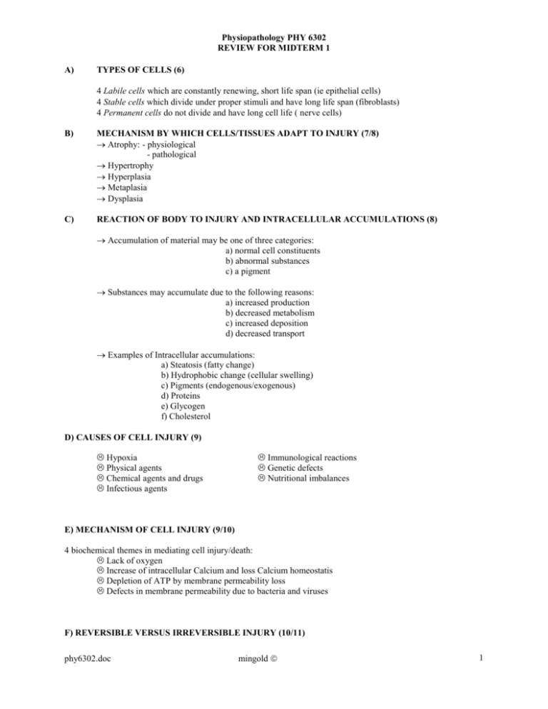
Physiopathology PHY 6302 REVIEW FOR MIDTERM 1 A) TYPES OF CELLS (6) Labile cells which are constantly renewing, short life span (ie epithelial cells) Stable cells which divide under proper stimuli and have long life span (fibroblasts) Permanent cells do not divide and have long cell life ( nerve cells) B) MECHANISM BY WHICH CELLS/TISSUES ADAPT TO INJURY (7/8) Atrophy: - physiological - pathological Hypertrophy Hyperplasia Metaplasia Dysplasia C) REACTION OF BODY TO INJURY AND INTRACELLULAR ACCUMULATIONS (8) Accumulation of material may be one of three categories: a) normal cell constituents b) abnormal substances c) a pigment Substances may accumulate due to the following reasons: a) increased production b) decreased metabolism c) increased deposition d) decreased transport Examples of Intracellular accumulations: a) Steatosis (fatty change) b) Hydrophobic change (cellular swelling) c) Pigments (endogenous/exogenous) d) Proteins e) Glycogen f) Cholesterol D) CAUSES OF CELL INJURY (9) Hypoxia Physical agents Chemical agents and drugs Infectious agents Immunological reactions Genetic defects Nutritional imbalances E) MECHANISM OF CELL INJURY (9/10) 4 biochemical themes in mediating cell injury/death: Lack of oxygen Increase of intracellular Calcium and loss Calcium homeostatis Depletion of ATP by membrane permeability loss Defects in membrane permeability due to bacteria and viruses F) REVERSIBLE VERSUS IRREVERSIBLE INJURY (10/11) phy6302.doc mingold 1 Physiopathology PHY 6302 REVIEW FOR MIDTERM 1 REVERSIBLE INJURY observable characteristics: cellular swelling Fatty change IF INJURIOUS AGENT NOT REMOVED THEN WE PROCEED TO: IRREVERSIBLE INJURY observable characteristics (seen with E.M.) vacuolization rupture lysosomes Nuclear changes (pyknosis; shrinking, karyorrhexis; fragmentation, karyolysis; nucleus digested) G) TWO CASUES OF IRREVERSIBLE INJURY (11/12) HYPOXIC & ISCHEMIC INJURY [up to this point may be reversible] FREE RADICAL INDUCED INJURY involves lipid peroxidation/oxidative protein modification/DNA lesions 1. 2. 3. 4. 5. IMPORTANT FREE RADICAL Oxygen (Strongest) OH hydroxyl (biggest imune response) CCL3 (found in liver damage) H202 (least noxious) NO Nitric Oxide (inflammatory response) H) DYSTROPHIC AND METASTATIC CALCIFICATION (12) Dystrophic only occurs in dead (necrotic) tissue Metatstatic inovles deposition in normal tissue due to hypercalcemia I) NECROSIS (13) Necrosis refers to morphological changes following cell death in living tissue Sum of protein denaturation and enzymatic digestion of organelles TYPES OF NECROSIS 1. 2. 3. 4. 5. Coagulative involving protein denaturation. Caused by ischemic injury to tissue such as heart. Can still visualize cell pattern. Liquefactive is caused by bacteria and can’t recognize the tissue you are visualizing. Gangrenous is caused by both ischemia and microbial infection and deals particularly with lower extremeties Caseous is specific to tuberculosis and looks like white cheezy curds. Enzymatic Fat is injury to pancreas by chronic alcoholism and LIPASE attacks fat cells J) APOPTOSIS (14) phy6302.doc mingold 2 Physiopathology PHY 6302 REVIEW FOR MIDTERM 1 Programmed cell destruction and is characterized by: cell shrinkage apoptotic bodies chromatin condensation phagocytosis of apoptotic cells by healthy cells May lead to neoplasm Heat Shock Proteins are naturally occuring proteins which play important role in cell metabolism. Without them cells die when exposed to stressors HSP 60/70 (Chaperonins) help fold and target proteins to final destination Ubiquitin help breakdown of proteins K) INFLAMMATION A GENERAL OVERVIEW (15/16) Inflammation sets in motion series of events that heal and reconstitute tissue Repair involves cell regeneration and scar formation ACUTE INFLAMMATION RESPONSE - Hallmark is neutrophila - clinically rapid onset, short lived vague symptoms CHRONIC INFLAMMATION RESPONSE - Hallmark is macrophage but also involves fibroblast and lymphocytes - Clinically slow onset, long lived and symptoms appear later but are well defined. Cardinal signs of inflammation are: Calor - heat Rubor - redness Dolor - pain Tumor - swelling L) INFLAMMATORY RESPONSE (17) 3 major components of inflammation are: Vascular wall permeability changes Vasodilation White blood cell movement CELLULAR EVENTS OF ACUTE INFLAMMATION (18/19) Margination Adhesion/Pavimentation - Selectins permit Rolling - Immunoglobins permit Transmigration - Integrins permit adhesion Emigration/Migration via chemotaxis Aggregation around the inflammation site Phagocytosis involving 3 stages - recognition (opsonization) - engulfment - degradation M) PLASMA MEDIATORS OF INFLAMMATION (20/21/22) 3 families exist within the plasma mediators (all consist of Plasma Protease) phy6302.doc mingold 3 Physiopathology PHY 6302 REVIEW FOR MIDTERM 1 Each family can activate itself and also the other families if need be. FAMILY OF PLASMA MEDIATOR KININ (involves Hageman Factor) COMPLEMENT SYSTEM (triggers immune system) CLOTTING SYSTEM ACTIVE INGREDIENT AND FUNCTION - Prekallikrein can reactivate Hageman Factor - Bradykinin (smallest protein) vasodilation, vascular permeability, causes pain, rapidly degraded 20 proteins involving a Classical pathway and Alternative pathway. Classical involves antigen-antibody complex Alternative involves immunoglobin complex, venoms etc. - Anaphylatoxins: C3a, C4a, C5a (Vasodilation, vascular permeability & bronchoconstriction) C5a is chemotaxic - C3b is “opsonin” - C5b-C9 punch holes in bacteria - activated by Hageman Factor XII - Thrombin vascular permeability, WBC adherance to endothelium, fibroblasts - Plasmin cleaves C3 into C3a & C3b & splits fibrin Also activates Complement system N) CELL DERIVED MEDIATORS OF INFLAMMATION (22/23/24/25) FAMILY OF CELL DERIVED MEDIATORS phy6302.doc ACTIVE INGREDIENT AND FUNCTION mingold 4 Physiopathology PHY 6302 REVIEW FOR MIDTERM 1 VASOACTIVE AMINE ARACHIDONIC ACID METABOLITES CYTOKINES NITRIC OXIDE (endothelial derived relaxing factor) PLATELET AGGREGATING FACTOR - Histamine - Serotonin made by platelets LEUKOTRIENES LTB4 - chemotaxis LTC4 LTD4 - permeability & vasoconstriction LTE4 CYCLOOXYGENASE PGI2 (prostacyclin) - vasodilation, inhibits platelet aggregation TXA2 (thromboxane A2) - vasoconstriction, promotes platelet aggregation PGD2 - pain and edema PGE2 - vasodilation, permeability, inhibits inflammatory cell function produced by lymphocytes and macrophages IL1, IL2, IL6, IL8 & TNF mediate inflammation cytokines induce endothelial adhesion molecules, induce synthesis of nitric oxide, induce neutrophil aggregation, induce systemic acute phase reactions, chemotactic neutrophils (IL8) allows for normal blood vessel tone vasodilation by muslce relaxation cytotoxic as a free radical derived from neurons, vascular endothelial and macrophage cells has a short half life Constitutive form from endothelial cells Inducible form from macrophage potent versatile inflamatory mediator derived from phospholipids of granulocytes, monocytes, endothelial cells (essentially all cells) functions to vascular permeability vasodilation platelet aggregation & degranulation arachidonic acid metabolism leukocyte adherence to endothelium O) CELLULAR PARTICIPANTS IN ACUTE INFLAMMATION (26/27) Neutrophils: - PMN that comprise 60-70 percent of leukocytes - arise from bone marrow stem cells phy6302.doc mingold 5 Physiopathology PHY 6302 REVIEW FOR MIDTERM 1 - granulated and release PAF Eosinophils: - Similar to PMN but not as effective - cytoplasm stains bright pink - Common in allergic reactions - contains histaminase Basophils: - Involved type I allergic reactions - granules contain histamine and heparin Macrophages: - Key player in last stages of acute inflammation - Major player in Chronic inflammation P) POTENTIAL OUTCOMES OF ACUTE INFLAMMATION (29) Complete Resolution - no sequelae Healing by fibrosis/organisation - scarring Abscess formation - maybe staph/strep Q) CELLULAR PARTICPANTS IN CHRONIC INFLAMMATION (27/28) Lymphocytes which are broken down into two categories: 1) T Lymphocytes 2) B Lymphocytes - majority of lymphocytes this kind - responsible humoral immunity - cell mediated immunity - IgG highest [ ] - CD4 are helpers, secrete cytokines - IgD may bind to B cells - CD8 are cytotoxic/suppressor T cells - IgM is very big cell - NK killer cells - IgA have secretory fct and mucous - IgE most common in allergic rx. - Antibodies neutralize, trigger complement activation system and opsonize for phagocytosis Plasma cells are activated B cells that synthetize immunoglobulins The produce antibodies directed against persistent antigen or altered tissue compound Monocytes: - Release PAF and leukotrienes and prostaglandins. - In tissue become known as macrophages and given special names: kupffer cells alveolar macrophages histiocytes microglial cells osteoclasts Chronic inflammation is assosciated with: - continued monocyte recruitment - local proliferation of macrophages at inflammatory site - immobilization of macrophages within site of inflammation TISSUE DESTRUCTION BY MACROPHAGES IS HALLMARK OF CHRONIC INFLAMMATION R) CHRONIC INFLAMMATION(29/30) Chronic inflammation is prolonged duration (weeks/months) in which active inflammation, tissue destruction and healing attempts occur simultaneously. Chronic inflammation may arise without acute inflammation being present. phy6302.doc mingold 6 Physiopathology PHY 6302 REVIEW FOR MIDTERM 1 1) persistent microbial infections create “granulomatous inflammation” 2) prolonged exposure to exogenous/endogenous agents such as silica 3) autoimmune diseases such as rheumatoid arthritis and lupus The hallmark of chronic inflammation is the macrophage but plasma cells are also involved. Fibrosis is characteristic of Chronic Inflammation S) GRANULOMATOUS INFLAMMATION (30) predominant cell is the activated macrophage (epithelioid like appearance) Multinucleated giant cells, lymphocytes and necrosis are present. A GRANULOMA is a focal area of granulomatous inflammation with epitheloid cells surrounded by a of mononuclear leukocytes. Two types exist: 1) Foreign body and 2) immune granulomas. Specific causes of granulomas include: - tuberculosis - sarcoidosis - leprosy - cat scratch disease - fungal infection - syphilis collar - suture material - lipids T) MORPHOLOGICAL PATTERNS IN ACUTE AND CHRONIC INFLAMMATION (30/31) Serous Inflammation (Acute) - Thin transudate fluid from blood serum or mesoepithelial cell secretions. Low in protein. - example is a blister Fibrinous Inflammation: - Thick and Gooey and found in pericardium and pleura - Amorphous coagulum or eosionphilic meshwork - example is fibrinous pericarditis Suppurative or Purulent Inflammation: - Thick & Gooey with large amounts of pus made up of neutrophils, necrotic cells & edema fluid. - Produced often by “pyogenic bacteria such as Staph. - example is acute appendicitis Ulcers: - localized defect on surface of organ or tissue when shedding of inflammatory necrotic tissue. - example is decubitis ulcer (aka bed sore) U) REPAIR OF DAMAGED TISSUE (32) total reconstruction partial reconstruction universal repair by fibrous scar Repair by Connective tissue phy6302.doc mingold 7 Physiopathology PHY 6302 REVIEW FOR MIDTERM 1 - produces fibrosis or scarring - Four components to tissue repair by scarring are: a) angiogenesis b) migration and proliferation of fibroblasts c) deposition of extracellular matrix (ie collagen, laminin) d) remodelling by replacing young scar tissue Granulation tissue is the special tissue which is hallmark of healing by fibrosis Granulation tissue is composed of:- proliferating small capillaries - proliferating fibroblasts - extracellular fluid - macrophages Granulation tissue function is to: - fill in tissue “gaps” - remove debris - aid in wound contracture - form a “pre-scar” V) SKIN WOUND HEALING may be by primary or secondary intention PRIMARY INTENTION (MINIMAL TISSUE SEPARATION) Blood clots fill incsion Neutrophil infiltration (24-48 hrs) epithelial continuity restored macrophage infiltration (3 days) granulation tissue fills in (about 5 days) progressive collagenization leads to scar Type III collagen replaced by Type I; strength in 1014 days SECONDARY INTENTION (LARGE TISSUE DEFECTS) large amounts of granulation tissue Inflammatory reaction is prolonged More debris & inflammatory exudate to clean up Greater scar formation & contracture Complications of normal wound healing include the following: - proud flesh: excessive granulation tissue interfereing with repair - keloid scar: which is excesive scar tissue and is genetically predisposed - wound dehiscence: wound bursting/defective scar formation (vit c deficiency) - excessive wound contracture: puckering of wound phy6302.doc mingold 8
