Chapter 15_GI - V14-Study
advertisement
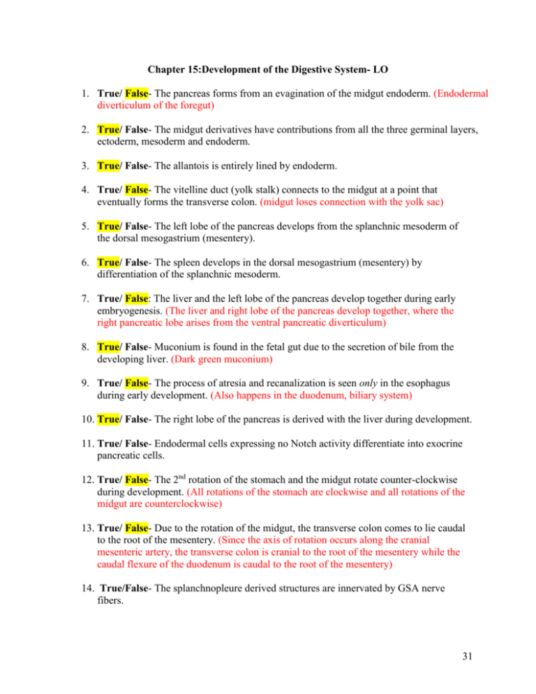
Chapter 15:Development of the Digestive System- LO 1. True/ False- The pancreas forms from an evagination of the midgut endoderm. (Endodermal diverticulum of the foregut) 2. True/ False- The midgut derivatives have contributions from all the three germinal layers, ectoderm, mesoderm and endoderm. 3. True/ False- The allantois is entirely lined by endoderm. 4. True/ False- The vitelline duct (yolk stalk) connects to the midgut at a point that eventually forms the transverse colon. (midgut loses connection with the yolk sac) 5. True/ False- The left lobe of the pancreas develops from the splanchnic mesoderm of the dorsal mesogastrium (mesentery). 6. True/ False- The spleen develops in the dorsal mesogastrium (mesentery) by differentiation of the splanchnic mesoderm. 7. True/ False: The liver and the left lobe of the pancreas develop together during early embryogenesis. (The liver and right lobe of the pancreas develop together, where the right pancreatic lobe arises from the ventral pancreatic diverticulum) 8. True/ False- Muconium is found in the fetal gut due to the secretion of bile from the developing liver. (Dark green muconium) 9. True/ False- The process of atresia and recanalization is seen only in the esophagus during early development. (Also happens in the duodenum, biliary system) 10. True/ False- The right lobe of the pancreas is derived with the liver during development. 11. True/ False- Endodermal cells expressing no Notch activity differentiate into exocrine pancreatic cells. 12. True/ False- The 2nd rotation of the stomach and the midgut rotate counter-clockwise during development. (All rotations of the stomach are clockwise and all rotations of the midgut are counterclockwise) 13. True/ False- Due to the rotation of the midgut, the transverse colon comes to lie caudal to the root of the mesentery. (Since the axis of rotation occurs along the cranial mesenteric artery, the transverse colon is cranial to the root of the mesentery while the caudal flexure of the duodenum is caudal to the root of the mesentery) 14. True/False- The splanchnopleure derived structures are innervated by GSA nerve fibers. 31 15. True/ False- The duodenum is supplied by both the celiac and cranial mesenteric arteries, implying its derivation from foregut and midgut. 16. True/ False- The urorectal septum divides cloacal membrane into anal and buccopharyngeal membranes. (separates the allantois from the rectum) 17. True/False- Hox genes determine the non-overt segmentation of gut into different parts of small and large intestines. 18. True/ False- Hindgut is supplied by the vagus nerve during early development. 19. True/ False- Septum transversum contributes to various ligaments of the spleen during development. (contributes to various ligaments of the liver) 20. True/ False- The liver in domestic animals is attached to diaphragm because of developmental relationships. 21. True/ False- Kupffer cells of the liver are derived from pleuropericardial folds. (Derived from the septum transversum) 22. True/ False- Renal agenesis may result in hydroamnios. (actually results in oligohydramnios, or lack of amniotic fluid) 23. True/ False- Atretic esophagus may contribute to oligohydramnios. (Results in hydramnios, where amniotic fluid is not absorbed) 24. True/False- The cardiac primordium induces the stomach formation. (cardiogenic mesoderm has inductive influence over liver formation) 25. True/False- The allantoic stalk becomes the Meckel’s diverticulum. (A congenital diverticulum or bulge of the ileum of the small intestine present as a remnant of the vitelline duct or yolk stalk) 26. Anterior (cranial) and posterior (caudal) intestinal portals. Folding of the embryo (largely the yolk sac in the beginning) results in the formation of a primitive gut separated into 3 distinct parts: foregut, midgut, and hindgut. The midgut is connected to the yolk sac via the yolk stalk (vitelline duct, omphalomesenteric). The transitions between the open-floored midgut and the tubular foregut and tubular hindgut are called the cranial intestinal portal and caudal intestinal portal, respectively. The endoderm at these cranial and caudal junctions express a site-specific signaling molecule, Shh. Differences in expression of other signaling factors between the cranial and caudal portals results in the ability for regional differentiation and the formation of the digestive system. 32 27. Differentiate between cloacal membrane versus stomodeum. In the head region, the foregut comes into contact with the ectoderm to form the buccopharyngeal membrane. As the head region enlarges, this membrane comes to lie at the floor of an ectodermally-lined pit called the stomodeum, which forms the oral cavity. The hindgut is also sealed off from the outside by opposing surface of the ectoderm and endoderm, resulting in the formation of the cloacal membrane. 28. Transdifferentiation. A rare process where a fully differentiated cell transforms into another cell type. During development of the esophagus from the foregut, local splanchnic mesoderm surrounds the esophagus to form a smooth muscle coat (tunica muscularis). Smooth muscle is the major type of muscle formed in the wall of the esophagus. However, the smooth muscle is transformed into skeletal muscle via transdifferentiation, a process mediated by the expression of myogenic regulatory factor myf-5 and meltrin (fuses skeletal myoblasts to form myotubes). Depending on the species, the tunica muscularis has various proportions of skeletal and smooth muscle. 29. What is Omental bursa and how it is formed?. The omental bursa is a pancreatico-enteric recess formed by the coalescing of clefts within the dorsal mesogastrium. In relation to the developing stomach, the dorsal mesentery is called the dorsal mesogastrium and the ventral the ventral mesogastrium. As the primitive gut first rotates along its long axis (90°), the dorsal mesogastrium expands, forming the great omentum. This results in the elongation of the dorsal mesogastrium, resulting in the formation of the omental bursa. Multiple Choices- Select the correct response: 30. During development, the liver is associated with all of the following except: a. b. c. d. e. Hematopoiesis Ventral mesentery Septum transversum Ventral pancreas Dorsal pancreas 33 31. Further development of the lining of the stomach results in the formation of all of the following, except: a. b. c. d. e. Gastric pits Beta cells Parietal cells Chief cells Argentaffin cells 32. Select the wrong item: a. The spleen develops from the splanchnic mesoderm of dorsal mesogastrium b. Kupffer cells of the liver are from the septum transversum c. All domestic animals develop a cystic diverticulum during the formative stages of the liver (absent in horses and camelids, which lack a gallbladder) d. The triangular ligaments of the liver are developed at the site of contact with septum transversum e. Fetal hepatic cells are functionally active and form bile (by mid-gestation) 33. List structures derived from the midgut: Jejunum, ileum, cecum, ascending colon, transverse colone, and most of the duodenum 34. What is physiological hernia? The migration of the midgut into the extraembryonic coelom is called a physiological hernia, and occurs due to the shortage of space inside the abdomen due to the relatively large size of the developing liver and kidneys. 35. Where is the approximate demarcation of foregut and midgut in the adult animal? 36. Name any 2 contributions of the hindgut? All the contributions of the hindgut are the left colic flexure, descending colon, rectum, bladder, and urethra 34 37. Describe the 270 degree rotation of the midgut segment. Due to rapid growth of the cranial limb of the “U”-shaped midgut tube, the midgut rotates 90° counterclockwise around the axis of the cranial mesenteric artery, resulting in the cranial limb of the midgut loop to the right side and the caudal limb to the left. As the abdomen enlarges, the relative sizes of the kidneys and liver decrease and the twisted midgut is withdrawn from the abdomen (reversal of physiological hernia). As the midgut returns to the abdomen, it undergoes another 180° counterclockwise rotation along its axis. This results in the elongation of the mesentery supporting the jejunum, and final placement of the duodenum, pancreas, and colon (initial part) to the right side of the midline. 38. What is cloaca? In hindgut development, the allantois is connected to an ampulla-like enlargement at the caudal part of the hindgut, called the cloaca (“sewer” in Latin). The cloaca is lined by gut endoderm joins a surface ectoderm invagination to form the cloacal membrane. 39. What is the developmental significance of the anorectal line? In differentiation of the hindgut into urogenital segments and the rectum, there is an anorectal line that clearly demarcates between the endoderm ectodermal pit inside the anorectal canal. 40. What is portosystemic shunt? What are some clinical signs exhibited by affected patient? Abnormal direct connection between portal and systemic circulation where blood bypasses the liver (i.e. persistent ductus venosus). Symptoms can vary from no symptoms to anorexia, lethargy, vomiting, hepatic encephalopathy, elevated serum bile acids, and neurological signs (blindness, ataxia, behavioral changes, coma). 41. Why high protein diet exacerbates symptoms of portosystemic shunt defects (Read Dr. Beth Allegretto’s note on TUSK)? Liver catabolizes amino acids (derived from dietary protein) to urea and excreted urea in urine. The liver is the primary site of protein metabolism. If amino acids and their byproducts (ammonia) are prevented access to the liver, hepatic encephalopathy can result from the buildup of ammonia and other compounds, leading to CNS disorders, coma, or death. Two specific mechanisms follow: 35 Portosystemic shunts lead to an increase of ammonia in the blood because ammonia formed in GI system (from bacteria breaking down protein) cannot be excreted by the liver. Ammonia crosses the blood-brain barrier, is cytotoxic, causes neuronal cell death, and can lead to depression (increased formation of GABA). Hepatic bypass also leads to elevated AAAs in the blood compared to BCAAs (b/c liver isn’t utilizing AAAs). This results in the formation of “pseudoneurotransmitters”, which cause neurological disorders. 42. How the ventral mesentery in relation to stomach undergoes further division? 36
