Pedigree Charts & Genetic Inheritance Worksheet
advertisement
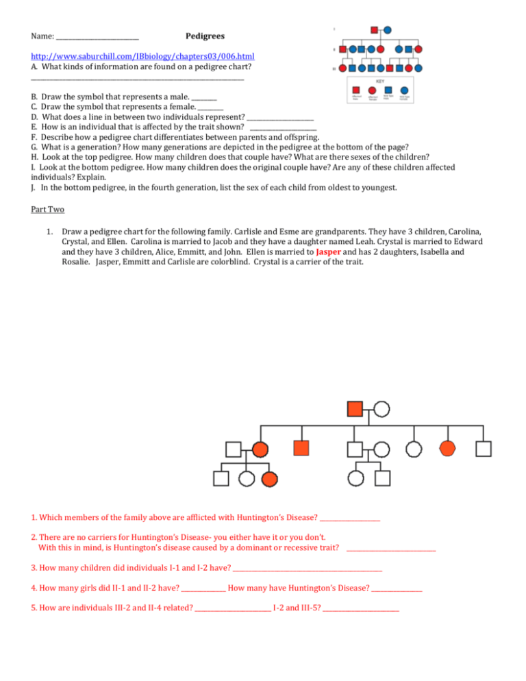
Name: __________________________ Pedigrees http://www.saburchill.com/IBbiology/chapters03/006.html A. What kinds of information are found on a pedigree chart? ___________________________________________________________________ B. Draw the symbol that represents a male. ________ C. Draw the symbol that represents a female. ________ D. What does a line in between two individuals represent? _____________________ E. How is an individual that is affected by the trait shown? _____________________ F. Describe how a pedigree chart differentiates between parents and offspring. G. What is a generation? How many generations are depicted in the pedigree at the bottom of the page? H. Look at the top pedigree. How many children does that couple have? What are there sexes of the children? I. Look at the bottom pedigree. How many children does the original couple have? Are any of these children affected individuals? Explain. J. In the bottom pedigree, in the fourth generation, list the sex of each child from oldest to youngest. Part Two 1. Draw a pedigree chart for the following family. Carlisle and Esme are grandparents. They have 3 children, Carolina, Crystal, and Ellen. Carolina is married to Jacob and they have a daughter named Leah. Crystal is married to Edward and they have 3 children, Alice, Emmitt, and John. Ellen is married to Jasper and has 2 daughters, Isabella and Rosalie. Jasper, Emmitt and Carlisle are colorblind. Crystal is a carrier of the trait. 1. Which members of the family above are afflicted with Huntington’s Disease? ___________________ 2. There are no carriers for Huntington’s Disease- you either have it or you don’t. With this in mind, is Huntington’s disease caused by a dominant or recessive trait? ____________________________ 3. How many children did individuals I-1 and I-2 have? _______________________________________________ 4. How many girls did II-1 and II-2 have? ______________ How many have Huntington’s Disease? ________________ 5. How are individuals III-2 and II-4 related? ________________________ I-2 and III-5? ________________________ 2. Answer the following questions using the next website: http://www.ygyh.org/hemo/whatisit.htm and go to “How is it inherited”? A. How does a boy get hemophilia?_____________________ B. How does a girl become a carrier? _______________________ 3. If a woman is a carrier and the male does not have hemophilia, draw a Punnett Square and indicate the possible outcome. Karyotyping Activity In this exercise you will simulate human Karyotyping using digital images from actual genetic studies. Go to http://learn.genetics.utah.edu A) Click on “Chromosomes and Inheritance” on the left hand side of the page B) Click on “Make a Karyotype” on the right hand side of the page What is a Karyotype? How is a Karyotype constructed? Now read the directions and construct the Karyotype. Was that of a male or female? How do you know? Go back to Chromosomes and Inheritance and click on “Using Karyotypes to predict genetic disorders” What is an autosome? How many do humans have? What is a sex chromosome and how many do humans have? How do disorders come about? Click on Play and watch how normal and abnormal meiosis occurs Go to www.biology.Arizona.edu/ This will take you to the University of Arizona’s “The Biology Project” page On the left Activities column click on Karyotyping Carefully read though the Introduction, G-Banding and Your Assignment. Go to “Patient Histories” Follow the directions to complete the each of the three Karyotypes. PATIENT A: 1. Normal or abnormal Karyotype? Explain 2. Is your Karyotype that of a Male or female? 3. What notation would you use to characterize Patient A’s Karyotype? 4. What diagnosis would you give Patient A? PATIENT B: 6. Normal or abnormal karyotype? Explain 7. Is your Karyotype that of a Male or female? 8. What notation would you use to characterize Patient B’s Karyotype? 9. What diagnosis would you give Patient B? PATIENT C: 10. Normal or abnormal Karyotype? Explain 11. Is your Karyotype that of a Male or female? 12. What notation would you use to characterize Patient C’s Karyotype? 13. What diagnosis would you give Patient C?
