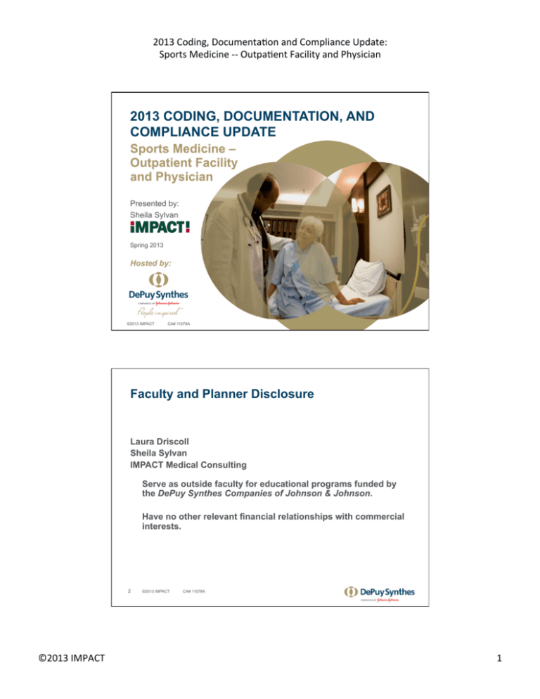
2013 Coding, Documenta4on and Compliance Update: Sports Medicine -­‐-­‐ Outpa4ent Facility and Physician 2013 CODING, DOCUMENTATION, AND
COMPLIANCE UPDATE
Sports Medicine –
Outpatient Facility
and Physician
Presented by:
Sheila Sylvan
Spring 2013
Hosted by:
1
©2013 IMPACT
CA# 11078A
Faculty and Planner Disclosure
Laura Driscoll
Sheila Sylvan
IMPACT Medical Consulting
Serve as outside faculty for educational programs funded by
the DePuy Synthes Companies of Johnson & Johnson.
Have no other relevant financial relationships with commercial
interests.
2
©2013 IMPACT ©2013 IMPACT
CA# 11078A
1 2013 Coding, Documenta4on and Compliance Update: Sports Medicine -­‐-­‐ Outpa4ent Facility and Physician Copyright and Disclaimer
Copyright ©2013 Innovative Medical Practice Advisors, Consultants, Trainers, Inc. All rights reserved.
No part of this publication may be reproduced, stored in a retrieval system, or transmitted, in any form or
by any means, electronic, mechanical, photocopy, recording, or otherwise, without the prior written
permission of the author, except that copies of the forms included herein may be made for the sole use
of your organization. Printed in the United States. All CPT™ codes and definitions cited are copyrighted
by the American Medical Association.
The reimbursement information contained in this program is provided for informational purposes only
and represents no statement, promise, or guarantee by DePuy Synthes Mitek Sports Medicine, DePuy
Synthes Joint Reconstruction, and DePuy Synthes Spine, all of which are divisions of DePuy
Orthopaedics, Inc., concerning levels of reimbursement, payment, or charge. Similarly, all CPT and
HCPCS codes are supplied for informational purposes only and represent no statement, promise, or
guarantee by these companies that these codes will be appropriate or reimbursement will be made. The
information presented is not intended to increase or maximize reimbursement by any payor. We
strongly recommend that you consult your payor organization with regard to its reimbursement policies.
This publication is designed to provide accurate and authoritative information on the subject matter
covered, and is for guidance and reference purposes only. It is of a general informational and
educational nature and warrants of merchantability or fitness for a particular purpose are specifically
excluded. It is produced with the understanding that the publisher is not engaged in rendering legal,
medical, accounting, or other professional service in specific situations.
Although prepared for
professionals, this publication should not be utilized as a substitute for professional service in specific
situations. If legal or medical advice is required, the services of a professional should be sought. The
companies referenced above may not have products approved for certain indications/procedures
discussed, but all products discussed in this publication are approved/cleared for commercial marketing
in the U.S. No product uses discussed in this publication are considered investigational or off-label.
Please refer to the package insert for a complete description of indications and contraindications for
each medical device used.
3
©2013 IMPACT
CA# 11078A
OVERVIEW – OUTPATIENT FACILITY AND
PHYSICIAN CODING AND DOCUMENTATION
ISSUES
©2013 IMPACT
©2013 IMPACT CA# 11078A
2 2013 Coding, Documenta4on and Compliance Update: Sports Medicine -­‐-­‐ Outpa4ent Facility and Physician Procedural Coding Concepts
•
•
•
HCPCS Coding Family
Symbols of CPT
Bundling Issues:
•
•
•
•
•
•
•
5
CPT Definitions
The Correct Coding Initiative (CCI)
Private Software
Multiple Procedures
Surgical Package Concepts
Groupers / APC Packaging
Payor Coverage Criteria
©2013 IMPACT
CA# 11078A
Case Scenario
A question was posed whether a provider
would receive separate payment for a
combination of procedures and, if so,
whether that payment would be at a
reduced amount.
This question is not as "simple" as it
originally appeared.
6
©2013 IMPACT ©2013 IMPACT
CA# 11078A
3 2013 Coding, Documenta4on and Compliance Update: Sports Medicine -­‐-­‐ Outpa4ent Facility and Physician Case Scenario – Issues to Consider
• Some procedure codes are paid separately at full rate.
• Some are subject to the multiple procedure payment reduction.
• Some are ancillary or packaged into other services, either
always, or in combination only with specific other codes.
• Some services are clearly not separately reimbursed, based
upon the CPT code definitions and/or notes.
• It may vary for open surgery vs. arthroscopic procedures.
• Hospital outpatient rules may differ from ASC.
• It will almost certainly vary by payor type / contract.
7
©2013 IMPACT
CA# 11078A
Surgical Package Modifiers
Included Services
Exceptions
Modifiers
Incidental anesthesia
and cognitive services
(E/M) in preparation for
surgery, within 24
hours in advance
Significant and
Separate
-25
0/10
Decision for Surgery
-57
90
Typical postoperative
follow-up care
Return to O.R. for
Related Procedure
-78
Unrelated E/M
-24
Unrelated Procedure
-79
Staged or Related
-58
Some payors have historically varied from these interpretations, but this
may be changing.
8
©2013 IMPACT ©2013 IMPACT
CA# 11078A
4 2013 Coding, Documenta4on and Compliance Update: Sports Medicine -­‐-­‐ Outpa4ent Facility and Physician New Coverage Criteria Trends
•
Healthcare payor entities have been adopting
increasingly stringent coverage criteria.
•
These guidelines may be more conservative than
the clinical standards of care developed by
specialty medical societies.
•
The stricter criteria lead to a need for more specific
documentation of patient history and other findings
to support medical necessity.
9
©2013 IMPACT
CA# 11078A
ORTHOPAEDIC SURGERY PROCEDURES
©2013 IMPACT
©2013 IMPACT CA# 11078A
5 2013 Coding, Documenta4on and Compliance Update: Sports Medicine -­‐-­‐ Outpa4ent Facility and Physician CPT Musculoskeletal System -- General
•
Wound Exploration
•
Injections
•
Tendon sheath or origin/insertion (each)
•
Trigger point(s) - # muscles
•
Arthrocentesis - size of joint
•
Application of Fixation Systems
•
Grafts or Implants
•
Other Procedures
11
©2013 IMPACT
CA# 11078A
Case Study #1 -- Viscosupplementation
Patient presents with chronic knee pain and reduced mobility due
to osteoarthritis. Initial treatments include anti-inflammatory
medication(s), home exercises, and physical therapy.
719.46
719.76
715.36
Over time, symptoms have become more severe, and an evaluation
of joint function is performed. Following the exam, there is a
discussion with the patient regarding options. The decision is
made to proceed with injections of ORTHOVISC® High Molecular
Weight Hyaluronan, and informed consent obtained.
Each knee is cleansed with alcohol, an injection of lidocaine is
administered to numb the knee, followed by injection of
hyaluronan into the joint capsule. The patient is instructed on
post-procedure care and encouraged to contact the practice if
there are any significant side effects. An appointment is
scheduled in one week for follow-up and sequential injection.
12
©2013 IMPACT ©2013 IMPACT
CA# 11078A
6 2013 Coding, Documenta4on and Compliance Update: Sports Medicine -­‐-­‐ Outpa4ent Facility and Physician Case Study #1 -- Viscosupplementation
Week #1 – Initial Treatment
Significant and Separate E/M Service with decision for
surgery and review of comorbidities
992xx-25
Major joint injection, bilateral
20610-50
Hyaluronan or derivative, ORTHOVISC, for intra-articular
injection, per dose
J7324 x 2
Week #2 & #3* – Planned injections
Major joint injection, bilateral
20610-50
Hyaluronan or derivative, ORTHOVISC, for intra-articular
injection, per dose
J7324 x 2
13
©2013 IMPACT
CA# 11078A
Case Study #1 -- Viscosupplementation
CASE NOTES:
•
The initial E/M service during which the decision for surgery is made
and any cormorbid conditions which may affect recovery are
assessed may be reported, so long as it represents a "significant
and separate" E/M service – modifier -25 will apply. Depending upon
circumstances, this may be office visit new patient (9920x),
established patient (9921x), or outpatient consultation (9924x), at the
level of service appropriate for extent of encounter and
documentation.
•
Some payors may require a separate diagnosis code for the E/M, or
have other limitations on reporting of E/M and minor surgery on the
same day.
•
Local anesthetic, if used, is included in 20610.
14
©2013 IMPACT ©2013 IMPACT
CA# 11078A
7 2013 Coding, Documenta4on and Compliance Update: Sports Medicine -­‐-­‐ Outpa4ent Facility and Physician Case Study #1 -- Viscosupplementation
CASE NOTES:
•
For subsequent injections, no E/M service is typically reported, as the
reason for the encounter is the minor surgery, which was planned
prospectively.
•
For some payors, no additional surgical modifiers apply to
subsequent injections, as code 20610 is assigned a zero-day global
period by CMS. However, variations exist between different payors:
• Plans may attempt to bundle any type of injection into an E/M
service the same day
• Some may consider weekly injections to be overlapping surgical
periods, and require use of modifier -58.
•
15
Payor coverage may require documentation of failed conservative
treatment for a specified period of time.
©2013 IMPACT
CA# 11078A
Example – Florida Medicare LCD
Indications for viscosupplementation therapy for the knee when ALL of
the following conditions are met:
•
•
•
The patient is symptomatic.
•
If the first course of treatment produces relief, subsequent courses of
treatment if symptoms return, at least six (6) months after the last
injection of a previous course of treatment.
•
Knee arthroplasty is not being considered as a current treatment
option.
•
Imaging visualization to provide guidance for needle placement will
not be covered.
Diagnosis is supported by radiologic evidence of osteoarthritis.
The patient has failed at least three months of conservative therapy,
including aspiration and intra-articular corticosteroid injection therapy
when inflammation is a component.
Excerpted from Local Coverage Determination (LCD) for Viscosupplementation Therapy for Knee (L29307); http://
www.cms.gov/medicare-coverage-database/search/advanced-search.aspx Enter “L29307” in Search by Document ID,
and effective date of 01/01/2010 on second search screen to access a copy of the complete LCD document.
16
©2013 IMPACT ©2013 IMPACT
CA# 11078A
8 2013 Coding, Documenta4on and Compliance Update: Sports Medicine -­‐-­‐ Outpa4ent Facility and Physician Case Study #2 –
Removal of External Fixation Device
PRE-OP DIAGNOSIS:
POST-OP DIAGNOSIS:
Left distal radius fracture.
Same.
PROCEDURE PERFORMED:
Removal of external fixator as
well as two radial styloid pins,
left wrist.
SURGEON:
ASSISTANT:
Dr. X
None.
ANESTHESIA:
TOURNIQUET TIME:
COMPLICATIONS:
DRAINS:
Monitored anesthesia care.
No tourniquet.
None.
None.
17
©2013 IMPACT
813.42
V54.89
V54.12
CA# 11078A
Case Study #2 –
Removal of External Fixation Device
OPERATIVE REPORT: The patient was taken to the operating
room and after the induction of adequate IV analgesia, the left
upper extremity was prepped with povidone-iodine. The
small external fixator frame was removed from the four pins.
This was followed by removal of the pins using the small AO
external fixator set wrench. This was then followed by
20694-58(?)
removal of the two radial styloid pins.
Once this was done, the wounds were irrigated and sterile
dressings were applied, the patient tolerated the procedure
well and went to the recovery room in stable condition.
18
©2013 IMPACT ©2013 IMPACT
CA# 11078A
9 2013 Coding, Documenta4on and Compliance Update: Sports Medicine -­‐-­‐ Outpa4ent Facility and Physician Musculoskeletal System
Standard Subheadings Within Each Body Region
•
•
•
•
•
•
•
•
•
19
Incision
Excision
Introduction or Removal
Repair, Revision, and/or Reconstruction
Fracture and Dislocation
Manipulation
Arthrodesis
Amputation
Other Procedures
©2013 IMPACT
CA# 11078A
Repair, Revision, and/or
Reconstruction
• Repair of:
• Single tendon, multiple tendons, or each tendon
• Primary vs. secondary
• May specify through same incision
• Codes may reflect repair, transfer, advancement,
lengthening or shortening, or release of a specified
muscle, tendon, or ligament
• Some services may be reported separately
• Grafts
• Arthroplasty
20
©2013 IMPACT ©2013 IMPACT
CA# 11078A
10 2013 Coding, Documenta4on and Compliance Update: Sports Medicine -­‐-­‐ Outpa4ent Facility and Physician Examples:
27650
27652
Repair, primary, open or percutaneous, ruptured
Achilles tendon;
with graft (includes obtaining graft)
27654
Repair, secondary, Achilles tendon, with or without
graft
27685
Lengthening or shortening of tendon, leg or ankle;
single tendon (separate procedure)
27686
multiple tendons (through same incision) each
(Source: CPT Professional 2012, pg. 134)
21
©2013 IMPACT
CA# 11078A
Example of Cumulative Descriptors
25000
Incision, extensor tendon sheath, wrist
25001
Incision, flexor tendon sheath, wrist
25020
Decompression fasciotomy, forearm and/or wrist, flexor
OR extensor compartment, without debridement of
nonviable muscle and/or nerve
25023
25024
25025
with debridement of nonviable muscle and/or nerve
Decompression fasciotomy, forearm and/or wrist, flexor
AND extensor compartment, without debridement of
nonviable muscle and/or nerve
with debridement of nonviable muscle and/or nerve
(Source: CPT Professional 2012, pg. 116)
22
©2013 IMPACT ©2013 IMPACT
CA# 11078A
11 2013 Coding, Documenta4on and Compliance Update: Sports Medicine -­‐-­‐ Outpa4ent Facility and Physician Example – Variations on a Theme
29290
Correction, hallux valgus (bunion), with or without
sesamoidectomy; simple exostectomy (eg, Silver type
procedure)
28292
Keller, McBride, or Mayo type procedure
28293
resection of joint with implant
28294
with tendon transplants (eg, Joplin type procedure)
28296
with metatarsal osteotomy (eg, Mitchell, Chevron,
or concentric type procedure)
28297
Lapidus-type procedure
28298
by phalanx osteotomy
28299
by double osteotomy
(Source: CPT Professional 2012, pp. 138-140)
23
©2013 IMPACT
CA# 11078A
Case Study #3 –
ACL Reconstruction with Tendon Graft
Preoperative Diagnosis: ACL deficient left knee
Postoperative Diagnosis: Same
Surgeon: Dr. X
Procedure:
844.2 /
717.83
Asst: Dr. Y
Left ACL reconstruction using patellar
tendon graft
Surgery time: Approximately 103 minutes
Anesthesia: Epidural
Operative Report: The patient was taken to the operating room
and after the induction of adequate epidural analgesia, the left
lower extremity was prepped and draped in the usual sterile
fashion. An Esmarch was used to exsanguinate the limb and
the tourniquet was inflated to 300 mm. Mercury.
24
©2013 IMPACT ©2013 IMPACT
CA# 11078A
12 2013 Coding, Documenta4on and Compliance Update: Sports Medicine -­‐-­‐ Outpa4ent Facility and Physician Case Study #3 –
ACL Reconstruction with Tendon Graft
At this point in time, a 5-6 cm incision was made over the
patellar tendon in the midline, taken down through the skin and
subcutaneous tissues until the peritenon was identified. The
peritenon was split longitudinally thus exposing the patellar
tendon. After this was done, the central third of the patellar
tendon along with bone plugs from both the inferior pole of the
patella and the proximal tibia were harvested without difficulty.
The graft was then placed on another table and prepared for
implantation into the knee. Visual inspection of the medial and
lateral compartments showed no significant meniscal pathology.
There were no chondral lesions wither on the tibia plateau of the
femoral condyles, however, there was evidence of a disruption
of the ACL. The PCL looked intact and normal in configuration.
25
©2013 IMPACT
CA# 11078A
Case Study #3 –
ACL Reconstruction with Tendon Graft
At this point in time, a shave was introduced into the joint. The
remnants of the ACL as well as part of the fat pat were debrided.
This was then followed by a notchplasty that was done using a
combination of a burr and the (Brand) wand. Once adequate
notchplasty was performed, the (Brand) tibial guide was placed
into the joint and a tibial tunnel was drilled in the appropriate
fashion. This was then followed by placement of the over the top
(Brand) tibial guide followed by drilling of a femoral tunnel. Both
tunnels were deemed to be in adequate position arthroscopically.
Once this was done, the graft was then drawn from the tibial
tunnel into the femoral tunnel under direct guidance and secured
in the femoral tunnel with a 7 x 25 interference screw as well as 7 x
20 screw in the tibial tunnel.
29888
26
©2013 IMPACT ©2013 IMPACT
CA# 11078A
13 2013 Coding, Documenta4on and Compliance Update: Sports Medicine -­‐-­‐ Outpa4ent Facility and Physician Case Study #3 –
ACL Reconstruction with Tendon Graft
The knee was put through a range of motion. There is no
evidence of the graft impinging in flexion or extension, the wound
was thoroughly irrigated and then the patellar tendon was closed
in layers initially with the tendon and peritenon followed by suture
of the subcutaneous tissues. This was followed by instillation of
local anesthetic into the joint as well as placement of a drain. The
initial arthroscopic portals were closed as well and went to the
Recovery Room in a stable condition.
Notes: Code 20924 is defined as tendon graft, from a distance ;
the tendon graft is not documented as obtained through a
separate incision, so it is not separately reported.
Operative report does not specify new vs. old injury. However, as
current surgical repair, the acute injury code 844.2 may be
reported even if initial injury was some time earlier.
27
©2013 IMPACT
CA# 11078A
Case Study #3 – Lateral Epicondylitis
PREOPERATIVE DIAGNOSIS: Lateral epicondylitis.
POSTOPERATIVE DIAGNOSIS: Lateral epicondylitis.
726.32
OPERATIONS PERFORMED: Lateral release with
lengthening of the ECRB tendon.
DESCRIPTION OF PROCEDURE: With the patient under
adequate anesthesia, the upper extremity was prepped and
draped in a sterile manner. The arm was exsanguinated and
the tourniquet elevated to 290 mm/Hg. An incision was
made over the lateral aspect of the right elbow anterior to
lateral epicondyle and centered on the lateral epicondyle.
Blunt dissection exposed the antebrachial fascia. The
interval between the extensor carpi radialis longus and
anterior edge of the extensor aponeurosis was identified.
28
©2013 IMPACT ©2013 IMPACT
CA# 11078A
14 2013 Coding, Documenta4on and Compliance Update: Sports Medicine -­‐-­‐ Outpa4ent Facility and Physician Case Study #3 – Lateral Epicondylitis
The antebrachial fascia was then incised and further
blunt dissection developed the interval between ECRL
and extensor aponeurosis from the lateral epicondyle
distal to the joint line. The ECRL was then released by
sharp dissection and retracted anterior to expose the
origin of the extensor carpi radialis brevis. Medial
dissection further delineated the origin of the ECRB. The
origin of the ECRB was then further inspected and gross
pathological changes of grayish tissue were identified
and this friable tissue was then excised. The joint
capsule was then incised. The radial capitellar joint was
inspected and found to be without pathological changes.
29
©2013 IMPACT
24358
CA# 11078A
Case Study #3 – Lateral Epicondylitis
The wound was then copiously irrigated. The joint
capsule was closed with interrupted 3-0 sutures.
The ECRB was then lengthened and then sutured to the
undersurface of the ECRL using 3-0 sutures. The
antebrachial fascia was closed with a running 3-0 suture
with a Krackow technique. The wound was then
infiltrated with 10 cc of .025% bupivicaine HCl.
24305
The skin was closed in a layered fashion. Sterile
dressings were applied. The tourniquet was deflated.
The patient was awakened from anesthesia and returned
to the recovery room in satisfactory procedure having
tolerated the procedure well.
30
©2013 IMPACT ©2013 IMPACT
CA# 11078A
15 2013 Coding, Documenta4on and Compliance Update: Sports Medicine -­‐-­‐ Outpa4ent Facility and Physician Case Study #4: Open Plantar Fasciotomy
PREOPERATIVE DIAGNOSIS: Chronic plantar fasciitis, right foot.
POSTOPERATIVE DIAGNOSIS: Chronic plantar fasciitis, right
foot.
PROCEDURE: Open plantar fasciotomy, right foot.
728.71
ANESTHESIA: Local infiltrate with IV sedation.
INDICATIONS FOR SURGERY: The patient has had a longstanding
history of foot problems. The foot problem has been progressive
in nature and has not been responsive to conservative care
despite multiple attempts at conservative care. [Informed Consent
Discussion]. The patient requested for surgical repair since the
problem has reached a point to interfere with normal daily
activities. The purpose of the surgery is to alleviate the pain and
discomfort.
31
©2013 IMPACT
CA# 11078A
Case Study #4: Open Plantar Fasciotomy
DETAILS OF THE PROCEDURE: The patient was given (drug) for
antibiotic prophylaxis 30 minutes prior to the procedure. The
patient was brought to the operating room and placed in the
supine position. Following a light IV sedation, a posterior tibial
nerve block and local infiltrate of the operative site was performed
with (medications). The lower extremity was prepped and draped
in the usual sterile manner. Balance anesthesia was obtained.
PROCEDURE: Plantar fasciotomy, right foot. The plantar medial
tubercle of the calcaneus was palpated and a vertical oblique
incision, 2 cm in length with the distal aspect overlying the
calcaneal tubercle was affected. Blunt dissection was carried out
to expose the deep fascia overlying the abductor hallucis muscle
belly and the medial plantar fascial band.
32
©2013 IMPACT ©2013 IMPACT
CA# 11078A
16 2013 Coding, Documenta4on and Compliance Update: Sports Medicine -­‐-­‐ Outpa4ent Facility and Physician Case Study #4: Open Plantar Fasciotomy
A periosteal elevator did advance laterally across the
inferior aspect of the medial and central plantar fascial
bands, creating a small and narrow soft tissue tunnel.
Utilizing a Metzenbaum scissor, transection of the medial
two-third of the plantar fascia band began at the junction
of the deep fascia of the abductor hallucis muscle belly
and medial plantar fascial band, extending to the lateral
two-thirds of the band. The lateral plantar fascial band
was left intact. Visualization and finger probe confirmed
adequate transection. The surgical site was flushed with
normal saline irrigation.
33
©2013 IMPACT
28008
CA# 11078A
Case Study #4: Open Plantar Fasciotomy
The deep layer was closed with 3-0 suture and the skin
edges coapted with combination of 1 horizontal mattress
and simples. Non-adhering wound dressing, 4x4, and an
elastic wrap to provide mild compression were applied.
The patient tolerated the procedure and anesthesia well,
and left the operating room to recovery room in good
postoperative condition with vital signs stable and arterial
perfusion intact. A walker boot was dispensed and
applied. The patient will be allowed to be full
weightbearing to tolerance, in the boot to encourage
physiological lengthening of the release of plantar fascial
band. After a short recuperative period, the patient was
discharged home with vital signs stable and in no acute
distress.
34
©2013 IMPACT ©2013 IMPACT
DME?
CA# 11078A
17 2013 Coding, Documenta4on and Compliance Update: Sports Medicine -­‐-­‐ Outpa4ent Facility and Physician Case Study #5 –
Multiple Digit Capsular Release, Hand
PREOPERATIVE DIAGNOSIS: Contractures right hand
including MP and IP joints.
POSTOPERATIVE DIAGNOSIS: Contractures right
hand including MP and IP joints.
INDICATIONS FOR PROCEDURE: The patient is a
pleasant 41-year-old female with a severe crush injury
to the right hand. She now has extension
contractures of the MP and IP joints of the right hand
as well as retained hardware in the small finger. She
has had a recent rotational osteotomy of the small
finger.
The patient is taken to the OR today for dorsal capsular
release of the MP and PIP joints as well as extensor
tenolysis and removal of hardware of the small finger.
35
©2013 IMPACT
727.81
905.8
718.44
V54.01
Modifier?
CA# 11078A
Case Study #5 –
Multiple Digit Capsular Release, Hand
DESCRIPTION OF PROCEDURE: The patient was taken to the
operating room, placed supine upon the operating table, after
an adequate general anesthesia, her right upper extremity
was prepped and draped in the standard surgical fashion. I
exsanguinated the arm with ___ dressing, inflated the
tourniquet to 250 mm Hg.
I began with the small finger. I made a longitudinal
incision over the dorsum of the small finger. I used
the previous longitudinal incision that was used for
the osteotomy. I incised sharply through the skin and
dermis and identified the extensor mechanism. The
extensor mechanism was split longitudinally down
the middle, identified the MP joint and made a
transverse incision over the MP joint capsule.
36
©2013 IMPACT ©2013 IMPACT
26445-F9
26520-F9
CA# 11078A
18 2013 Coding, Documenta4on and Compliance Update: Sports Medicine -­‐-­‐ Outpa4ent Facility and Physician Case Study #5 –
Multiple Digit Capsular Release, Hand
I identified the buried K-wires and these were both
removed without difficulty. These were buried into
the proximal phalanx. I then had to flex the MP joint,
I was able to flex it to 100 degrees. An extensor split
was continued to the proximal interphalangeal joint,
and the level of the proximal interphalangeal joint I
performed a dorsal capsulotomy. I left the central
slip intact on the dorsum of the middle phalanx. I
released the collateral ligament. I recessed the
collateral ligament on both sides of the proximal
interphalangeal joint and once this was
accomplished I was able to flex the PIP joint to about
95 degrees. I copiously irrigated the incision, closed
the extensor split with a running 4-0 suture, closed
the small finger incision with a running nylon suture.
37
©2013 IMPACT
20680-F9
26525-F9
CA# 11078A
Case Study #5 –
Multiple Digit Capsular Release, Hand
I then turned my attention to the ring finger, where I
made a longitudinal incision over the ring finger,
incising sharply through skin and dermis, identified
the extensor mechanism, split the extensor
mechanism longitudinally. Over the MP joint I made a
transverse incision through the capsule. I then
manipulated the MP joint. I was able to obtain 100
degrees of flexion. I then identified the proximal
interphalangeal joint, made a transverse capsulotomy
over the PIP joint. I left the central slip intact. I
recessed the collateral ligaments and then I was able
to flex the PIP joint to approximately 95 degrees. I
then copiously irrigated the incision. The extensor
tendon was repaired with a running suture and the
skin was repaired with interrupted nylon.
38
©2013 IMPACT ©2013 IMPACT
26445-F8
26520-F8
26525-F8
CA# 11078A
19 2013 Coding, Documenta4on and Compliance Update: Sports Medicine -­‐-­‐ Outpa4ent Facility and Physician Case Study #5 –
Multiple Digit Capsular Release, Hand
26445-F6
26445-F7
26520-F6
26520-F7
26525-F6
26525-F7
The same procedure was repeated on the long finger
and the index finger in the same fashion and again
long finger MP joint flexion was to 100, PIP joint was
to 95, index finger MP joint to 100, PIP joint was
approximately to 95 and again simultaneous full
flexion was not obtained. However, no intrinsic
tightness was identified in any of the digits. They all
had signs of extrinsic tightness.
When we were finished, a sterile dressing was applied
followed by a dorsal splint to keep the wrist in the
most extension it could be, which was approximately
10 degrees and to keep the fingers flexed. The patient
tolerated the procedure well. There were no
complications.
39
©2013 IMPACT
CA# 11078A
Case Study #5 –
Multiple Digit Capsular Release, Hand
PROCEDURES PERFORMED:
1.
2.
3.
4.
5.
6.
7.
8.
9.
10.
11.
12.
13.
40
©2013 IMPACT Capsulotomy, index finger, MP joint.
Capsulotomy, index finger, PIP joint.
Extensor tenolysis index finger.
Capsulotomy, long finger, MP joint.
Capsulotomy, long finger, PIP joint.
Extensor tenolysis long finger.
Capsulotomy, ring finger, MP joint.
Capsulotomy, ring finger, PIP joint.
Extensor tenolysis ring finger.
Capsulotomy, small finger, MP joint.
Capsulotomy, small finger, PIP joint.
Extensor tenolysis small finger.
Removal of hardware, small finger.
©2013 IMPACT
26520-F6
26525-F6
26445-F6
26520-F7
26525-F7
26445-F7
26520-F8
26525-F8
26445-F8
26520-F9
26525-F9
26445-F9
Bundled
CA# 11078A
20 2013 Coding, Documenta4on and Compliance Update: Sports Medicine -­‐-­‐ Outpa4ent Facility and Physician Case Study #5 –
Multiple Digit Capsular Release, Hand
CASE NOTES:
•
Level II modifiers clarify coding for digits on hands and feet -reduces likelihood of denials for duplicate procedures.
Alternative option is number of units.
•
2013 CCI states, “If a superficial or deep implant (e.g., buried
wire, pin, rod) requires surgical removal (CPT codes 20670
and 20680), it is not separately reportable if it is performed as
an integral part of another procedure.
•
Operative note stated recent rotational osteotomy -- if
within 90 days of either this osteotomy or initial treatment,
modifiers -58, -78, and/or -79 may apply.
41
©2013 IMPACT
CA# 11078A
Fracture Care
•
•
•
•
Closed treatment:
Open treatment
1) without manipulation
2) with manipulation
3) with or without traction
Percutaneous skeletal fixation
Manipulation
TRACTION DEVICES
•
•
•
Skeletal traction
Skin traction
External fixation
(Source: CPT Professional 2012, pg. 88)
42
©2013 IMPACT ©2013 IMPACT
CA# 11078A
21 2013 Coding, Documenta4on and Compliance Update: Sports Medicine -­‐-­‐ Outpa4ent Facility and Physician Case Study #6 –
Open Reduction and Internal Fixation
DIAGNOSIS: Left distal radius fracture
813.42
TITLE OF OPERATION: Left distal radius open reduction and
internal fixation and application of external fixator.
PROCEDURE IN DETAIL: After the patient was identified and
after adequate anesthesia, he was positioned supine on the
operating table. The left arm was placed on the arm table. It
was prepped and draped in a sterile fashion. The arm was
exsanguinated with an Esmarch bandage and then tourniquet
was inflated to 250 mmHg. A volar approach was done, distal
modified Henry approach. Care was taken to protect the
superficial radial nerve and also the radial artery and its venae
comitantes. The pronator quadratus was incised
longitudinally and elevated off the bone. The fracture was
identified, reduced, and the joint was also visualized.
43
©2013 IMPACT
CA# 11078A
Case Study #6 –
Open Reduction and Internal Fixation
Once the fracture had been reduced, it was plated with a
(brand) plate. Using AO technique, the plate was applied.
The fracture was controlled in both AP and lateral planes
using image intensifier. It was found to be well reduced,
and the screws and the tips of the screws were not
impinging any vital structures and were extra-articular.
After that was done, the wound was irrigated copiously
with antibiotic normal saline solution. It was closed in
layers using 2-0 absorbable and 3-0 nylon sutures. Next,
an external fixator was placed dorsolaterally in order to
provide additional stability. Dressings were applied
without difficulty again using standard AO technique.
The incision was closed with 3-0 nylon after it had been
irrigated copiously. A dressing was applied and the
patient was aroused from anesthesia without any
complications.
44
©2013 IMPACT ©2013 IMPACT
25607
73100
20690
CA# 11078A
22 2013 Coding, Documenta4on and Compliance Update: Sports Medicine -­‐-­‐ Outpa4ent Facility and Physician Endoscopy / Arthroscopy
•
Diagnostic arthroscopy is included in
surgical arthroscopy.
•
Some surgical arthroscopies are also
bundled.
•
G-code for knee arthroscopy:
G0289
Arthroscopy, knee, surgical, for removal of loose body,
foreign body, debridement/shaving of articular cartilage
(chondroplasty) at the time of other surgical knee
arthroscopy in a different compartment of the same knee
•
Diagnostic arthroscopy followed by open treatment, report
arthroscopy with modifier 51.
•
If no arthroscopy code exists, do not report as open surgery -use unlisted arthroscopy.
45
©2013 IMPACT
CA# 11078A
Case Study #7 –
Knee Arthroscopy with Meniscectomy
OPERATIVE REPORT
Preoperative diagnosis: Medial compartment
arthritis with torn medial meniscus.
Postoperative diagnosis: Same with torn lateral
meniscus and synovitis.
715.36
836.0
836.1
727.00
Operation(s): Arthroscopy, partial medial menisectomy, partial
lateral menisectomy, abrasion arthroplasty and synovectomy.
Description of procedure: After the administration of general
anesthesia the patient s knee was examined. There was a large
effusion and a mild varus deformity, crepitus upon range of
motion.
46
©2013 IMPACT ©2013 IMPACT
CA# 11078A
23 2013 Coding, Documenta4on and Compliance Update: Sports Medicine -­‐-­‐ Outpa4ent Facility and Physician Case Study #7 –
Knee Arthroscopy with Meniscectomy
The knee was then prepped and draped in the normal fashion
and secured to a leg holding device. Through a superior and
medial stick an inflow cannula was inserted and 25 ccs of joint
fluid were returned via the inflow cannula.
The arthroscope was introduced in the inferolateral portal and
the arthroscopy was begun. The suprapatellar pouch area
showed a lot of synovial hypertrophy. The undersurface
of the patella showed mostly grade II changes of
717.7
chondromalacia. The medial joint space was entered
and one could immediately see there was a large degenerative
tear of the medial meniscus and an area of bare bone 1.5cm in
diameter on the tibia, and matching area of the medial femoral
condyle.
717.3
47
©2013 IMPACT
CA# 11078A
Case Study #7 –
Knee Arthroscopy with Meniscectomy
Small medial arthrotomy incision was made and a meniscal probe
was introduced. The tear of the meniscus was then delineated. Then
with a series of instruments, including a meniscus scissors, grasping
forceps, biopsy forceps and motorized meniscal cutter, the entire torn
portion of the meniscus was removed, leaving a well-balanced and
stable rim.
Attention was then directed to the intercondylar notch
area, where a lot of synovial hypertrophy was debrided
with the motorized synovectomy tool. The lateral joint
space was entered and the lateral meniscus was well
visualized. There was a tear at the posterior horn region,
which was quite soft, and this was removed with the
motorized meniscus cutter.
48
©2013 IMPACT ©2013 IMPACT
717.43
29880
CA# 11078A
24 2013 Coding, Documenta4on and Compliance Update: Sports Medicine -­‐-­‐ Outpa4ent Facility and Physician Case Study #7 –
Knee Arthroscopy with Meniscectomy
The lateral joint space looked much better, the medial joint
space with only a small area of grade I chondromalacia.
Areas of synovitis were then debrided in the medial and
lateral compartments and the undersurface of the patella
was smoothed off. Following this with a small abrader, the
area of the bare bone in the medial joint compartment was
burred. The abrasion extended intracortically to a depth of
about 1 mm until there was bare bleeding bone.
29880
29879-59
or G0269
Following this the knee was irrigated and suctioned off.
Sterile dressing was applied and the procedure was
terminated. Patient tolerated the procedure well ad was
taken to the recovery room in good condition.
49
©2013 IMPACT
CA# 11078A
Case Study #8 –
Arthroscopic Rotator Cuff Repair
OPERATIVE REPORT
Preoperative diagnosis: Right rotator cuff tear.
Right subacromial impingement.
Postoperative diagnosis: Right rotator cuff tear.
Right subacromial impingement.
Acromioclavicular joint arthritis.
727.61
726.10
715.31
Procedure: Diagnostic arthroscopy with arthroscopic rotator
cuff repair. Arthroscopic subacromial decompression.
Arthroscopic distal clavicle excision.
Indications: This patient has persistent pain within his right
shoulder. It has been ongoing. He has failed conservative
regimen and is in need of the above procedure.
50
©2013 IMPACT ©2013 IMPACT
CA# 11078A
25 2013 Coding, Documenta4on and Compliance Update: Sports Medicine -­‐-­‐ Outpa4ent Facility and Physician Case Study #8 –
Arthroscopic Rotator Cuff Repair
OPERATIVE PROCEDURE: Following preoperative
informed consent the patient was brought to the
operating room. He underwent general anesthesia and
interscalene block. The right shoulder was prepped and
draped in sterile manner. We first did a manipulation
under anesthesia and indeed he did have some
adhesions present going into external rotation as well as
with internal rotation.
23700
29805
We then went into the shoulder and did a diagnostic
arthroscopy. You could see some bleeding where we
had lysed adhesions. We cleaned those up gently via an
anterior portal. We then looked up and there was a
726.13
small, almost full thickness tear of the supraspinatus
behind the biceps tendon. We debrided this area out
and then completed the tear via the bursal side.
51
©2013 IMPACT
CA# 11078A
Case Study #8 –
Arthroscopic Rotator Cuff Repair
We marked the area of significant partial tear and then
went into the subacromial space. We then debrided
out the bursa and you could see the almost full
thickness tear. We completed it at that point, removing
the marking stitch. There was a small tear present.
We then debrided the subacromial bursa and went up
and noticed the fairly significant subacromial spur and
we debrided this back to stable rim using an oval burr.
We then went over the AC joint and I took off the
undersurface of the AC joint and it was really, really
arthritic. It hung down over onto the rotator cuff
musculature. I debrided the undersurface of that and
then went ahead via the anterior portal and debrided
out the entire distal clavicle. We removed about the
distal 8 mm of the clavicle.
52
©2013 IMPACT ©2013 IMPACT
29822/
29823
29824
CA# 11078A
26 2013 Coding, Documenta4on and Compliance Update: Sports Medicine -­‐-­‐ Outpa4ent Facility and Physician Case Study #8 –
Arthroscopic Rotator Cuff Repair
There was good free motion at that point after that. We
then went back to the tear and freshened the greater
tuberosity up and then placed a [brand] anchor there.
We placed the anchor off the articular surface.
We then brought the two suture ends up through a small
tear. I didn't think it needed a double row repair. We
went ahead at that point and placed two sutures without
difficulty. We then tied simple sutures and this gave a
good solid repair there.
29827
We then thoroughly irrigated the wound and removed
the arthroscope. Sterile dressing was applied. The
patient was taken to the recovery room in stable
condition.
53
©2013 IMPACT
CA# 11078A
2013 CCI Notes
•
NEW FOR 2013: CMS considers the shoulder joint to be a single
anatomic structure. An NCCI procedure to procedure edit code pair
consisting of two codes describing two shoulder joint procedures
should never be bypassed with an NCCI-associated modifier when
performed on the ipsilateral shoulder joint. This type of edit may be
bypassed only if the two procedures are performed on contralateral
joints.
•
When it is necessary to perform skeletal/joint manipulation under
anesthesia to assess range of motion, reduce a fracture or for any
other purpose during another procedure in an anatomically related
area, the corresponding manipulation is not separately reportable.
54
©2013 IMPACT ©2013 IMPACT
CA# 11078A
27 2013 Coding, Documenta4on and Compliance Update: Sports Medicine -­‐-­‐ Outpa4ent Facility and Physician Case Study #8 – Arthroscopic Rotator Cuff Repair
PROCEDURES
Arthroscopy, shoulder; with rotator cuff
repair
Arthroscopy, shoulder; distal
claviculectomy
Arthroscopy, shoulder; debridement.
limited (29822) vs. extensive (29823)
Diagnostic arthroscopy of shoulder
(Bundled)
Manipulation of shoulder under anesthesia
(Bundled)
2012
PROCEDURE
CODES
2013
PROCEDURE
CODES
29827
29827
29824
29824
29822/
29823-59
29822/
29823
29805
29805
23700
23700
DIAGNOSIS
CODES
727.61
726.10
715.31
726.13
CASE NOTES: 29827 is the primary procedure. Neither 29824 nor 29823 is
bundled into 29827; however, 29823 is bundled into 29824 per CCI — and therefore
must be dropped with this new instruction. Previously, it might have been
reported separately with modifier -59 when performed in distinct part of shoulder.
55
©2013 IMPACT
CA# 11078A
Case Study #9 – Arthroscopic Debridement &
Labral Repair
PREOPERATIVE DIAGNOSIS: Femoroacetabular impingement.
POSTOPERATIVE DIAGNOSIS: Femoroacetabular impingement.
719.85
OPERATIONS PERFORMED
1. Left hip arthroscopic debridement.
2. Left hip arthroscopic femoral neck osteoplasty.
3. Left hip arthroscopic labral repair.
ANESTHESIA: General.
OPERATION IN DETAIL: The patient was taken to the operating room,
where he underwent general anesthetic. His bilateral lower extremities
were placed under traction on the Hana table. His right leg was placed
first. The traction post was left line, and the left leg was placed in traction.
Sterile skin cleansing and alcohol prep and drape were then undertaken.
56
©2013 IMPACT ©2013 IMPACT
CA# 11078A
28 2013 Coding, Documenta4on and Compliance Update: Sports Medicine -­‐-­‐ Outpa4ent Facility and Physician Case Study #9 – Arthroscopic Debridement &
Labral Repair
A fluoroscopic localization was undertaken. Gentle traction was
applied. Narrow arthrographic effect was obtained. Following this,
the portal was made under the fluoro visualization, and then, a
direct anterolateral portal made and a femoral neck portal made
Bundled
under direct visualization. The diagnostic arthroscopy showed the
articular surface to be intact with a moderate anterior lip articular
cartilage delamination injury that propagated into the acetabulum.
For this reason, the acetabular articular cartilage was taken down
and stabilized. This necessitated takedown of the anterior lip of
the acetabulum and subsequent acetabular osteoplasty
debridement with associated labral repair. The labrum was
repaired using absorbable [brand] anchors with a sliding SMC
knot. After stabilization of the labrum and the acetabulum, the
ligamentum teres was assessed and noted to be stable.
57
©2013 IMPACT
29915
29916
Bundled
w/ 29915
CA# 11078A
Case Study #9 – Arthroscopic Debridement &
Labral Repair
The remnant articular surface of the femoral head and acetabulum
was stable. The posterior leg was stable. The traction was left half
off, and the anterolateral aspect of the head and neck junction was
identified. A stable femoral neck decompression was accomplished
starting laterally and proceeding anteriorly. This terminated with
the hip coming out of traction and indeterminable flexion. A
combination of burrs and shavers was utilized to perform a stable
femoral neck osteoplasty decompression. The decompression was
completed with thorough irrigation of the hip.
29914
The cannula was removed, and the portals were closed using interrupted
nylon. The patient was placed into a sterile bandage and anesthetized
intraarticularly with 10 mL of [medication] and subcutaneously with 20 mL
of [medication] and at this point was taken to the recovery room. He
tolerated the procedure very well with no signs of complications.
58
©2013 IMPACT ©2013 IMPACT
CA# 11078A
29 2013 Coding, Documenta4on and Compliance Update: Sports Medicine -­‐-­‐ Outpa4ent Facility and Physician Nervous System
•
The group of nervous system codes most applicable to general
orthopaedic surgery are the neuroplasty procedures, which
describe decompression.
•
Specific codes (63000s) exist for decompression of spinal
nerves.
•
Additional code groups which may be utilized include:
59
•
Excision of neuromas or neurofibromas -- codes 64774 64792.
•
Neurorrhaphy, suturing of a nerve -- codes 64831 – 64876.
©2013 IMPACT
CA# 11078A
Case Study #10 – Tenosynovectomy &
Corticosteroid Injections
PREOPERATIVE DIAGNOSES
1. EMG-proven left carpal tunnel syndrome.
2. Tenosynovitis of the left third and fourth fingers at the
A1 and A2 pulley level.
POSTOPERATIVE DIAGNOSES
1. EMG-proven left carpal tunnel syndrome.
2. Tenosynovitis of the left third and fourth fingers at the
A1 and A2 pulley level.
PROCEDURE: Left carpal tunnel release with flexor
tenosynovectomy; steroid injections of trigger fingers, left
third and fourth fingers.
354.0
727.03
ANESTHESIA: Local plus IV sedation (MAC).
ESTIMATED BLOOD LOSS: Zero.
SPECIMENS: None.
DRAINS: None.
60
©2013 IMPACT ©2013 IMPACT
CA# 11078A
30 2013 Coding, Documenta4on and Compliance Update: Sports Medicine -­‐-­‐ Outpa4ent Facility and Physician Case Study #10 – Tenosynovectomy &
Corticosteroid Injections
PROCEDURE DETAIL: Patient brought to the operating room.
After induction of IV sedation the left hand was anesthetized
suitable for carpal tunnel release; 10 cc of (anesthetic) was
injected in the distal forearm and proximal palm suitable for carpal
tunnel surgery. Routine prep and drape was employed. Arm was
exsanguinated by means of elevation of Esmarch elastic
tourniquet and tourniquet inflated to 250 mmHg pressure. Hand
was positioned palm up in the lead hand-holder.
A short curvilinear incision about the base of the thenar eminence
was made. Skin was sharply incised. Sharp dissection was
carried down to the transverse carpal ligament and this was
carefully incised longitudinally along its ulnar margin. Care was
taken to divide the entire length of the transverse retinaculum
including its distal insertion into deep palmar fascia in the
midpalm.
61
©2013 IMPACT
CA# 11078A
Case Study #10 – Tenosynovectomy &
Corticosteroid Injections
Proximally the antebrachial fascia was released for a distance of
2-3 cm proximal to the wrist crease to insure complete
decompression of the median nerve. Retinacular flap
was retracted radially to expose the contents of the
64721
carpal canal. Median nerve was identified, seen to be
locally compressed with moderate erythema and mild
narrowing. Locally adherent tenosynovium was present and this
was carefully dissected free. Additional tenosynovium was
dissected from the flexor tendons, individually stripping and
peeling each tendon in sequential order so as to debulk the
contents of the carpal canal. Epineurotomy and partial
epineurectomy were carried out on the nerve in the area of mild
constriction to relieve local external scarring of the epineurium.
When this was complete retinacular flap was laid loosely in place
over the contents of the carpal canal and skin only was closed
with interrupted 5-0 nylon horizontal mattress sutures.
62
©2013 IMPACT ©2013 IMPACT
CA# 11078A
31 2013 Coding, Documenta4on and Compliance Update: Sports Medicine -­‐-­‐ Outpa4ent Facility and Physician Case Study #10 – Tenosynovectomy &
Corticosteroid Injections
A syringe with 2 cc of (corticosteroid) and 2 cc of 1%
(anesthetic) using a 25 gauge short needle was then
selected; 1 cc of this mixture was injected into the
third finger A1 and A2 pulley tendon sheaths using
standard trigger finger injection technique; 1 cc was
injected into the fourth finger A1 and A2 pulley
tendon sheaths using standard tendon sheath
injection.
20550-F2
20550-59-F2
20550-F3
20550-59-F3
Routine postoperative hand dressing with wellpadded, well-molded volar plaster splint and lightly
compressive Ace wrap was applied. Tourniquet was
deflated. Good vascular color and capillary refill were
seen to return to the tips of all digits. Patient
discharged to the ambulatory recovery area and from
there discharged home.
63
©2013 IMPACT
CA# 11078A
Radiology
• Professional and
technical components
• Component coding
• Code order in Diagnostic
Radiology
• Specialized subcategories
of X-ray
• Documenting radiology
services
• ASC Packaging / Rates
64
©2013 IMPACT ©2013 IMPACT
CA# 11078A
32 2013 Coding, Documenta4on and Compliance Update: Sports Medicine -­‐-­‐ Outpa4ent Facility and Physician DIAGNOSIS CODING ISSUES
©2013 IMPACT
CA# 11078A
Diagnostic Coding Issues
• Annual code changes are effective Oct. 1st.
• ICD-9-CM is published as a three-volume set
•
•
•
Volume 1 -- Tabular List of Diseases
Volume 2 -- Alphabetic Index
Volume 3 -- Procedures
• There are sixteen basic guidelines for physicians and
outpatient services.
• A copy of the complete guidelines are available from
the Center for Healthcare Statistics:
http://www.cdc.gov/nchs/icd9.htm
• ICD-9-CM Changes for FY 2013 -- NONE
66
©2013 IMPACT ©2013 IMPACT
CA# 11078A
33 2013 Coding, Documenta4on and Compliance Update: Sports Medicine -­‐-­‐ Outpa4ent Facility and Physician ICD-10-CM
On January 16, 2009, the Department
of Health and Human Services (HHS)
published a Final Rule for the adoption of
ICD-10-CM and ICD-10-PCS, with a compliance
date of October 1, 2013 (now 2014).
Under the electronic health transaction
standards final rule, also issued on January 16, 2009,
covered entities must comply with Version 5010 (for some
health care transactions) and Version D.0 (pharmacy
transactions) on January 1, 2012 (extended to July 1).
An updated release of ICD-10-CM and ICD-10-PCS is available
for view at http://www.cms.gov/ICD10.
However, the codes in ICD-10 are not currently valid for any
purpose or use in the United States.
67
©2013 IMPACT
CA# 11078A
ICD-10-CM
• ICD-10 is the international standard to report and monitor
diseases and mortality, with U.S. implementation
scheduled for October 2013.
• ICD-10-CM reflects advances in medicine and medical
terminology.
• ICD-10-CM provides codes to allow comparison of mortality
and morbidity data.
• ICD-10 provides better data for:
68
©2013 IMPACT •
Measuring care furnished
to patients;
•
Designing payment
systems;
•
Processing claims;
©2013 IMPACT
•
•
•
Making clinical decisions;
•
Conducting research.
Tracking public health;
Identifying fraud and
abuse; and
CA# 11078A
34 2013 Coding, Documenta4on and Compliance Update: Sports Medicine -­‐-­‐ Outpa4ent Facility and Physician ICD-9-CM vs. ICD-10-CM
•
•
Some codes do have direct translations from ICD-9-CM to ICD-10-CM.
•
For some categories, terms may be defined in different ways, or whole
chapters are organized along a different axis of classification, such
that the mapping is only only a series of approximations or possible
compromises.
•
There are cases where ICD-9 contains more detail than ICD-10, where a
clinical concept or axis of classification is no longer deemed essential
information.
•
ICD-9 may also contain more detail than ICD-10 when ICD-9-CM
captured information on issues relating to procedures, which ICD-10
does not consider an appropriate element of the diagnosis code.
69
Some ICD-10 diagnosis codes combine multiple presentations or
facets of a condition into a single code – such as incorporating
underlying cause, concurrent condition, or complication as a
subclassification -- which in ICD-9-CM requires 2 or more codes.
©2013 IMPACT
CA# 11078A
Sample Code Comparisons
And many of the ICD-10 categories offer a much greater degree of
specificity / granularity than is possible with ICD-9, such as more precise
anatomic site, laterality, and/or episode of care.
ICD-9-CM
ICD-10-CM
727.xx Other disorders of
synovium, tendon & bursa
ICD-9-CM 4th digit choices:
.0 synovitis and tenosynovitis [8]
.1 bunion
.2 specific bursitides, occupational
.3 other bursitis
.4 ganglion and cyst [5]
.5 rupture of synovium [3]
.6 nontraumatic rupture of
tendon [10]
.8 other [4]
.9 unspecified
ICD-10 has 565 codes for disorders of synovium,
tendons, and bursa which includes:
• Synovium and tendons are a separate code
range from bursa
• Specific anatomic sites, including laterality
• Abscess vs. other infective
• Type of tendon affected (flexor, extensor, other)
• Transient vs. chronic
• Greatly expanded
• For example, there are 16 distinct codes just
for trigger finger – which is a single code
(727.03) in ICD-9.
For a total of 34 codes.
70
©2013 IMPACT ©2013 IMPACT
CA# 11078A
35 2013 Coding, Documenta4on and Compliance Update: Sports Medicine -­‐-­‐ Outpa4ent Facility and Physician Coding and Documentation
Improvement
•
With complete information in the record, coders can effectively
analyze, code, and report necessary information for claims
and for quality measures
•
•
•
•
71
Physician review / sign all facility documentation
Make sure key elements are captured – query when needed
Specificity of diagnosis documentation, including documentation
for POA indicators
Without such documentation,
the application of all coding
guidelines is a difficult, if not
impossible, task – and accuracy
of reimbursement is affected
©2013 IMPACT
CA# 11078A
Coding and Documentation
Improvement
•
Health care is increasingly data driven
•
Cross functional skill sets support evolving
activities
•
Enhanced roles of HIM and Coding Department staff
in quality of information
•
Education and open communication are key
•
Work Smart
72
©2013 IMPACT ©2013 IMPACT
CA# 11078A
36 2013 Coding, Documenta4on and Compliance Update: Sports Medicine -­‐-­‐ Outpa4ent Facility and Physician Questions?
73
©2013 IMPACT
CA# 11078A
THANK YOU ALL FOR PARTICIPATING!
Presented by:
Sheila Sylvan
Spring 2013
Hosted by:
74
©2013 IMPACT
©2013 IMPACT CA# 11078A
37


