How Much Do We Know about Atopic Asthma: Where Are We Now?
advertisement
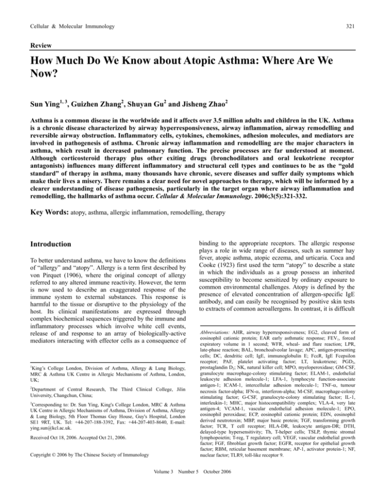
Cellular & Molecular Immunology 321 Review How Much Do We Know about Atopic Asthma: Where Are We Now? Sun Ying1, 3, Guizhen Zhang2, Shuyan Gu2 and Jisheng Zhao2 Asthma is a common disease in the worldwide and it affects over 3.5 million adults and children in the UK. Asthma is a chronic disease characterized by airway hyperresponsiveness, airway inflammation, airway remodelling and reversible airway obstruction. Inflammatory cells, cytokines, chemokines, adhesion molecules, and mediators are involved in pathogenesis of asthma. Chronic airway inflammation and remodelling are the major characters in asthma, which result in decreased pulmonary function. The precise processes are far understood at moment. Although corticosteroid therapy plus other exiting drugs (bronchodilators and oral leukotriene receptor antagonists) influences many different inflammatory and structural cell types and continues to be as the “gold standard” of therapy in asthma, many thousands have chronic, severe diseases and suffer daily symptoms which make their lives a misery. There remains a clear need for novel approaches to therapy, which will be informed by a clearer understanding of disease pathogenesis, particularly in the target organ where airway inflammation and remodelling, the hallmarks of asthma occur. Cellular & Molecular Immunology. 2006;3(5):321-332. Key Words: atopy, asthma, allergic inflammation, remodelling, therapy Introduction To better understand asthma, we have to know the definitions of “allergy” and “atopy”. Allergy is a term first described by von Pirquet (1906), where the original concept of allergy referred to any altered immune reactivity. However, the term is now used to describe an exaggerated response of the immune system to external substances. This response is harmful to the tissue or disruptive to the physiology of the host. Its clinical manifestations are expressed through complex biochemical sequences triggered by the immune and inflammatory processes which involve white cell events, release of and response to an array of biologically-active mediators interacting with effector cells as a consequence of 1 King’s College London, Division of Asthma, Allergy & Lung Biology, MRC & Asthma UK Centre in Allergic Mechanisms of Asthma, London, UK; 2 Department of Central Research, The Third Clinical College, Jilin University, Changchun, China; 3 Corresponding to: Dr. Sun Ying, King's College London, MRC & Asthma UK Centre in Allergic Mechanisms of Asthma, Division of Asthma, Allergy & Lung Biology, 5th Floor Thomas Guy House, Guy's Hospital, London SE1 9RT, UK. Tel: +44-207-188-3392, Fax: +44-207-403-8640, E-mail: ying.sun@kcl.ac.uk. Received Oct 18, 2006. Accepted Oct 21, 2006. Copyright © 2006 by The Chinese Society of Immunology Volume 3 Number 5 binding to the appropriate receptors. The allergic response plays a role in wide range of diseases, such as summer hay fever, atopic asthma, atopic eczema, and urticaria. Coca and Cooke (1923) first used the term “atopy” to describe a state in which the individuals as a group possess an inherited susceptibility to become sensitized by ordinary exposure to common environmental challenges. Atopy is defined by the presence of elevated concentration of allergen-specific IgE antibody, and can easily be recognised by positive skin tests to extracts of common aeroallergens. In contrast, it is difficult Abbreviations: AHR, airway hyperresponsiveness; EG2, cleaved form of eosinophil cationic protein; EAR early asthmatic response; FEV1, forced expiratory volume in 1 second; WFR, wheal- and flare reaction; LPR, late-phase reaction; BAL, bronchoalveolar lavage; APC, antigen-presenting cells; DC, dendritic cell; IgE, immunoglobulin E; FcεR, IgE Fcepsilon receptor; PAF, platelet activating factor; LT, leukotriene; PGD2, prostaglandin D2; NK, natural killer cell; MPO, myeloperoxidase; GM-CSF, granulocyte macrophage-colony stimulating factor; ELAM-1, endothelial leukocyte adhesion molecule-1; LFA-1, lymphocyte function-associate antigen-1; ICAM-1, intercellular adhesion molecule-1; TNF-α, tumour necrosis factor-alpha; IFN-α, interferon-alpha; M-CSF, macrophage-colony stimulating factor; G-CSF, granulocyte-colony stimulating factor; IL-1, interleukin-1; MHC, major histocompatibility complex; VLA-4, very late antigen-4; VCAM-1, vascular endothelial adhesion molecule-1; EPO, eosinophil peroxidase; ECP, eosinophil cationic protein; EDN, eosinophil derived neutrotoxin; MBP, major basic protein; TGF, transforming growth factor; TCR, T cell receptor; HLA-DR, leukocyte antigen-DR; DTH, delayed-type hypersensitivity; Th, T-helper cells; TSLP, thymic stromal lymphopoietin; T-reg, T regulatory cell; VEGF, vascular endothelial growth factor; FGF, fibroblast growth factor; EGFR, receptor for epithelial growth factor; RBM, reticular basement membrane; AP-1, activator protein-1; NF, nuclear factor; TLR9, toll-like receptor 9. October 2006 322 Atopic Asthma Clinical models of allergy After natural exposure or provocation by allergen, atopic allergic individuals can exhibit immediate responses in the lung (early asthmatic response, EAR) such as airway obstruction, immediate fall in FEV1 (forced expiratory volume in 1 second), skin (wheal and flare reactions, WFR, and oedema), nose (nasal itching, sneezing, and blockage), and eyes (tearing, oedema and itching) (conjunctiva). The immediate responses are usually maximal at 15 minutes and spontaneously resolved within 2 to 3 hours. Symptoms may recur in a proportion of individuals, starting at 3 to 12 hours after the initial exposure. This delayed-in time response is Volume 3 Number 5 +25 A FEV1 (%) Normal saline 0 -25 Allergen -50 -75 0 1 2 3 4 5 6 24 Time (hour) 80 Size (mm) to define asthma precisely. Most definitions are operational, i.e. they just describe clinical manifestations. The most important feature is airflow obstruction which is reversible either spontaneously or as the result of treatment. However, there is a reversible component in other airway diseases, such as cystic fibrosis, chronic bronchitis, and atopic rhinitis (1, 2). Asthma is also characterized by increased airway hyperresponsiveness (or hyperreactivity) (AHR) to physical, chemical, pharmacological and/or immunological stimuli. The major clinical symptoms of asthma are the episodic occurrence of coughing, dyspnea, wheezing and chest tightness, alone or in combination (1, 2). Atopy and asthma are closely related. Atopy may predispose asthma, although about 30% asthmatics are non-atopic (3). It has been known for many years that airway inflammation is a feature of asthmatic deaths, but whether this is a result of the disease or a feature of terminal illness is uncertain. The characteristic histologic pattern at autopsy includes denudation of airway epithelium, increased collagen deposition beneath the basement membrane, and infiltration of the tissue with eosinophils and lymphocytes (4, 5). With the advent of bronchoscopy as a research tool in asthma it was appreciated that many of these changes could be found even in patients with mild asthma (6, 7). Attention has more recently focussed on the lymphocyte and the interaction between activated T-cells and eosinophils. For example, irregularly shaped lymphocytes expressing CD25 markers (IL-2 receptor) as well as EG2+ (cleaved form of eosinophil cationic protein) eosinophils were found in bronchial mucosal biopsies of symptomatic but mild asthmatics (7, 8). Additionally, increases in the numbers of activated CD4+ helper T lymphocytes (CD25+) and eosinophils (EG2+) were also predominant features of biopsies obtained from allergen-induced late phase nasal and skin reactions (LPR) (9, 10). The number of eosinophils closely followed nasal symptoms and the diameter of late phase cutaneous reaction, respectively (11, 12) and there was predominance of “memory” T lymphocytes (CD4+/CD45RO+) after allergen exposure (13, 14). All these observations support the concept that: a) there is an inflammatory component in baseline clinical asthma and in models of asthma (i.e., LPR’s); b) the products of inflammatory cells play a critical role in asthma pathogenesis. B 60 40 20 0 Dil 15' 30' 1h 3h 6h 24 h 48 h 72 h Times Figure 1. Comparison of allergen-induced early and late phase responses in asthmatic airways (A) and skin of atopic subjects (size of late phase reaction) (B). FEV1, forced expiratory volume in 1 second termed the “late phase reaction” (LPR) (15). The peak of the reactions is usually at 6-12 hours post-challenge which gradually diminishes over the next 12-24 hours. LPR is, therefore, different in its kinetics from classical delayed-type (tuberculin) hypersensitivity which peaks at 24-48 hours. LPR is characterized by an antigen dose-dependent erythema and induration in the skin, and mucosal swelling in the respiratory tract, which results in decreased airflow (2). A comparison of allergen-induced airway response and skin reaction is shown in Figure 1. In human allergen-provoked skin, initial infiltration of neutrophils occurs within 1-2 hours of allergen-challenge (peaked at 6 hours) followed by prominent eosinophil accumulation, increasing over the next 12-48 hours. A variable increase in the number of CD4+ lymphocytes, predominantly CD45RO+ memory lymphocytes, with a minority expressing CD25+, was observed within 6 to 48 hours after allergen challenge. No accumulation of these inflammatory cells was found in October 2006 Cellular & Molecular Immunology 323 biopsies from diluent challenge sites (13, 16). Similar cellular infiltration of neutrophils, eosinophils, and CD4+/CD25+ cells was observed in nasal biopsies 24 hours after allergen provocation (17). In the lung, bronchoalveolar lavage (BAL) obtained 18-24 hours after allergen challenge showed little immediate cellular response but an allergen dose-dependent accumulation of neutrophils, CD4+/CD25+ lymphocytes and eosinophils (18, 19). Similar observation has been found in the bronchial mucosa in isocyanate-induced asthma (20). Collectively, the observations described above are similar to those findings in many patients with atopic dermatitis, atopic rhinitis, and atopic asthma. These studies confirm that LPR is a good model for researches and possibly a reason of chronic atopic allergic diseases, including atopic asthma. In symptomatic asthma, the percentages of mast cells, eosinophils, and epithelial cells in BAL fluid are found to be higher than that in healthy controls. When compared with normal controls, increases in activated eosinophils (EG2+), and T lymphocytes (CD4+/CD25+) have also been observed in both BAL fluid and bronchial biopsies from patients with mild or asymptomatic asthma (8, 21, 22). In addition, the degree of bronchial hyperresponsiveness is correlated with the increase in the proportions of the infiltrated inflammatory cells and their mediators. These observations indicate that the airways of asthmatic subjects undergo a number of priming inflammatory changes which may play an important role in priming cells for the subsequent release of mediators in acute attacks. Mechanisms of Allergy and Allergic Asthma As described above, allergy is an exaggerated immune response. When an allergen (like other antigens) enters the body, it is processed by macrophages and other antigenpresenting cells (APCs), such as dendritic cells (DCs). Degraded fragments from antigen (or allergen) are then presented to T lymphocytes which in turn help B lymphocytes to produce allergen-specific immunoglobulin E (IgE) antibodies. These antibodies attach to IgE Fcepsilon receptors (FcεRI) on mast cells in tissue as well as circulating basophils. This step is called “sensitization”. FcεRI is also expressed on epidermal langerhans cells, monocytes, eosinophils and smooth muscle (23, 24, 25). Once the relative allergen encounters the sensitized cell, the allergen molecules bind to IgE antibodies on the cell membrane. The binding of allergen and IgE on mast cells/basophils leads to the release of mediators, which include histamine, platelet-activating factor (PAF), leukotrienes (LT), and prostaglandin D2. These constrict bronchial airways, dilate blood vessels, increase permeability of small blood vessels, stimulate nerve endings and secretion of mucus in airway, resulting in the symptoms of allergic diseases (2). These events occur rapidly (seconds to 30 min) and resolve quickly. Mediators may also participate in the infiltration of eosinophils and other inflammatory cells which contribute to pathogenesis of allergy and asthma. Traditionally, mast cells were believed to be the major cells in the pathogenesis of Volume 3 Number 5 allergic disease. However, as already explained it is likely that other cells are involved, particularly in chronic allergic inflammation. In addition to FcεRI, lower-affinity IgE receptors (FcεRII) are expressed on various inflammatory cells, such as eosinophils, B lymphocytes, monocytes and other cell types (26). Mediators such as leukotrienes and prostaglandins are produced by several types of inflammatory cells, including eosinophils, platelets, and monocytes by IgE-dependent mechanisms (27). This is a further reason for believing that other cells, beside mast cells, are involved in IgE-dependent inflammatory mechanisms. Inflammatory cells in airway allergic inflammation Airway chronic inflammation is the feature of asthma and many cells are possibly involved in allergic inflammation include neutrophils, eosinophils, lymphocytes, monocytes/ macrophages, basophils, mast cells, natural killer (NK) cells, DCs, and platelets. Additionally, fibroblasts, endothelial, epithelial and smooth muscle cells may also participate under appropriate circumstances (i.e., remodelling, see below). Neutrophils Neutrophils possess three types of granules. The primary, azurophilic granule contains myeloperoxidase (MPO), neutral proteases (for example, elastase), acid hydrolyses and cationic proteins. The secondary “specific” granule contains lactoferrin, lysozyme, vitamin B12-binding protein, complement 5 (C5) cleaving enzymes, and specific collagenase. The tertiary granule is similar to the conventional lysosomes and contains acid hydrolases. After activation of neutrophils, their granule contents are secreted by exocytosis. Neutrophils also produce PAF and LTB4 from lipid membrane. Agents, such as GM-CSF (granulocyte macrophage-colony stimulating factor), enhance neutrophil effector function and other biological properties such as adhesion and migration. For instance, integrins such as endothelial leukocyte adhesion molecule 1 (ELAM-1), lymphocyte function-associate antigen-1 (LFA-1), and intercellular adhesion molecule-1 (ICAM-1) are involved in adhesion of neutrophils to endothelium and in their migration to local inflammatory sites. Neutrophils are also important source of cytokines such as TNF-α (tumour necrosis factor alpha), IFN-α (interferon alpha), M-CSF (macrophage-colony stimulating factor) G-CSF (granulocyte colony stimulating factor), IL-1 (interleukin), IL-1 receptor antagonist, IL-3, GM-CSF, IL-6, IL-8 and other chemokines (28). Although neutrophils clearly play an important role in inflammation (29), their role in allergy and asthma is still unclear. The precise role of neutrophils in the LPR and their contribution to atopic asthma remains to be confirmed although a few studies have reported that these also appear early in tissue after allergen challenge of skin and nose and in BAL after inhalational challenge (12, 17, 30). In steady state on-going asthma their numbers are no different from controls (8). October 2006 324 Atopic Asthma Monocytes/macrophages Monocytes/macrophages are part of the mononuclear phagocyte series. They are derived from the bone marrow. Promonocytes differentiate in the blood to monocytes, and finally reside the tissues as mature macrophages. However, monocytes and macrophages differ in both morphology and certain functions. For instance, the monocyte is relatively less adherent, and less “activated”. In contrast, tissue macrophages have a higher capacity for adherence, and have efficiency at phagocytosis and antigen presentation to T lymphocytes. Monocytes and macrophages both express receptors for the Fc portion of IgG1, IgG3, IgA, IgM, IgE as well as complement receptor CR1, CR3 and MHC (major histocompatibility complex) class II antigens. These cells also contain a very wide variety of enzymes and mediators including eicosanoids, lysosomal enzymes, interleukins, complements and coagulation factors which are involved in various inflammatory reactions. Thus, macrophages are major phagocytic cells with a special ‘scavenger role’ in tissue repair. A number of cytokines can activate macrophages or modulate their functions, such as IL-2, IL-3, IL-4, GM-CSF, TNF-α, M-CSF and IFN-γ. Of these, IFN-γ is particularly effective at activating macrophages. It has been suggested that activated macrophages may contribute to the airway inflammation of asthma through their capacity to secrete performed mediators, oxidative burst metabolites, arachidonate metabolites, PAF and other cytokines. Usually, more than 80% of the cells in BAL fluid are macrophages. Compared to controls, the numbers of lung FcεRII+ macrophages and peripheral blood FcεRII+ monocytes were increased in atopic individuals. Furthermore, macrophages release LTB4 and LTC4 following treatment with anti-IgE antibody. Surprisingly, most studies of BAL have suggested that the number of macrophages in asthmatics was not increased compared with normal controls. Some previous reports have suggested that alveolar macrophages possess potent down-regulatory functions in both mitogen- and antigen-stimulated T- and B-cell proliferation as well as in vitro antibody responses. Elimination of alveolar macrophage was associated with an increase in the pulmonary immune response in mice. Alveolar macrophages of mice suppress the in vitro generation of IgM, IgA, IgG and IgE. These observations suggest that macrophages, or at least alveolar macrophages, may have a normal physiological role by such potent inhibitory function. However, the precise function of monocytes/macrophages in allergic inflammation remains to be determined. A very recent study showed that deficient induction of interferon-λ by rhinovirus in asthmatic primary bronchial epithelial cells and alveolar macrophages, which was highly correlated with severity of rhinovirus- induced asthma exacerbation and virus load in experimentally infected human volunteers (31). This suggests that some products from macrophages might be benefit for asthmatics. Eosinophils Eosinophilia in peripheral blood and local tissues is a feature of asthma. In 1879, Paul Ehrlich firstly identified a particular type of blood cell which had the remarkable ability to bind Volume 3 Number 5 acidic dye eosin and termed this as the “eosinophil”. Eosinophils are non-dividing bone marrow-derived cells. In health, they constitute less 5% of the circulating white cells. Eosinophil accumulation is associated with the pathology and symptomatology of several diseases, such as pulmonary eosinophilic syndrome, helminth-induced inflammation, certain chronic digestive diseases, and allergic disorders, such as allergic bronchial asthma, allergic rhinitis and atopic dermatitis (1, 2). In patients with the rare idiopathic hyper eosinophilic syndrome, there is a predominance of hypodense eosinophils, as shown by separation on density gradients. These low-density cells have fewer granules in their cytoplasm, and increased oxygen consumption, phagocytic and cytotoxic capacity and generation of lipid mediators. These characteristics suggest that the hypodense eosinophils are activated. Eosinophils carry surface receptors for IgG (FcγRII, CD32), and for the complement fragments (CR1 and CR2). They also express integrins such as VLA-4 (very late antigen) and LFA-1. The integrins and their ligands (adherence molecules) are involved in immigration of eosinophils, basophils and T cells, particularly VLA-4VCAM-1 (vascular endothelial adhesion molecule) pathways (32). Any factor upregulating these integrins/ligands might induce eosinophil migration, which might represent the mechanism whereby eosinophils infiltrate into local tissue. Of these various agents, some CC chemokines, particularly eotaxin 1-3 specifically upregulate eosinophil chemotaxis (28, 33, 34). CD34+ early eosinophil precursors differentiate mature under the influence of IL-3, GM-CSF and IL-5. Terminal differentiation is mediated by IL-5. Once activated, eosinophils are primed for enhanced release of both granuleassociated and membrane-derived mediators following contact with large surface opsonized by immunoglobulin and/or complement. These mediators are released from eosinophils by either IgE- or IgA-dependent mechanisms. Eosinophils are readily recognized by their bilobed nucleus and many granules. The major granule population in mature cells (secondary granules) consists of large membrane-bound structures with two substructural compartments, crystalline core and matrix-within which the granule proteins, such as EPO (eosinophil peroxidase), ECP (eosinophil cationic protein), EDN (eosinophil derived neutrotoxin) and MBP (major basic protein) are compartmentalized. There is a second less numerous primary granule devoid of central crystalline cores and contains the major store of Charcot-Leyden crystal (CLC) protein. There are also small granules which contain hydrolytic enzymes and numerous endocytic structures. Additionally, mature tissue eosinophils display increased numbers of lipid bodies rich in arachidonic acid. The eosinophil secondary granule proteins, ECP, EDN, MBP and EPO, have been isolated and purified and their biological properties have also been characterized. Basic proteins, such as MBP, ECP, EDN and EPO, are cytotoxic to parasites, bacteria, and mammalian cells. Because highaffinity FcεRI also expressed on eosinophils from hypereosinophilic patients, and receptor-binding enhances the release of EPO, suggesting that FcεRI may play a major part role in immune defence against parasites. EDN and ECP in October 2006 Cellular & Molecular Immunology 325 particular are potent ribonuclease and display neurotoxic properties. EPO and ECP are also cytotoxic to epithelium in vitro. In particular, MBP is toxic to epithelial cells in concentration as low as 9 × 10⁻⁷ M and MBP deposition was found in the bronchial wall and mucus plugs in asthma deaths (2). The sites of MBP deposition coincided with widespread epithelial damage characteristic of the airways in severe bronchial asthma. In addition, PAF and LTC4 produced by activated eosinophils may enhance the allergic response due to their abilities of causing vasodilation and increased vascular permeability, and inducing eosinophil chemotaxis. Corresponding this, there is increasing evidence to implicate eosinophils in tissue damage characteristic of asthma. For example, bronchial hyperresponsiveness is associated with increased amounts of eosinophils and their products in BAL fluid. Furthermore, accumulation and infiltration with activated eosinophils (EG2+) have been observed in asthmatic bronchial biopsies, and cutaneous and nasal late phase responses following antigen challenge in atopic subjects (2). Additionally, activated eosinophils are also important source of cytokines and chemokines, including TGF-α (transforming growth factor-alpha), TGF-β1, IL-1α, IL-3, IL-5, IL-6, IL-8, IL-10, IL-13, IL-25, TNF-α, GM-CSF, some CC and CXC chemokines (35). These observations, taken together, suggest that eosinophils and their products may play a direct or indirect effector role in allergic inflammation, especially in asthma. However, they may also act as repair cells by remodelling (see below) possibly through elaboration of TGF-β (35). Our and other recent studies also indicate that eosinophils are effector cells in process of remodelling (see below) in chronic inflammation including asthmatic airways (36-38). Basophils and mast cells Basophils and mast cells are important effector cells in pathogenesis of asthma and other allergic diseases. Basophils and mast cells have common properties. They both contain large intracytoplasmic histamine-containing metachromatic granules, and express membrane FcεRI. When cross-linked by anti-IgE or, by binding of specific antigen to IgE-bound to these receptors, both cell types can be triggered to release granule-derived, pre-formed products and lipid mediators (39). However, there are a number of important differences between mast cells and basophils. Basophils are smaller than mast cells, comprising around 0.1% of the total white cell count. Compared with mast cells, basophils have a multilobed nucleus and contain a few large more electrondense granules. Basophils are attracted to tissues in certain inflammatory and immune reactions, e.g., a particular type of delayed reactions called cutaneous basophil hypersensitivity (40). IL-3 appears to be a major factor for the growth, differentiation and maturation of basophils from myelocyte progenitors and committed precursors. After activation, basophils release numerous mediators such as histamine, chondroitin sulphate, major basic protein, and LTC4. Mast cells are usually distributed throughout normal mucosa and connective tissues, especially in the skin, respiratory system, gastrointestinal and genitor-urinary tracts. In man, mast cells Volume 3 Number 5 are a heterogeneous population which are according to their content of the neutral proteases, tryptase and chymase. Thus, mucosal mast cells which contain tryptase are designated McT, while connective tissue mast cells which contain tryptase and chymase are designated McTC (41). IL-3, stem cell growth factor and IL-4 appear to be involved in the growth and activation of both types of mast cells. The activated mast cells can rapidly release large amounts of histamine, neutral proteases, heparin, chondroitin sulphates, lipid mediators including PGD2, PAF, LTB4, and LTC4. The release of PGD2 and LTC4 requires about 15 minutes whereas histamine release is completed within 2-5 minutes. These mediators may cause smooth muscle contraction, vessel dilatation, capillary permeability, mucosal oedema, cellular infiltration and mucus hyper-secretion (39, 42). Mast cells and basophils, therefore, have been considered to participate in IgE-mediated immediate type hypersensitivity. It has been reported that there are increases in the concentrations of histamine and other mediators in BAL fluid during the early asthmatic responses provoked by allergen, suggesting that mast cells may dominate the process underlying early phase allergic reactions. Basophils appear to be mainly associated with the late phase responses, Naclerio et al. (43) demonstrated that in the early response following nasal allergen challenge, the levels of histamine, LTC4 and PGD2 are increased, while in the late phase response, increases in histamine and LTC4, but not PGD2, were observed. This finding was also supported by the observations in skin blister and bronchial allergen challenge models (18). The mechanisms of basophil recruitment in allergic diseases are still unclear. A number of studies have suggested that cytokines such as IL-3, IL-5, GM-CSF and some CC chemokines are potential chemotactic factors for basophils (44). In addition to immuno-stimulation, numerous nonimmunological agents also cause mast cell or basophil degranulation, including C3a, C5a, certain drugs such as codeine, certain cytokines, chemokines, a number of physical stimuli, and several neuropeptides, i.e., substance P. These studies indicate that the events associated with the activation and degranulation of mast cells and basophils are complex and not confined to atopic allergic diseases. Same as other cell types, basophils and mast cells are potential sources of cytokines and chemokines (39). It is possible that mast cell-derived cytokines, chemokines and other mediators implicate the relevant cells in the initiation of late phase responses. Collectively, basophils and mast cells may serve as a potent source of growth, regulatory and inflammatory agents. Their mediators, including cytokines, may serve as autocrine growth factors and influence other cells involved in allergic inflammation. Lymphocytes As far as we knew, B cell-derived IgE is an important mediator in pathophysiology of asthma. IgE is directly associated with allergic inflammation and elevated concentration of total and specific IgE is characteristic of many allergic diseases. As described above, IgE binds to mast cells/basophils via the high affinity receptor (FcεRI). October 2006 326 Atopic Asthma Following exposure to specific antigen, allergen-IgE complexes lead to the release of mediators from the sensitized cells. This is the basis of immediate hypersensitivity reactions. The exposure of B lymphocytes to antigen and direct contact with “helper” (CD4+) T cells are critical for IgE production since they provide critical co-factors, particularly IL-4 and soluble CD23. In addition, other soluble mediators produced by T cell, such as IL-2, IL-5, IL-6, IL-9, IL-13 and IFN-γ, can also influence the regulation of IgE production. B cells themselves can produce cytokines such as TNF-α, IL-6, IL-10 and TGF-β in vitro after stimulation. Apart of B cells, T lymphocytes are another kind of “memory cells”. Upon a primary antigen exposure, T cells differentiate from “virgin” or “naïve” to memory T cells. Following secondary antigenstimulation, T cells mature into effector cells and accumulate in local tissues. T cells recognize foreign antigen via the T cell receptor (TCR) as the antigen is processed and presented on the surface of accessory cells in association with MHC class I or class II molecules. Cells termed APC perform this function. Several cell types can act as APC’s, including mononuclear phagocytic series (macrophages, Langerhans cells, and dendritic cells) as well as B cells, endothelial and epithelial cells and even eosinophils. The interaction of T cells and APC leads to T cell differentiation, proliferation and activation. The activated cell is then endowed with effector functions and secretes cytokines. In man, the majority of T cells can be divided into two subsets (CD4+ and CD8+). CD4+ T cells perform “helper” function in antibody production and exhibit an antigen response restricted to MHC class II molecules. CD8+ T cells, originally described as “suppressor” cells in immunoglobulin production are “cytotoxic” for antigen-bearing target cells, and their antigen recognition is class I MHC-restricted. Thus, CD4+ T cells perform most of their functions via the action of secreted cytokines and also interact with B cells to induce proliferation and antibody production. CD8+ cells, on the other hand, “kill” MHC class I-bearing neoplastic and virally infected cells expressing novel antigens. Αctivated T lymphocytes can express a number of surface molecules that can serve as makers of activation. These include CD25 (IL-2 receptor), VLA-1, human leukocyte antigen-DR (HLA-DR) and CD69. It is currently believed that T lymphocytes and their soluble products (1, 2) may mediate the characteristic cell pathology of allergic inflammation (i.e., eosinophil infiltration). Increased CD4+ T cells were also observed in BAL fluid 48 hours after allergen challenge and this was accompanied with decreases in peripheral blood CD4+ cells (45). This suggested that CD4+ T cells might have been recruited from intravascular space into BAL fluid after antigen provocation. It was then observed that atypical, irregular lymphocytes (CD25+ presumed to be activated T cells) were a prominent feature of asthmatic bronchial biopsies examined by electron microscopy (7, 8, 19, 20). This was in agreement with earlier observations involving allergen-induced late phase responses in the skin (12). This was also in agreement with observations in peripheral blood, where activated CD4+ cells (i.e., IL-2R+, HLA-DR+, and VLA-1+) were observed in the patients with acute, severe Volume 3 Number 5 asthma. After therapy, the percentage of activated T cells decreased and clinical symptoms resolved (47). In a mouse model of delayed-type hypersensitivity reaction (DTH) to picryl chloride in the lung, the induced tracheal hyperreactivity and increased pulmonary resistance were shown to be T cell dependent. Airway hyperresponsiveness was observed after challenge of mice passively sensitized by intravenous injection of lymphoid cells from sensitized donor mice. After the depletion of T cells from the donor mice, the passively transferred hyperresponsiveness was significantly suppressed (48). Thus, in this model, it seems that mainly T cells mediated airway hyperresponsiveness and altered lung function. In another mouse model, antigen-induced eosinophil recruitment into the mouse trachea was shown to be mediated by CD4+ T cells and IL-5. Administration of recombinant IFN-γ prevented antigen-induced eosinophil infiltration and also decreased antigen-induced CD4+ T cell infiltration into the trachea (49, 50). Taken together, these observations indicate that lymphocytes, particularly activated CD4+ T cells, may play a critical role in allergic inflammation, not only by “helping” B cells to produce IgE, but also by releasing cytokines which may act on the effector cells which amplify the inflammatory response. Th1 and Th2 cytokines Following the establishment of techniques of cell cloning, it became clear that there might be distinct subsets in T lymphocytes. In 1986, Mosmann and colleagues suggested that murine CD4+ T helper (Th) cell clones could be divided into at least two distinct subsets, termed Th1 and Th2, based on their different pattern of cytokine secretion (45, 46, 51, 52). Th1 cells elaborated IL-2, IFN-γ and TNF-β (but not IL-4 and IL-5), whereas Th2 secreted IL-4, IL-5, IL-6, IL-10, IL-13 (but not IL-2 and IFN-γ). Both subsets secreted IL-3, GM-CSF and TNF-α. Cytokines produced by both subsets appear to downregulate the function of each other. A precursor of Th1 and Th2 cells that produces all of these cytokines was termed Th0. A major interest of this T cell dichotomy related to the different types of pathology associated with the murine Th1 and Th2 response. Th1 responses were produced by immunization with tuberculin and were associated with classical delayed type hypersensitivity. In contrast, infection with helminths gave a Th2 pattern together with IgE and eosinophilia. The cytokines produced by Th1 cells can induce expression MHC antigens on macrophages and activate these cells to kill intracellular pathogens. Th1 (but not Th2) cells mediate delayed-type hypersensitivity, while IL-4 induces proliferation of Th2 cells that were effective cells in helping B-lymphocytes to produce IgG1 and IgE. Thus, Th2 cells are believed to participate mainly in humoral immune reactions through their ability to promote antibody synthesis. IL-4 plays important role in IgE isotype switching, the upregulation of vascular cell adhesion of eosinophils, T cells and basophils, increasing Th2 cell commitment in the October 2006 Cellular & Molecular Immunology 327 airways of allergic asthmatic subjects and possibly contributing to allergen-induced airway eosinophilia. IL-5 is very specific cytokine involved in the development, differentiation, recruitment, activation, and survival of eosinophils. Additionally, IL-13, shared same receptor with IL-4, induces mucous hyperplasia that may be necessary for allergen-induced airway hyperresponsiveness, at least in mouse models (53). Increasing data suggest that Th1/Th2 imbalance result in the clinical expression of allergy and asthma. We and others have demonstrated predominant expression of Th2 cytokines in asthmatic airways and in some allergic models (54-68). How CD4+ T helper lymphocytes differentiate into distinct subsets such as Th2 is not clear. It has been proposed that several factors may be involved in this process, including the nature and concentration of antigen, the type of APC, the tissue microenvironment, and route of immunization during priming and the clonal expansion or effector stage of response. In vitro, TSLP (thymic stromal lymphopoietin)“educated” DC selectively drives development of Th2 responses through its actions on CD11c+ dendritic cell (69). Underlining the clinical relevance of TSLP, we observed that expression of TSLP and Th2-attracting chemokines was elevated in the airway of asthmatics as compared with normal controls, and that expression of TSLP correlated both with that of Th2-attracting chemokines and with clinical lung function (70). Organ-specific expression of a TSLP transgene induced a Th2-type T cell-mediated atopic dermatitis-like phenotype and airways inflammation and hyperreactivity in mouse models (71, 72). Furthermore, TSLPR gene-deleted mice exhibit strong Th1 response (73). TSLP-exposed DCs not only induce a robust expansion of CD4+ Th2 memory cells, but also maintain their central memory phenotype and Th2 commitment (74). These observations suggest that TSLP favours the development of Th2 responses to allergen through its effects on dendritic cells, which in turn release Th2-attracting chemokines and prime naïve T cells forwards to Th2 commitment. It worth notes that T regulatory (T-reg) cells may protect against the development of atopic allergic diseases, including asthma, possibly through IL-10 although there are still many unclear events to be known (75, 76). Compared with Th2 cytokines, Th1 cytokines (i.e., IFN-γ and TNF-α) may play less role in mild and moderate asthma but may maintain chronic inflammation (77). Airway Remodelling In asthma, inflammation occurs throughout the tracheobronchial tree, even in the small airways, and is the underlying component to be treated. Remodelling in the airways results from the chronic inflammation associated with asthma that disrupts the normal repair process. It involves thickness of the airway walls accounting for mucous hyperplasia, mucosal neovascularisation, smooth muscle hyperplasia, deposition of fibrous and other extracellular matrix protein, myofibroblast hyperplasia, epithelial hypertrophy, and mucus gland and goblet cell hyperplasia. Volume 3 Number 5 Although these phenomena have been observed for long time, most processes are far understood. Excessive mucus production is a cardinal clinical feature of asthma (78). In animal models, the cytokines IL-13 and IL-9 have been directly implicated in causing mucous hyperplasia (79, 80), but the contribution of this phenomenon to incompletely reversible airway obstruction is unknown. Compelling evidence suggests that mucosal neovascularisation occurs in chronic asthma (81, 82). Studies have implicated growth factors including vascular endothelial growth factor (VEGF) and fibroblast growth factor 2 (FGF2) derived from eosinophils, macrophages and smooth muscle cells in its causation (83). Inverse correlation of mucosal vascularity with airway calibre (82) suggests that neovascularisation may contribute to airways obstruction. However, the contribution of neovascularisation to partial irreversibility of airway obstruction is unknown. Increased airway smooth muscle mass (increase in myocyte numbers and possibly also size) has long been documented in chronic severe asthma (84). Thickening of the airway wall caused by increased smooth muscle mass may cause a greater degree of airway narrowing for a given degree of muscle contraction (85); nevertheless the contribution of smooth muscle hyperplasia to partially reversible airways obstruction is unknown. In asthma, deposition of fibrous and matrix proteins such as collagen types I, III and V, tenascin and fibronectin beneath the epithelial basement membrane (lamina reticularis) and within the submucosa of the airways is again a pathognomonic feature of asthma (86, 87). Mesenchymal cells, including myofibroblasts and smooth muscle cells produce such proteins. There is good evidence that this process, somewhat analogously to that of scar formation, attenuates airway compliance as measured, for example, by distensibility (88), but the contribution of this phenomenon to partly irreversible airway obstruction is not clear. Fibroblast activation and myofibroblast differentiation and proliferation also occur in asthmatic airways. Activation of fibroblasts, with differentiation to myofibroblasts results in lay down of fibrous proteins in the airways. This is regulated by the production of fibrogenic growth factors produced by epithelial cells (partly as a result of exposure to fibrogenic cytokines such as IL-13 and IL-9) and infiltrating inflammatory cells such as eosinophils and mast cells (89). The most important of these is TGF-β. As with fibrous protein deposition per se, this phenomenon has not so far been implicated in causing airway obstruction which is not fully reversible. Mucosal epithelial cell perturbation is a common feature of asthmatic airway. Mucosal epithelial cells are activated in asthma and produce mediators relevant to remodelling such as TGF-β, as well as other pro- inflammatory mediators such as IL-8 and GM-CSF (89). Although the question of whether or not they are “damaged” by products of inflammatory cells in asthma remains controversial (90), they certainly show a marked “repair” response by upregulating the receptor for epithelial growth factor (EGFR, c-erb B1) (91), although one early single study (92) did not suggest that they undergo rapid mitosis. Nevertheless epithelial cell activation, and possible turnover, is likely a key driving stimulus for airway October 2006 328 Atopic Asthma Allergen IgE B cell Mast cell Mediators IL-4 IL-5 IL-13 DC IL-4 Th0 cell Chemokines IL TG -4, I F- L-1 β 3 P T ch h2em ok in es + T SL C chem C okin es ? ? Th2 IL-5 TGF-β, IL-13 Immediate reactions GFs, Smooth muscle Chronic inflammation Growth factors Fibrogenic factors Late reactions ongoing asthma Airway remodelling Eosinophil Figure 2. Mechanisms in atopic asthma. A Th0 (naïve helper T cell) is stimulated from an antigen (allergen) presenting cell (APC, here is a dendritic cell, DC) and IL-4, to become a Th2 cell. Th2 cell produces Th2 cytokines, induce B cell to produce IgE which will binding to mast cell through IgE FcεR (sensitization). When the same allergen enters body again, it will directly bind to IgE bound on mast cell and result in releasing of mediators causing immediate reactions. Th2 cell also promotes eosinophilia by IL-5. Activated eosinophil produces TGF-β, IL-13 and other growth factors (GFs) participating in airway remodelling. Th2 cell might directly induce smooth muscle construction and late reactions of asthma. Chemokines, derived from DC and other cell types, contribute to accumulation of Th2 cells and eosinophils into local tissue. Additionally, thymic stromal lymphopoietin (TSLP)-educated DC favours differentiation of Th0 to Th2 cell. remodelling, but whether or not it can be implicated in causing airway obstruction which is not fully reversible is unknown. Current mechanism of atopic asthma is diagrammed in Figure 2. Common and potential treatments of asthma Current asthma therapy with corticosteroid plus bronchodilators and oral leukotriene receptor antagonists is aimed at controlling airway inflammation and relief disease symptoms. However, a subset of asthma patients remains symptomatic despite optimal therapy indicating that an unmet medical need exists for these patients. Also anti-airway remodelling may be particularly useful for benefits of asthmatics. We just list some potential molecular targets for consideration for developing further therapeutics in asthma. Anti-IgE monoclonal antibody (omalizumab) and IgE receptor antagonists IgE is important initial mediator in pathogenesis of allergic inflammation and atopic asthma. Thus, anti IgE is possibly Volume 3 Number 5 useful treatment for these patients. Using recombinant humanized monoclonal IgG anti-IgE antibody (omalizumab), we have shown that omalizumab-treated patients had a progressive reduction in the LPR that was significantly greater than its effect on the EPR (93). There are two other clinical trails also showed that omalizumab reduced moderate and severe asthmatic exacerbations and reduced the dose of corticosteroids and β-agonist needed (94-96). The costing and safety seem to be the major question as common usage of this treatment. Modulate Th2- and enhance Th1-cytokines Because Th2 cells play role in atopic asthma and Th1 cells inhibit function of Th2 cells, it is possible that re-balance of Th1/Th2 may get some benefits for asthmatics. At moment, there is no convincing data showing any benefits obtained from anti IL-4 antibodies or antagonists of IL-4 receptor (97). In a double-blind, placebo-controlled study, targeting IL-5 with monoclonal antibody (mepolizumab) in asthmatics showed that at baseline, airway eosinophil infiltration and basic protein deposition was increased in the reticular basement membrane (RBM) of asthmatics compared with October 2006 Cellular & Molecular Immunology 329 nonasthmatic controls (37). Treating asthmatics with anti-IL-5 antibody, which specifically decreased airway eosinophil numbers, significantly reduced the expression of tenascin, lumican, and procollagen III in the bronchial mucosal RBM when compared with placebo. In addition, anti-IL-5 treatment was associated with a significant reduction in the number and percentage of airway eosinophils expressing mRNA for TGF-β1 and the concentration of TGF-β1 in BAL fluid, suggesting that eosinophils may contribute to tissue remodeling processes in asthma by regulating the deposition of eosinophil basic proteins (37). Although some promising data have been obtained from targeting IL-13 in murine studies (53, 98), this still needs to be confirmed in human studies. Attentively, enhancing Th1 cytokines such as IFN-α and γ, IL-12, IL-18 and IL-23 may inhibit Th2-mediated responses. However, formal clinical trail will need for convincing these treatments. Although some effects were shown in a double blind study with anti-human CD4 antibody (99), some risk still exists due to lack of target specificity and infection of HIV. Additionally, some effects were shown in severe asthma treated with anti-TNF-α antibodies and antagonist of soluble TNF-α receptor (77). Anti-TNF-α treatment was originally used in patients with rheumatoid arthritis, a typical Th1-mediated disease. The benefits of such treatment in asthma suggest that TNF-α and other Th1 cytokines may be responsible in some severe steroid-resistant asthma. Blocking accumulation of inflammatory cells by targeting chemokines and receptors or adhesion molecules As mentioned before, eosinophilia and predominance of Th2 cells are the characters of atopic asthma, blocking accumulation of these cells may relieve asthmatic attack. Some effects have been shown in animal models of asthma by usage of blocking chemokines and their receptors as well as targeting adhesion molecules such as LFA-1, VCAM, and VLA-4 (100, 101). Again, there is still long way to go because many chemokines and adhesion molecules are involved in cellular accumulation and single blocking may not be sufficient to treat asthma. Targeting key factors in signal transduction and gene transcription It is reasonable for that specifically targeting some key factors such as in signal transduction and gene transcription pathways may be effective in asthma treatment because these factors play important roles either in up- or down-stream of functions of certain molecules. Using specific inhibitors or nucleic-acid based technologies such as antisense oligonucleotides, ribozymes, DNAzymes, decoys oligonucleotides and RNA interference, blocking action of STAT1 and STAT6 in signal transduction and activator protein-1 (AP-1) or nuclear factor (NF-κB) in gene transcription could be helpful in reducing asthmatic symptoms, at least in mouse models of asthmatic inflammation (102, 103). The major questions are how to employ these techniques to human and how to minimise or avoid the side effects derived from targeting these factors. Volume 3 Number 5 Immunotherapy In a previous study, we showed that immunotherapy was associated with suppression of allergen-induced CD4+ T lymphocyte infiltration, but among the cells that are recruited, there is upregulation of CD25 and HLA-DR as well as enhancement of Th1 cytokines (without affecting Th2 cytokines), suggesting that immunotherapy may possibly be working through induction of T cell tolerance (60). Oral administration of an allergen favours the development of tolerance in which T-reg secreting TGF-β is involved. Administration of high-dose allergen immunotherapy by means of subcutaneous injection also induces the development of T-reg, with evidence that the secretion of both IL-10 and TGF-β is important in the mechanism of tolerance (104). It has also been shown that bacterial DNA with CpG oligodeoxynucleotides capable to stimulate Th1 cell but inhibits Th2 cell responses through interacting with toll-like receptor 9 (TLR9). These molecules, therefore, can be used alone or with allergen-specific immunotherapy (105). Thus, immunotherapy might be profitable for asthmatics and other atopic diseases. Mediator antagonists Selective mediator antagonists are the potential drugs for asthma treatment. A successful example is cysteinyl leukotriene antagonists such as montelukast which has been used as profitable medication for chronic asthma (106). Other potential drugs, including inhibitors of thromboxane and prostaglandin D2 (PGD2) might be also useful for treatment of asthma (107, 108). Inhibitors for airway remodelling It was reported that overexpressing VEGF caused neovascularization and Th2-like inflammation in genetransgenic mice (109). However, at moment, there is still no promising evidence for singly targeting which could be useful for asthmatic therapy (110). Conclusion Asthma care cost the huge mount of money annually, not to mention the economic and psychosocial implications of loss of time from work and school. Although atopic asthma has been known for a quite long time, the pathogenesis is still far known, particularly late phase reactions and remodelling. Remodelling could be the results from chronic inflammation or parallel exist from the beginning. We do not know if the persistent structural changes still remain without allergen exposure or inflammation either. Many factors contributing to these processes and various clinical subtypes of asthma may limit our development of therapy. The data from animal models that cannot be fully translated to human also limit our knowledge of asthma. Thus, any potential treatments in late phase responses and airway remodelling will be great helpful, not only in subgroup of asthmatics, but also in other airway obstructive diseases. October 2006 330 Atopic Asthma References 1. McFadden ER Jr, Gilbert IA. Asthma. N Engl J Med. 1992;327: 1928-1937. 2. Kay AB. Allergy and allergic diseases. First of two parts. N Engl J Med. 2001;344:30-37. 3. Humbert M, Menz G, Ying S, et al. The immunopathology of extrinsic (atopic) and intrinsic (non-atopic) asthma: more similarities than differences. Immunol Today. 1999;20:528-533. 4. Dunnill MS, Massarella GR, Anderson JA. A comparison of the quantitative anatomy of the bronchi in normal subjects, in status asthmatics, in chronic bronchitis, and in emphysema. Thorax. 1969;24:176-179. 5. Cutz E, Levison H, Cooper DM. Ultrastructure of airways in children with asthma. Histopathol. 1978;2:407-421. 6. Laitinen LA, Heino M, Laitinen A, Kava T, Haahtela T. Damage of the airway epithelium and bronchial reactivity in patients with asthma. Am Rev Respir Dis. 1985;131:599-606. 7. Jeffery PK, Wardlaw AJ, Nelson FC, Collins JV, Kay AB. Bronchial biopsies in asthma. An ultrastructural, quantitative study and correlation with hyperreactivity. Am Rev Respir Dis. 1989;140:1745-1753. 8. Azzawi M, Bradley B, Jeffery PK, et al. Identification of activated T lymphocytes and eosinophils in bronchial biopsies in stable atopic asthma. Am Rev Respir Dis. 1990;142:14071413. 9. Durham SR, Ying S, Varney V, et al. Cytokine messenger RNA expression for the IL-3, IL-4, IL-5 and GM-CSF in the nasal mucosa after local allergen provocation: relationship to tissue eosinophilia. J Immunol. 1992;148:2390-2394. 10. Kay AB, Ying S, Varney V, et al. Messenger RNA expression of the cytokine gene cluster, IL-3, IL-4, IL-5 and GM-CSF in allergen-induced late-phase cutaneous reaction in atopic subjects. J Exp Med. 1991;173:775-778. 11. Pipkorn U, Karlsson G, Enerback L. The cellular response of the human allergic mucosa to natural allergen exposure. J Allergy Clin Immunol 1988;82:1046-1054. 12. Frew AJ, Kay AB. The relationship between infiltrating CD4+ lymphocytes, activated eosinophils, and the magnitude of the allergen-induced late phase cutaneous reaction in man. J Immunol. 1988;141:4158-4164. 13. Frew AJ, Kay AB. UCHL1+ (CD45RO+) “memory” T cells predominate in the CD4+ cellular infiltrate associated with allergen-induced late-phase skin reactions in atopic subjects. Clin Exp Immunol. 1991;84:270-274. 14. Hellquist HB, Karlsson MG. Nasal memory T lymphocytes capable of producing IL-4 in the allergic reaction. Allergy. 1992; 47:334-336. 15. Booij-Nord H, Orie NGM, de Vries K. Immediate and late bronchial obstructive reactions to inhalation of house dust and protective effects of disodium cromoglycate and prednisolone. J Allergy Clin Immunol. 1972;48:344-354. 16. Zweiman B. The late-phase reaction: role of IgE, its receptor and cytokines. Curr Opin Immunol. 1993;5:950-955. 17. Varney VA, Jacobson MR, Sudderick RM, et al. Immunohistology of the nasal mucosa following allergen-induced rhinitis. Identification of activated T lymphocytes, eosinophils, and neutrophils. Am Rev Respir Dis. 1992;146:170-176. 18. Sedgwick JB, Calhoun WJ, Gleich GJ, et al. Immediate and late airway response of allergic rhinitis patients to segmental antigen challenge. Am Rev Respir Dis. 1991;144:1274-1281. 19. Robinson DS, Ying S, Bentley AM, et al. Relationships among numbers of bronchoalveolar lavage cells expressing messenger ribonucleic acid for cytokines, asthma symptoms, and airway Volume 3 Number 5 20. 21. 22. 23. 24. 25. 26. 27. 28. 29. 30. 31. 32. 33. 34. 35. 36. 37. methacholine responsiveness in atopic asthma. J Allergy Clin Immunol. 1993;92:397-403. Bentley AM, Maestrelli P, Saetta M, et al. Activated T-lymphocytes and eosinophils in the bronchial mucosa in isocyanateinduced asthma. J Allergy Clin Immunol. 1992;89:821-829. Walker C, Kaegi MK, Braun P, Blaser K. Activated T cells and eosinophilia in bronchoalveolar lavages from subjects with asthma correlated with disease severity. J Allergy Clin Immunol. 1991;88:935-942. Wardlaw AJ, Dunnette S, Gleich GJ, Collins JV, Kay AB. Eosinophils and mast cells in bronchoalveolar lavage in subjects with mild asthma. Relationship to bronchial hyperreactivity. Am Rev Respir Dis. 1988;137:62-69. Ying S, Barata LT, Meng Q, et al. High affinity IgE receptor (FcεRI)-bearing eosinophils, mast cells, macrophages and Langerhans cells in allergen-induced late-phase cutaneous reactions in atopic subjects. Immunology. 1998;93:281-288. Smith SJ, Ying S, Meng Q, et al. Blood eosinophils from atopic donors mRNA for the α, β, and γ subunits of the high affinity IgE receptor (FcεRI) and intracellular, but not cell surface, α subunit protein. J Allergy Clin Immunol. 2000;105:309-317. Gounni AS. The high-affinity IgE receptor (FcεRI): a critical regulator of airway smooth muscle cells? Am J Physiol Lung Cell Mol Physiol. 2006;291:L312-321. Rosenwasser LJ, Meng J. Anti-CD23. Clin Rev Allergy Immunol. 2005;29:61-72. Nowak D. Management of asthma with anti-immunoglobulin E: A review of clinical trials of omalizumab. Respir Med. 2006; 100:1907-1917. Ying S. C-C chemokine expression in atopic and nonatopic asthma. Chemical Immunol. 2000;78:178-188. Ulter L, Persson CG, Erijefalt JS. Resolution of airway disease: removal of inflammatory cells through apoptosis, egression or both? Trends Pharmacol Sci. 2006;27:461-466. Smith HR, Larsen GL, Cherniack RM, et al. Inflammatory cells and eicosanoid mediators in subjects with late asthmatic responses and increases in airway responsiveness. J Allergy Clin Immunol. 1992;89:1076-1084. Contoli M, Message SD, Laza-Stanca V, et al. Role of deficient type III interferon-λ production in asthma exacerbations. Nat Med. 2006;12:1023-1026. Fernandes DJ, Bonacci JV, Stewart AG. Extracellular matrix, integrins, and mesenchymal cell function in the airways. Curr Drug Targets. 2006;7:567-577. Ying S, Meng Q, Zeibecoglou K, et al. Eosinophil chemotactic chemokines (eotaxin, eotaxin-2, RANTES, MCP-3, and MCP-4) and CCR3 expression in bronchial biopsies from atopic and non-atopic asthmatics. J Immunol. 1999;163:6321-6329. Menzies-Gow A, Ying S, Sabroe I, et al. Eotaxin (CCL11) and eotaxin-2 (CCL24) induce recruitment of eosinophils, basophils, neutrophils, and macrophages as well as features of early- and late-phase allergic reactions following cutaneous injection in human atopic and nonatopic volunteers. J Immunol. 2002;169: 2712-2718. Rothenberg ME, Hogan SP. The eosinophil. Annu Rev Immunol. 2006;24:147-174. Phipps S, Ying S, Wangoo A, Ong YE, Levi-Schaffer F, Kay AB. The relationship between allergen-induced tissue eosinophilia and markers of repair and remodeling in human atopic skin. J Immunol. 2002;169:4604-4612. Flood-Page P, Menzies-Gow A, Phipps S, et al. Anti-IL-5 treatment (mepolizumab) reduces deposition of extracellular matrix proteins in the bronchial subepithelial basement membrane of mild atopic asthmatics: evidence for a role for October 2006 Cellular & Molecular Immunology 38. 39. 40. 41. 42. 43. 44. 45. 46. 47. 48. 49. 50. 51. 52. 53. 54. 55. 56. 331 eosinophils in airways remodeling. J Clin Invest. 2003;112: 1029-1036. Kay AB, Phipps S, Robbinson DS. A role for eosinophils in airway remodelling in asthma. Trends Immunol. 2004;25:477482. Bradding P, Walls AF, Holgate ST. The role of the mast cell in the pathophysiology of asthma. J Allergy Clin Immunol. 2006; 117:1277-1284. Dvorak HF. Cutaneous basophil hypersensitivity. J Allergy Clin Immunol. 1979;58:229-240. Schwartz LB, Irani A-M A, Roller K, Castells MC, Schechter NM. Quantitation of histamine, tryptase, and chymase in dispersed human T and TC mast cells. J Immunol. 1987;138: 2611-2615. White MV, Kaliner MA. Mast cells and asthma. Lung Biol Health Dis. 1991;49:409-440. Naclerio RM, Proud D, Togias AG, et al. Inflammatory mediators in late-antigen-induced rhinitis. N Engl J Med. 1985; 313:65-70. Ying S, Robinson DS, Meng Q, et al. C-C chemokines in allergen-induced late phase cutaneous responses in atopic subjects: Association of eotaxin with early 6-hour eosinophils, and eotaxin-2 and MCP-4 with the later 24-hour tissue eosinophilia, and relationship to basophils and other C-C chemokines (MCP-3 and RANTES). J Immunol. 1999;163: 3976-3984. Kay AB. The role of T lymphocytes in asthma. Chem Immunol Allergy. 2006;91:59-75. Meiler F, Zimmermann M, Blaser K, Akdis CA, Akdis M. T-cell subsets in the pathogenesis of human asthma. Curr Allergy Asthma Rep. 2006;6:91-96. Corrigan CJ, Hartnell A, Kay AB. T lymphocyte activation in acute severe asthma. Lancet. 1988;1:1129-1132. Garssen J, Van Loveren H, Van Der Vliet H, Bot H, Nijkamp FP. T cell-mediated induction of airway hyperresponsiveness and altered lung functions in mice are independent of increased vascular permeability and mononuclear cell infiltration. Am Rev Respir Dis. 1993;147:307-313. Nakajima H, Iwamoto I, Tomoe S, et al. CD4+ T-lymphocytes and interleukin-5 mediate antigen-induced eosinophil infiltration into the mouse trachea. Am Rev Respir Dis. 1992;144:374-377. Iwamoto I, Nakajima H, Endo H, Yoshida S. Interferon γ regulates antigen-induced eosinophil recruitment into the mouse airways by inhibiting the infiltration of CD4+ T cells. J Exp Med. 1993;177:573-576. Mosmann TR, Cherwinski H, Bond MW, Giedlin MA, Coffman RL. Two types of murine helper T cell clone. I. Definition according to profiles of lymphokine activities and secreted proteins. J Immunol. 1986;136:2348-2357. Mosmann TR, Coffman RL. Th1 and Th2 cells: different patterns of lymphokine secretion to different functional properties. Annu Rev Immunol. 1989;7:145-173. Izuhara K, Arima K, Kanaji, Ohta S, Kanaji T. IL-13: a promising therapeutic target for bronchial asthma. Curr Med Chem. 2006;13:2291-2298. Hamid Q, Azzawi M, Ying S, et al. Expression of mRNA for interleukin-5 in mucosal bronchial biopsies from asthma. J Clin Invest. 1991;87:1541-1546. Barata LT, Ying, S, Meng Q, et al. Interleukin-4 (IL-4) and IL-5 positive T lymphocytes, eosinophils and mast cells in allergen-induced late-phase cutaneous reactions in atopic subjects. J Allergy Clin Immunol. 1998;101:222-230. Robinson DS, Hamid Q, Ying S, et al. Predominant Th2-type bronchoalveolar lavage T-lymphocyte population in atopic Volume 3 Number 5 asthma. N Engl J Med. 1992;326:298-304. 57. Tsicopoulos A, Hamid Q, Varney V, et al. Preferential mRNA expression of Th1-type cells (IFN-γ+, IL-2+), in classical delayed-type (tuberculin) hypersensitivity reaction in human skin. J Immunol. 1992;148:2062-2067. 58. Ying S, Durham SR, Barkans J, et al. T cells are the principal source of interleukin-5 mRNA in allergen-induced allergic rhinitis. Am J Respir Cell Mol Biol. 1993;9:356-360. 59. Robinson DS, Hamid Q, Ying S, et al. Prednisolone treatment in asthma is associated with modulation of bronchoalveolar lavage cell interleukin-4, interleukin-5, and interferon-γ cytokine gene expression. Am Rev Respir Dis. 1993;148:401-406. 60. Varney V, Hamid Q, Gaga M, et al. Influence of grass pollen immunotherapy on cellular infiltration and cytokine mRNA expression during allergen-induced late-phase cutaneous responses. J Clin Invest. 1993;92:644-651. 61. Ying S, Durham SR, Musuyama K, Jacobson MR, Kay A.B, Hamid Q. T cells are the principal source of interleukin-4 messenger RNA in the nasal mucosa in allergen-induced rhinitis. Immunology. 1994;82:200-206. 62. Robinson DS, Ying S, Taylor I, et al. Evidence of TH1-like bronchoalveolar T-cell subset and predominance of IFN-γ gene activation in pulmonary tuberculosis. Am J Respir Crit Care Med. 1994;149:989-993. 63. Ying S, Durham SR, Hamid Q, Kay AB. Phenotype of cells expressing mRNA for TH2-type (IL-4 and IL-5) and TH1-type (IL-2 and IFN-γ) cytokines in bronchoalveolar lavage and bronchial biopsies from atopic asthmatics and normal control subjects. Am J Respir Cell Mol Biol. 1995;12:477-487. 64. Durham SR, Ying S, Varney VA, et al. Grass pollen immunotherapy inhibits allergen-induced infiltration of CD4+ T lymphocyte and eosinophils in the nasal mucosa and increases the number of cells expressing messenger RNA for interferon-γ. J Allergy Clin Immunol. 1996;97:1356-1365. 65. Humbert M, Durham SR, Ying S, et al. Bronchial mucosal interleukin-4 (IL-4) and IL-5 expression is a feature of both atopic and non-atopic asthma. Am J Respir Crit Care Med. 1996;154:1497-1504. 66. Ying S, Meng Q, Barata LT, Robinson DS, Durham SR, Kay AB. Associations between IL-13 and IL-4 (mRNA and protein), VCAM-1 expression and the infiltration of eosinophils, macrophages and T cells in allergen-induced late-phase cutaneous reactions in atopic subjects. J Immunol. 1997;158: 5050-5057. 67. Ying S, Humbert M, Barkans J, et al. Expression of IL-4 and IL-5 mRNA and protein product by CD4+ and CD8+ T cells, eosinophils, and mast cells in bronchial biopsies obtained from atopic and non-atopic (intrinsic) asthmatics. J Immunol. 1997; 158:3539-3544. 68. Nakamura Y, Hoshino M. TH2 cytokines and associated transcription factors as therapeutic targets in asthma. Curr Drug Targets Inflamm Allergy. 2005;4:267-270. 69. Soumelis V, Reche PA, Kanzler H, et al. Human epithelial cells trigger dendritic cell mediated allergic inflammation by producing TSLP. Nat Immunol. 2002;3:6673-6680. 70. Ying S, O’Connor B, Ratoff J, et al. Thymic stromal lymphopoietin (TSLP) expression is increased in asthmatic airways correlates with expression of Th2-attracting chemokines and disease severity. J Immunol. 2005;174:8183-8190. 71. Yoo J, Omori M, Gyarmati D, et al. Spontaneous atopic dermatitis in mice expressing an inducible thymic stromal lymphopoietin transgene specifically in the skin. J Exp Med. 2005;202:541-549. 72. Zhou B, Comeau MR, De Smedt T, et al. Thymic stromal October 2006 332 73. 74. 75. 76. 77. 78. 79. 80. 81. 82. 83. 84. 85. 86. 87. 88. 89. 90. 91. Atopic Asthma lymphopoietin as a key initiator of allergic airway inflammation in mice. Nat Immunol. 2005;6:1047-1053. Al-Shami A, Spolski R, Kelly J, Keane-Myers A, Leonard WJ. A role for TSLP in the development of inflammation in an asthma model. J Exp Med. 2005;202:829-839. Wang YH, Ito T, Wang YH, et al. Maintenance and polarization of human TH2 central memory T cells by thymic stromal lymphopoietin-activated dendritic cells. Immunity. 2006;24: 827-838. Akdis M, Blaser K, Akdis CA. T regulatory cells in allergy: novel concepts in the pathogenesis, prevention, and treatment of allergic diseases. J Allergy Clin Immunol. 2005;116:961-968. Hawrylowicz CM. Regulatory T cells and IL-10 in allergic inflammation. J Exp Med. 2005;202:1459-1463. Cazzola M, Polosa R. Anti-TNF-α and Th1 cytokine-directed therapies for the treatment of asthma. Curr Opin Allergy Clin Immunol. 2006;6:43-50. Ordonez CL, Khashayar R, Wong HH, et al. Mild and moderate asthma is associated with airway goblet cell hyperplasia and abnormalities of mucin gene expression. Am J Respir Crit Care Med. 2001;163:517-523. Zhu Z, Lee CG, Zheng T, et al. Airway inflammation and remodelling in asthma. Lessons from interleukin 11 and interleukin 13 transgenic mice. Am J Respir Crit Care Med. 2001;164:S67-S70. Louahed J, Toda M, Jen J, et al. Interleukin-9 upregulates mucus expression in the airways. Am J Respir Cell Mol Biol. 2000;22: 649-656. Li X, Wilson JW. Increased vascularity of the bronchial mucosa in mild asthma. Am J Respir Crit Care Med. 1997;156:229-233. Salvato G. Quantitative and morphological analysis of the vascular bed in bronchial biopsy specimens from asthmatic and non-asthmatic subjects. Thorax. 2001;56:902-906. Hoshino M, Takahashi M, Aoike N. Expression of vascular endothelial growth factor, basic fibroblast growth factor and angiogenin immunoreactivity in asthmatic airways and its relationship to angiogenesis. J Allergy Clin Immunol. 2001;107: 295-301. Heard BE, Hossain S. Hyperplasia of bronchial muscle in asthma. J Pathol. 1973;110:319-331. Wiggs BR, Bosken C, Pare PD, James A, Hogg JC. A model of airway narrowing in asthma and chronic obstructive pulmonary disease. Am Rev Respir Dis. 1992;145:1251-1258. Boulet LP, Turcotte H, Laviolette M, et al. Airway hyperresponsiveness, inflammation and subepithelial collagen deposition in recently diagnosed versus long-standing mild asthma. Influence of inhaled corticosteroids. Am J Respir Crit Care Med. 2000;162:1308-1313. Laitinen A, Altraja A, Kampe M, et al. Tenascin is increased in airway basement membrane of asthmatics and decreased by an inhaled steroid. Am J Respir Crit Care Med. 1997;156:951-958. Ward C, Johns DP, Bish R, et al. Reduced airway distensibility, fixed airflow limitation and airway wall remodelling in asthma. Am J Respir Crit Care Med. 2001;164:1718-1721. Holgate ST, Holloway J, Wilson S, et al. Epithelialmesenchymal communication in the pathogenesis of chronic asthma. Proc Am Thorac Soc. 2004;1:93-98. Ordonez C, Ferrando R, Hyde DM, et al. Epithelial desquamation in asthma: artefact or pathology? Am J Respir Crit Care Med. 2000;162:2324-2329. Puddicombe SM, Polosa R, Richter A, et al. Involvement of the Volume 3 92. 93. 94. 95. 96. 97. 98. 99. 100. 101. 102. 103. 104. 105. 106. 107. 108. 109. 110. Number 5 epidermal growth factor receptor in epithelial repair in asthma. FASEB J. 2000;14:1362-1374. Demoly P, Simony-Lafontaine J, Chanez P, et al. Cell proliferation in the bronchial mucosa of asthmatics and chronic bronchitics. Am J Respir Crit Care Med. 1994;150:214-217. Ong YE, Menzies-Gow A, Barkans J, et al. Anti-IgE (omalizumab) inhibits late-phase reactions and inflammatory cells after repeat skin allergen challenge. J Allergy Clin Immunol. 2005;116:558564. Busse W, Corren J, Lanier BQ, et al. Omalizumab, anti-IgE recombinant humanized monoclonal antibody, for the treatment of severe allergic asthma. J Allergy Clin Immunol. 2001;108: 184-190. Soler M, Matz J, Townley R, et al. The anti-IgE antibody omalizumab reduces exacerbations and steroid requirement in allergic asthmatics. Eur Respir J. 2001;18:254-261. Chapman KR, Cartier A, Hebert J, McIvor RA, Schellenberg RR. The role of omalizumab in the treatment of severe allergic asthma. Can Respir J. 2006;13 suppl B:1B-9B. Woodfolk JA. Cytokines as a therapeutic target for allergic diseases: a complex picture. Curr Pharm Des. 2006;12:23492363. Wills-Karp M. Interleukin-13 in asthma pathogenesis. Immunol Rev. 2004;202:175-190. Kon OM, Sihra BS, Compton CH, Leonard TB, Kay AB, Barnes NC. Randomised, dose-ranging, placebo-controlled study of chimeric antibody to CD4 (keliximab) in chronic severe asthma. Lancet. 1998;352:1109-1113. Lukacs NW, Miller AL, Hogaboam CM. Chemokine receptors in asthma: searching for the correct immune targets. J Immunol. 2003;171:11-15. Bochnner BS. Adhesion molecules as therapeutic targets. Immunol Allergy Clin North Am. 2004;24:615-630. Leath TM, Singla M, Peters SP. Novel and emerging therapies for asthma. Drug Discov Today. 2005;10:1647-1655. Sel S, Henke W, Dietrich A, Herz U, Renz H. Treatment of allergic asthma by targeting transcription factors using nucleicacid based technologies. Curr Pharm Des. 2006;12:3293-3304. Cox LS, Linnemann DL, Nolte H, Weldon D, Finegold I, Nelson HS. Sublingual immunotherapy: a comprehensive review. J Allergy Clin Immunol. 2006;117:1021-1035. Norman PS. Immunotherapy. J Allergy Clin Immunol. 2004;113: 1013-1023. Drazen JM, Israel E, O’Byrne PM. Treatment of asthma with drugs modifying the leukotriene pathway. N Engl J Med. 1999; 340:197-206. Torisu K, Kobayashi K, Iwahashi M, et al. Development of prostaglandin D2 receptor antagonist: discovery of potent antagonists. Bioorg Med Chem. 2004;12:4685-4700. Ishizuka T, Matsui T, Okamoto Y, Ohta A, Shichijo M. Ramatroban (BAY u 3405): a novel dual antagonist of TXA2 receptor and CRTh2, a newly identified prostaglandin D2 receptor. Cardiovasc Drug Rev. 2004;2:71-90. Lee CG, Link H, Baluk P, et al. Vascular endothelial growth factor (VEGF) induces remodelling and enhances TH2mediated sensitization and inflammation in the lung. Nat Med. 2004;10:1095-1100. Das AM, Griswold DE, Torphy TJ, Li L. Biopharmaceutical therapeutics for asthma remodelling. Curr Pharm Des. 2006;12: 3233-3240. October 2006
