MIDDLE & HIGH SCHOOL
TEACHER’S GUIDE
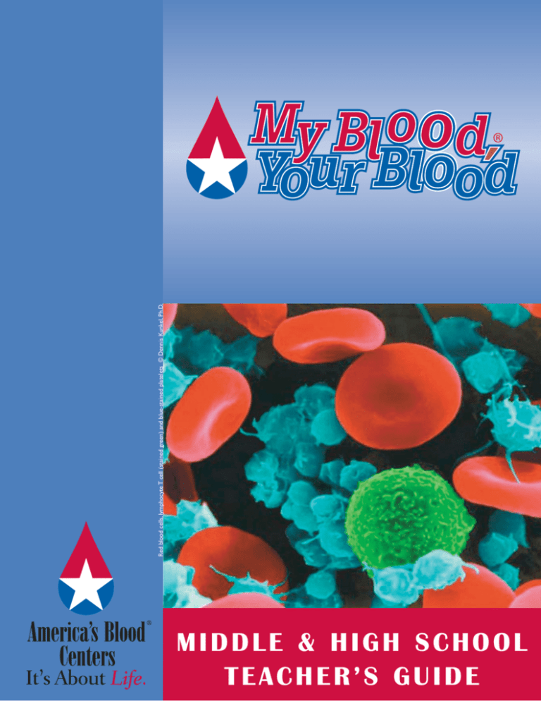
Red blood cells, lymphocyte T cell (stained green) and blue-stained platelets © Dennis Kunkel, Ph.D.
Contents
Part 1:
Blood is a Mixture
3
Lesson Plan
3
Demonstration: Blood is a Mixture
4-5
Teacher’s Guide to Healthy Hematocrit Lab
5
Answers to Worksheet and Lab Questions
Part 2:
Form and Function of Blood Cells
6
Lesson Plan
6
Demonstrations: Form and Function of Blood Cells
7
Teacher’s Guide to Cell Hunt Lab
7
Answers to Worksheet and Lab Questions
Part 3:
Arteries, Capillaries and Veins:The Roads Blood Cells Travel
8
Lesson Plan
8
Demonstration: Arteries, Capillaries and Veins:
The Roads Blood Cells Travel
9
Teacher’s Guide to Mystery Vessel Lab
9
Answers to Worksheet and Lab Questions
Part 4:
Oxygen and Carbon Dioxide Exchange
10
Lesson Plan
10
Demonstration: Oxygen and Carbon Dioxide Exchange
11
Teacher’s Guide to Bubbling Carbon Dioxide Lab
11
Answers to Worksheet Questions
Part 5:
Heart Cycle
12
Lesson Plan
12
Teacher’s Guide to Squirting and Filling Lab
12
Answers to Worksheet and Lab Questions
Part 6:
Blood Comes in Different Types
13-14 Lesson Plan
13-14 Teacher’s Guide to A Critical Decision Lab
14
Answers to Worksheet and Lab Questions
Part 7:
Researching For Cures
15
Lesson Plan
15
Demonstration: Sickle Cell Disease
15
Teacher’s Guide to Break the Sickle Cycle Lab
15
Answers to Worksheet and Lab Questions
Part 8:
16-17 Becoming Part of the Solution: Organizing a Blood
Drive at Your School (grades 11-12)
Worksheets & Lab Questions:
18-21 Blood is a Mixture
22-25 Form and Function of Blood Cells
26-27 Arteries, Capillaries and Veins:The Roads Blood Cells Travel
28-29 Oxygen and Carbon Dioxide Exchange
30-31 Heart Cycle
32-35 Blood Comes in Different Types
36-38 Researching For Cures
39
39
40
2
Glossary
Shopping List
Special Thanks & Resources
© 2004 All rights reserved. America’s Blood Centers
Introduction
America’s Blood Centers and the Foundation for
America’s Blood Centers are committed to increasing
public awareness about the need for blood donation to
help ensure that all Americans have access to a safe and
adequate blood supply. The creation of My Blood, Your
Blood underscores this commitment. While fostering
altruism and community spirit, this Teacher’s Guide
and the entire My Blood, Your Blood curriculum provide up-to-date information and creative strategies to
help teach the science of blood. Developed by a team
of physicians and educators, My Blood, Your Blood is
designed to be a turnkey education program easily
adapted to a variety of learning levels. America’s Blood
Centers hopes you and your students enjoy the learning activities. We encourage you to visit the My Blood,
Your Blood Web site at www.MyBloodYourBlood.org.
Our My Blood, Your Blood Guide follows current
National Science Education Standards:
The National Science Education Standards, developed by
the National Research Council and the National
Academy of Sciences, are criteria for the development
of curricula to increase science literacy of all students.
The My Blood, Your Blood Video and Teacher’s Guide
can assist educators in designing science lessons that
will aid student understanding in the following content
areas established by the National Science Education
Standards:
Life Sciences:
· Structure and Function in Living Organisms
(Level 5-8)
· The Cell and Matter, Energy, and Organization in
Living Systems (Level 9-12)
Science in Personal and Social Perspectives:
· Personal Health (Level K-8)
· Personal and Community Health (Level 9-12)
· Science and Technology in Local Challenges
and Society (Level K-12)
L E S S O N
P L A N
PART 1
Blood is a Mixture
Lesson Plan
Estimated Time: two class periods
· Show MBYB video segment (Start 00:00; End 11:00)
· Demonstration: Blood is a Mixture;Teach concepts presented in teacher background info (30 minutes)
· Use PowerPoint® slides 1-4 and Blood facts poster
· Students read worksheet and complete questions (in class or as homework)
· Lab: Healthy Hematocrit (30 minutes)
Demonstration: Blood is a Mixture
1. Create a mixture of red sequins (representing red blood cells (RBCs)) and glass or plastic beads (representing white blood
cells (WBCs) and platelets) to demonstrate the concept that blood is a mixture of cells suspended in plasma.
2. Plasma is a straw colored liquid.To simulate this color, add a few drops of yellow food color and a bit of cola to about
100 mL of water to get a pale straw color.
3. At a craft store, purchase red sequins in a large bag.You will need enough of these to fill 40 mL of a graduated cylinder.
4. Also purchase a small amount of glass or plastic spherical beads (glass beads with colored bits inside look very
realistic) to be leukocytes and some smaller, asymmetrical lumpy looking beads for platelets.The ratio should be
approximately 1,000 RBCs for every WBC and 100 platelets. No need to count them out, just estimate the ratio.
5. Mix the “cells” and the plasma in a clear plastic cup to show the students that the suspension looks red.
6. Then pour the mixture into a glass or clear plastic 100 mL graduated cylinder.
7. As the beads settle out, point out that the blood cells are suspended in the plasma.
8. After the beads have settled, discuss what the hematocrit is and how this measurement of blood cell volume is used in
medicine (see background info below).
Note:
You may wish to scale this up to a 1 L graduated cylinder to make it more visible for a large class.
A large 60 cc syringe can be used in place of a graduated cylinder for a more graphic approach.
100
100
90
90
80
80
70
70
60
60
50
50
40
40
30
30
20
20
10
10
MIXED
SETTLED
© 2004 All rights reserved. America’s Blood Centers
3
PART 1
Teacher’s Guide to Healthy Hematocrit
Overview
Students will measure
the hematocrit of a
patient’s blood by
centrifuging a blood
sample.The simulated
blood is actually redcolored sugar crystals
suspended in vegetable
oil.The simulated blood
can be centrifuged or
left to settle on its
own.The student then
determines the
hematocrit by using a
ruler to measure the
percent of “blood” that
is packed cells.
L A B
Teacher Background Information on Anemia and Hematocrit
In order to measure the proportion of the blood that is made up of red blood cells, a drop of
blood is drawn into a tiny capillary tube with a sealed end. The tube is centrifuged and the
percentage of blood that is red blood cells can easily be seen because the red blood cells (RBCs)
will have settled to the bottom leaving the yellowish plasma and the white blood cells and
platelets in the top portion of the tube. A normal hematocrit is from 37-45 percent blood cells.
If the hematocrit is too low, the person may be anemic.The word anemia means “lacking blood.”
Anemia is a sign of many conditions and not a disease itself. People may be anemic if they:
· Do not produce enough RBCs in the bone marrow because of disease,
radiation therapy, or toxins;
· Have red blood cells that do not survive for a normal period of time; or
· Have lost blood.
A hormone called erythropoietin is produced by the kidneys and stimulates the bone marrow
to produce RBCs. Recombinant human erythropoietin is now available to help those people
who have kidney disease and those whose bone marrow has been temporarily damaged by
chemotherapy.
Unfortunately, some long distance athletes abuse recombinant erythropoietin to artificially
increase the amount of RBCs so that they can carry more oxygen to their muscles. The
hematocrits of athletes are now routinely tested to determine if “blood doping” with
recombinant erythropoietin is suspected. A hematocrit over 50 percent is suspicious. Not only
is this practice unethical, and against the rules of competition, it is also dangerous. If the blood
becomes too thick with RBCs, it can turn to sludge, flowing sluggishly and forming clots.
Day before lab:
1. Purchase three containers of red colored sugar crystals per class from a grocery store. It
is usually in the section with the cake decorating supplies.You may be able to find larger
containers at a craft store that has a cake decorating section.
2. Purchase one bottle of corn or canola oil.
Day of or day before the lab:
1. Mix up a batch of “blood” by adding about 40 percent sugar crystals by volume to corn oil
(use a pale yellow colored oil to make it look like plasma).
2. Mix it up just before setting out samples of it in clear plastic cups or beakers at the lab
stations.
3. You may wish to make up different batches of blood to be different “patient” samples. A few
of the samples can be less than 40 percent sugar crystals and a few can be at 40 percent and
a few can be closer to 50 percent. Remember that the percentage is based on the total volume.
To make up blood with a 40 percent hematocrit reading, follow the recipe below.
4
© 2004 All rights reserved. America’s Blood Centers
PART 1
Normal Hematocrit Level Simulated Blood Recipe
1. Measure out 40 mL of sugar crystals into a dry graduated cylinder
2. Pour the sugar crystals into a 150 mL beaker
3. Add oil while stirring until the total volume reaches 100 mL
If you have access to a table top centrifuge:
If you are going to have the students centrifuge the samples, you may want to grind the
sugar up into smaller crystals with a mortar and pestle so that the mixture doesn’t settle
quickly on its own and it looks more like a homogeneous mixture to begin with.The small
crystals also make the blood look more realistic because there won’t be large, visible
particles sticking to the sides of the tube.
If you do not have a centrifuge:
Make up the simulated blood using the whole crystals.When the students put the blood
into their tubes, the crystals (cells) will settle out in 5-10 minutes.
Measuring the Hematocrit
Students can use a ruler to measure the height of the blood in the tube and then measure
the height of the cells. Use the following formula to determine the hematocrit.
(Cell level cm/Blood level cm) x 100 = percent blood cell volume = hematocrit
Note: If you use centrifuge tubes that have volumetric graduations, a ruler isn’t
necessary.
Answers to Worksheet Questions
1. Iron is needed to produce hemoglobin. Hemoglobin carries oxygen.
2. The answer is C. Either A (they do not have enough hemoglobin) or B (they do not
have enough RBCs) would cause someone to feel tired and weak.
3. Red blood cells outnumber white blood cells about 1,000:1.
4. Red blood cells would flow out of the wound, and then get stuck together by
platelets and fibrin. Platelets would patch up the wound by becoming sticky and
activating the process to produce fibrin.White blood cells would attack bacteria and
remove debris.
5. A person could become anemic by not eating enough iron or by losing blood.
6. Transfusion and iron supplements are both treatments for anemia.
7. Transfusion would be helpful to an accident victim.
8. The blood center will check to ensure that donors have enough iron in their blood
at the time of donation; donors should drink plenty of water before donating to
ensure their hematocrit measurement is accurate.
Suggested Answers to Lab Questions
1. Answers will vary depending on the hematocrit level.
2. Answers will vary depending on the student’s viewpoint.
© 2004 All rights reserved. America’s Blood Centers
5
PART 2
Form and Function of Blood Cells
L E S S O N
P L A N
Lesson Plan
Estimated Time: 1-2 class periods
· Replay MBYB video segment (Start 02:30; End 08:25)
· Demonstrations (red blood cell shape, white blood
cell and antibody interaction) (20 minutes)
· PowerPoint® slides 1-3, 9-11 and poster
· Students read and complete student worksheet
(15 minutes or as homework)
· Lab: Cell Hunt (30 minutes)
Demonstration: Form and Function of Blood Cells
A. Shape of red blood cells (RBCs)
1. Blow up a red balloon and tie it off.
2. Push in both sides of the balloon by shoving
Blue-stained macrophage and pink-stained monocyte attacking E.coli bacteria.
© Dennis Kunkel, Ph.D.
both fists into the balloon on opposite sides.
3. Explain that this is the shape of RBCs. Red
blood cells are basically sacks of hemoglobin.The concave sides mean that more of the hemoglobin in the cell is close to
the surface (in a sphere, a lot of the hemoglobin would be in the middle, far from the cell membrane). Platelets, on the
other hand, are irregularly shaped to provide more area when adhering to blood vessel walls to form clots.
Red blood cells, Lymphocyte T cell (green), Monocyte (gold) and Platelets.
© Dennis Kunkel, Ph.D.
6
© 2004 All rights reserved. America’s Blood Centers
B. Action of monocytes and antibodies
This is a fun activity that gives students a visual image of
how monocytes and antibodies work together to capture
microorganisms and viruses.
1. Give each student a small plate with a few spoonfuls
of crispy rice cereal on it.
2. Tell the students that they will be acting as
monocytes (the largest of the engulfing white blood
cells which mature into macrophages or “big eaters”).
Students lick their finger and pick up the crispies one
at a time and eat them.Time them for 30 seconds and
see how many they can eat this way.
3. Then explain how antibodies are like very specific
glue and bind to the micro-organisms clumping them
together.
4. Hand out rice crispy treats. Ask them what the
marshmallow represents. Now how many crispies
can the monocyte eat at one time? You may want to
have them estimate how many crispies are in a treat.
Teacher’s Guide to Cell Hunt
L A B
PART 2
Overview
Students will observe the shape and relative
abundance of red blood cells, white blood cells and
platelets.
A week or more before the lab:
Purchase or gather prepared slides of human blood smears
stained with Wright’s stain. You will need one slide for each team
of students.
Safety Note: there is no risk of blood-borne pathogens
because the blood is fixed and glued to the cover slip.
Classroom management tip: The students will be given a
description of each of the types of cells for which they will be
hunting. Once they find them, you may wish to verify that they
have found the correct cell type before they can check it off
their checklist. Once a student has found all the types, they can
be “certified” and enlisted to help you verify the cell findings
of the other students.
Two macrophages engulfing E.coli bacteria (stained green) in the lungs.
Notice the smooth type with filopodia, and the ruffled type. © Dennis Kunkel, Ph.D.
Answers to Worksheet Questions
1. Granulocytes and monocytes engulf bacteria. Lymphocytes produce antibodies which clump bacteria together so that the
phagocytic white blood cells can destroy them more efficiently.
2. Plasma transfusions or transfusions of blood factors isolated from plasma have helped hemophiliacs to live longer lives by
restoring the ability to form clots.
3. The blood can be separated. Platelets can be given to one patient, plasma proteins to another and red blood cells to a third patient.
Answers to Lab Questions
1. The chart should be filled in with appropriate functions of the blood cells.
2. Red blood cells are flexible and smooth to maneuver through tiny capillaries. Also, the concave sides mean that more of the
hemoglobin in the cell is close to the surface (in a sphere, a lot of the hemoglobin would be in the middle, far from the cell
membrane).
3. Platelets are irregularly shaped to provide more area when adhering to blood vessel walls.
4. Red blood cells should be the numerous.
5. Lymphocytes are generally the hardest to locate.
© 2004 All rights reserved. America’s Blood Centers
7
PART 3
Arteries, Capillaries and Veins:
The Roads Blood Cells Travel
L E S S O N
P L A N
Lesson Plan
Estimated Time: 1-2 class periods
· Show MBYB video segment on blood vessels and oxygen transport (Start 08:25; End 11:00)
· Students read worksheet and answer questions (15 minutes)
· PowerPoint® slides 1-11
· Demonstration - burn a peanut (5-10 minutes)
· Lab: Mystery Vessel (30 minutes)
Demonstration:
Arteries, Capillaries and Veins:The Roads Blood Cells Travel
Objective: To demonstrate what the body cells need in order to survive and have energy.
1. Bend a paperclip into a tripod shape.
2. Place a half a peanut or marshmallow on the end of the paperclip.
3. Ask students what gives your body cells energy. Most will say food some will say
oxygen.
4. Demonstrate the energy available in food by burning a half of a peanut and/or a
mini marshmallow. Most students understand a flame contains energy (heat
energy and light energy).
5. As the food is burning, explain that our cells use a series of chemical reactions to
release the energy out of the food in a controlled fashion. But what else is needed
for energy besides the food?
6. Place a glass jar over the burning peanut. As the flame goes out, ask students what
made the flame go out? Most students will know that the flame used up the
oxygen. Just like the energy being released by burning, our cellular energy
production requires food and oxygen. How do all of our cells get food and
oxygen? The blood transports food and oxygen to each cell of our body! Oxygen
and food molecule exchange occurs in the capillaries.
8
© 2004 All rights reserved. America’s Blood Centers
Teacher’s Guide to Mystery Vessel
PART 3
Overview
The students need to use the
information given in the
Arteries, Capillaries and Veins:
The Roads Blood Cells Travel
worksheet to identify the
different vessels.
L A B
CAPILLARY
A week or more in advance:
1. Purchase or gather
prepared slides of artery
and vein cross sections.
A day before the lab:
ARTERY
2. Place a square of white
paper over each label
and tape it in place.
Number the slides
1 and 2.
VEIN
Teacher Background Information on Anemia and Hematocrit
Classroom Management Tip: If there aren’t enough slides for each group to have a set, each group can have one to identify and
then go to other groups and see if they agree with their identification. The collaboration can be a great way for students to
communicate their understanding to their peers.
Answers to Worksheet Questions
1. Arteries should have the thickest muscular layer.
2. Veins should have the thinnest walls since they do not need to stretch or contract.
3. Body movements (mostly in the legs) help move blood from the feet to the heart. Much of the work is done by the pressure
from the other side of the capillaries: the movement created by the beating heart. Obviously, if you aren’t moving, blood still
gets from your feet to your heart. This movement can be important, though. People who sit for many hours on long airplane
trips can develop sluggish circulation or blood clots in the veins of their legs and feet - some doctors suggest doing exercises or
getting up to walk around once an hour when on a long flight.
Answers to Lab Questions
1. The arteries would change in diameter.
2. The arteries would become wider (dilate) to decrease blow pressure.
© 2004 All rights reserved. America’s Blood Centers
9
PART 4
Oxygen and Carbon Dioxide Exchange
L E S S O N
P L A N
Lesson Plan
Estimated Time: 1-2 standard periods
· Show MBYB video segment on oxygen and carbon dioxide exchange (Start 08:25; End 12:30)
· Students read and complete worksheet (10-15 minutes)
· PowerPoint® slides 9-11
· Demonstration: Oxygen diffusion in the alveoli (15 minutes)
· Lab: Bubbling Carbon Dioxide (30 minutes)
Demonstration:
Oxygen and Carbon Dioxide Exchange
Objective: This demonstration gives students a visual image of the diffusion of oxygen from the alveolus to the surrounding blood.
1. Fill an inexpensive sandwich-sized zip-top plastic bag with a solution of baking soda and water (about two tablespoons of baking
soda to 200 mL of water).
2. For quick results make a tiny hole in the bag with a fine needle. If no hole is punched, diffusion of bicarbonate will occur across
the semipermeable plastic into the surrounding liquid, but it will take several hours so the results won’t be visible until the next day.
Note: Do not tell students that there is bicarbonate in the “alveolus” because it will be confusing since they just learned about
the role of bicarbonate in the blood.
3. The “alveolus” (plastic bag with baking soda) is then placed into a clear bowl containing a solution of water, congo red and vinegar
(recipe below). As the baking soda diffuses into the bowl, the purple-red solution will turn bright red, simulating the color change
that occurs as hemoglobin becomes oxygenated.
Recipe for Congo Red Solution
1. Fill the bowl 2/3 full of water.
2. Add just enough congo red powder to get a bright red, but still transparent solution.This will require only a tiny dusting of
powder. Mix well.
3. Add vinegar to the bowl one drop at a time while stirring until the solution turns purplish.
10
© 2004 All rights reserved. America’s Blood Centers
Teacher’s Guide to Bubbling Carbon Dioxide
L A B
Overview
Students visually see that more carbon
dioxide is exhaled after exercise.
A few weeks prior
Locate or order bromthymol blue pH
indicator solution or powder
One day or more before the lab:
Purchase one straw, one large balloon
and one clear plastic cup for each
student
Day of the lab:
Prepare a dilute bromthymol blue
solution.
1. To 1 L of tap water add enough
bromthymol blue to obtain a
bright blue or green color
(depending on the pH of your
water).
2. If the solution above is blue, it is
ready. If it is green, you need to
stir in bleach or ammonia one
drop at a time just until the
solution turns blue. It may only
require one drop.
3. Divide the solution into several
smaller containers to be placed in
stations around the room.
Answers to Worksheet Questions
1. Hemoglobin can bind to carbon dioxide. Also, there is an enzyme in red
blood cells which converts carbon dioxide into bicarbonate which can
be stored in the plasma.
2. Cells need food and oxygen.
© 2004 All rights reserved. America’s Blood Centers
11
PART 5
Heart Cycle
L E S S O N
P L A N
Lesson Plan
Estimated Time: 2 standard periods
· Show MBYB video segment (Start 08:25; End 10:56)
· Students read worksheet and answer questions (20 minutes)
· Use PowerPoint® slides 4-8
· Lab: Squirting and Filling (30 minutes)
Teacher’s Guide to Squirting and Filling
L A B
Overview
Students measure their pulse rate and then listen to their heart sounds using stethoscopes.They determine the
rate of their heart sounds and compare it to their pulse rate to make the connection between pulse and heart
contraction.
Note: This lab can be done with a set of class stethoscopes or stethoscopes borrowed from a local blood
center or clinic. It can also be done with homemade stethoscopes. A funnel can be attached to rubber tubing
for a student to listen to their own heart beat by placing the wide end of the funnel on their chests.Toilet paper
or paper towel rolls can be used to listen to another person’s heartbeat. If you do this, girls prefer to have
female partners.
If you are using stethoscopes, sanitize the earpieces with alcohol soaked cotton.
Stopwatches make this lab easier, but are not necessary.
Answers to Worksheet Questions
1. Because your muscles are working harder, your heart has to pump the blood faster so that more oxygen and food (sugar,
etc.) can get to the cells and so that the waste product, carbon dioxide, can be released out of the lungs.
2. The muscles downstream of the clot would be stressed and then die if blood circulation wasn’t returned to that area quickly.
Second Section
3. You feel a pulse when the heart is squirting blood into the arteries.
4. Yes, each time your heart contracts, you should feel a pulse.
Answers to Lab Questions
1. No, but they are close.
2. We only measured our heart rate and pulse rate for six seconds, so this isn’t a very accurate measurement.
3. It is easier and more convenient to measure the pulse rate.
12
© 2004 All rights reserved. America’s Blood Centers
L E S S O N
P L A N
PART 6
Blood Comes In Different Types
Lesson Plan
· Show MBYB video segment on blood typing and blood donation (Start 10:56; End 12:17)
· PowerPoint® slide 12
· Students read worksheet and answer questions
· Lab: A Critical Decision (30-40 minutes)
A Critical Decision Lab Teacher’s Guide
Objective: Students learn how to type blood and learn the importance of blood donation.
Two weeks before the lab:
Order a simulated blood typing kit from a biological supply company
One day or more before the lab:
Allocate different types of simulated blood into micro-centrifuge tubes. Use the chart below to create the samples.
RECOMMENDED
BLOOD TO BE
TRANSFUSED
PATIENT
NUMBER
BLOOD
GROUP
BLOOD GROUP
PATIENT CAN ACCEPT
1
A-
A-, O-
A-
O-
2
O-
O-
O-
5 units
3
O+
O+, O-
O+
4
O+
O+, O-
O-
O+
5
B+
B+, B-, O+, O-
B+
6 units
6
B+
B+, B-, O+, O-
B+
7
A+
A+, A-, O+, O-
A+
A+
8
A-
A-, O-
A-
3 units
9
O+
O-,O+
O+
10
O+
O-, O+
O+
A-
11
A+
A-, A+, O-, O+
A-
2 units
12
AB+
AB+, AB-, O+, O-, B+, B-, A+, A-
A+
13
A-
A-,O-
O-
B+
14
B+
B+, B-,O-,O+
B-
2 units
15
O+
O+, O-
O+
16
O-
O-
O-
B-
17
O+
O+, O-
O+
2 units
18
A-
A-, O-
O-
19
B+
B+, B-, O+, O-
B-
20
O+
O+, O-
O+
(one solution to the
puzzle - other solutions
are possible)
BLOOD
AVAILABLE
© 2004 All rights reserved. America’s Blood Centers
13
PART 6
Blood Comes In Different Types
L E S S O N
P L A N
C O N T I N U E D
Answers to Worksheet Questions
RECIPIENT
DONOR
A
A
B
☺
☺
B
2.
3.
4.
5.
O
DONOR
☺
NEGATIVE (-)
☺
POSITIVE (+)
RECIPIENT
NEGATIVE (-) POSITIVE (+)
☺
☺
☺
☺
AB
O
AB
☺
☺
☺
☺
Group O Rh-negative blood is universal donor blood.
Group AB Rh-positive patients can receive blood of any group or type.
Yes. A person with B Rh-positive blood can safely receive O Rh-negative blood.
No, people who are Rh-negative produce anti-Rh antibodies.
Answers to Lab Questions
1. Group O Rh-negative blood is precious because it can be given to anyone.
2. You never know how much you will need or what group and type you will need.
3. Yes, there was enough blood to go around, but if the blood types were different, there could have been a crisis. For example, if
there were more group O Rh-negative people, there wouldn’t be enough group O Rh-negative blood to go around.
4. Give IV fluids to the people and helicopter to another facility or helicopter blood in from another facility.
5. It should be based on need; whoever is most critically injured and has a chance of recovering should be given first priority.
6. Organize a blood drive, donate money to a blood center, give blood regularly, encourage others to give blood.
4%
11%
45%
40%
Distribution of blood
types in the USA
14
© 2004 All rights reserved. America’s Blood Centers
L E S S O N
P L A N
PART 7
Researching for Cures
Lesson Plan
· Show MBYB video segment: science and research - saving lives
(Start 12:30; End 30:00)
· Students read worksheet and answer questions
· Lab: Break the Sickle Cycle
Demonstration: Sickle Cell Disease
Objective: In order for students to have a better understanding about how
sickle cell disease affects people, it is helpful to understand the structure of
the hemoglobin molecule.
Sickle cells (stained green) among healthy red blood cells.
© Dennis Kunkel, Ph.D.
1. Make a model of a hemoglobin molecule from pipe cleaners and
large buttons.You will need two pink pipe cleaners (alpha globin)
and two red pipe cleaners (beta globin).
2. In the center of each pipe cleaner, thread one red button.
3. Fold up each pipe cleaner to resemble the four globin chains of
hemoglobin. Hold all four globins together to form hemoglobin.
4. To make sickle hemoglobin, attach a small piece of masking tape
to both beta globin chains in approximately the same place.This
will represent the altered amino acid (glutamic acid to valine).
Teacher’s Guide to Break the Sickle Cycle Lab
The non-polar R group of valine (sickle cell hemoglobin substitution) on the beta
globin chain fits into the non-polar groove on the beta hemoglobin chains of an
adjacent hemoglobin molecule.This causes the hemoglobin to form stiff polymers
which make the red blood cell rigid and disfigured. Sometimes it causes the RBCs
to burst, causing anemia. More often, the irregular cells are removed from the
blood stream.
Key to model of deoxygenated sickle hemoglobin
Answers to Worksheet Questions
1. Low oxygen starts the crisis; then clotting makes it worse.
2. One amino acid is different on the beta globin chain.
3. There are two globin molecules on each hemoglobin molecule.
Answers to Lab Questions
1. Without the groove in the alpha chain, the hemoglobin won’t make a chain.
2. Normal hemoglobin doesn’t have the altered amino acid which fits into the groove.There won’t be anything to fit in the groove.
3. Normal red blood cells won’t sickle even in low oxygen so these normal cells can dilute out the effect of the sickled cells and
deliver oxygen.
© 2004 All rights reserved. America’s Blood Centers
15
PART 8
Becoming Part of the Solution:
O r g a n i z i n g
a
B l o o d
D r i v e
a t
Y o u r
S c h o o l
Your students have learned just how important blood is and how critical blood donation is to saving lives. Organizing a blood drive
at your school is a wonderful opportunity for students who are eager to make a difference.
Delegation of the tasks involved in a successful blood drive not only makes the job easier, it builds teamwork and gives more people
the opportunity to get involved. Create a committee by nominating four responsible students.These students in turn nominate other
students to help them.Visit www.MyBloodYourBlood.org for samples and templates of useful letters and other documents.
Blood Drive Committee
Faculty Advisor (you!)
Student Positions
Blood Drive Coordinator
Donor Recruitment Chairperson
Promotion Chairperson
Site Preparation Chairperson
Responsibilities of each Blood Drive Committee Member:
Faculty Advisor
· Choose the student leadership.
· Contact America’s Blood Centers at 1-888-USBLOOD or www.AmericasBlood.org to find out how to reach your local
community blood center.
· Meet with the representative of the local blood center and the principal of your school to set up a date for the
blood drive.
· Set up meetings with the blood drive committee.
· Check on the progress of the student leadership.
Blood Drive Coordinator
· Contact parents, faculty and staff and ask them to donate.
· Make announcements to students before the sign-up week and also prior to the blood drive as a reminder.
· Ensure that all chairpersons carry out their responsibilities.
Donor Recruitment Chairperson
· Form a team of students who will recruit student donors.Your team should know the importance of blood donation, be familiar
with the typical reasons why people do not donate and understand who can donate blood.
· When a student volunteers to donate blood, they need to make an appointment to donate blood during the blood drive. Get
the student’s home phone number so you can contact them the day before to remind them of their appointment.The blood
center will provide you with appointment cards for this purpose.
Promotion Chairperson
16
© 2004 All rights reserved. America’s Blood Centers
a
B l o o d
D r i v e
a t
Y o u r
S c h o o l
PART 8
O r g a n i z i n g
· Promote and publicize the blood drive through posters, school publications and announcements.
· Community blood centers will provide posters with the date and information for your blood drive printed on them. Find out
where you can place these posters at your school. Get permission from your principal before putting up posters or passing
out fliers.
· Obtain deadline for getting a story in the school newspaper. The blood center will give you personal stories from people whose
lives have been saved through blood donation.Weave these feature stories with facts about blood donation dispelling the
common myths, which can also be provided by your local community blood center. If possible, include quotes from donors.
· Coordinate with your principal and blood center to see if an assembly can be held to increase the awareness of the entire
student body for the need for blood donations.
· Encourage the PTA to get involved to solicit parent blood donations during the blood drive.
· As a group, brainstorm ways to encourage your friends who are of eligible age to donate blood. However, be aware that there
are many reasons an individual may not be able to donate blood (medications, medical conditions, high-risk behaviors, etc.) and
some of those reasons are very personal. You must recognize that if someone chooses not to donate, they should not be
pressured or coerced into donating because that could compromise the safety of the blood supply and because blood donation
is a personal decision which needs to be respected. Individuals who cannot or do not want to donate may contribute to the
blood drive in many other ways by educating others about the need, passing fliers, signing up donors for appointments, staffing
the blood drive canteen, etc.
· Ask your teachers if you can make an announcement before your classes or over the intercom about the blood drive. Make
the announcement personal: include what you have learned about the importance of blood donation. If possible, use snippets
from the My Blood,Your Blood video to add a human face to the need for blood.
Site Preparation Chairperson
· Make sure the area being used for the blood drive is ready.
· Arrange for parking for the blood center close to an entrance.
· Be sure that the area being used will be at a comfortable temperature (heating or air conditioning as needed).
· Provide a table and several chairs.
© 2004 All rights reserved. America’s Blood Centers
17
Worksheet: Blood is a Mixture
Name: ________________________________________________
What makes blood red?
Your blood contains cells called erythrocytes or red blood cells (RBCs). Red blood cells are packed full of a protein molecule called
hemoglobin. The heme in the hemoglobin is red in color. Each molecule of hemoglobin contains four iron atoms. The iron atoms in
the hemoglobin molecules bind to oxygen molecules.This is how your body transports oxygen from your lungs to all the cells of your
body. Each RBC contains millions of hemoglobin molecules, and each drop of blood contains millions of RBCs.
More than Red Blood Cells
But there is a lot more to blood than RBCs.There are two other types of cells in blood, white blood cells (leukocytes or WBCs) and
platelets (thrombocytes). Red blood cells outnumber WBCs 1,000:1. White blood cells are an important part of our immune or
defense system. They fight infection from bacteria and viruses. Platelets are responsible for creating a clot to stop bleeding from a
wound. The RBCs, WBCs and platelets float around in pale yellow liquid called plasma. Plasma is more than just water. It contains
important proteins, hormones and salts. Red blood cells live for only four months. Therefore, your bone marrow is always at work
producing new blood cells. As you heard in the video, your bone marrow makes 4-5 billion RBCs every hour! Red blood cells make
up 40-45 percent of your blood volume.
What causes anemia?
In order to make hemoglobin, you need to eat enough protein, iron and certain vitamins. Causes of anemia include an iron-poor diet
or chronic disease.
Sometimes the cause of anemia is that the bone marrow is not producing enough RBCs.This can be caused by the effects of cancer
treatment, radiation, toxins or kidney failure as well as by iron deficiency. Healthy kidneys produce a hormone called erythropoietin.
This hormone stimulates the bone marrow to produce RBCs.
Anemia can also be caused by a loss of blood. Women who have very heavy menstrual periods or people who have lost significant
blood due to an injury or surgery can become anemic.
How can people with low numbers of red blood cells recover?
If an injury or surgery has caused a loss of blood, a transfusion will correct the problem immediately. A common treatment for mild
anemia is to take supplemental iron in the form of pills or infusions. Chronic anemia is treated by identifying and eliminating its cause.
A blood transfusion can bring relief to patients while they are waiting for a proper diagnosis. Depending on the underlying cause of
anemia, other treatments may correct the problem so that no further transfusions are necessary.
Can donating blood cause the donor to become anemic?
The answer is usually no. Before donating blood, a person’s hematocrit is measured. Only those with healthy hematocrit levels can
donate blood.The red blood cells lost in a blood donation are quickly replaced in a healthy person in about four to six weeks.
18
© 2004 All rights reserved. America’s Blood Centers
Questions
Name: ________________________________________________
1. Why is it important to get enough iron in your diet?
___________________________________________________________________________________________________
___________________________________________________________________________________________________
___________________________________________________________________________________________________
2. Without oxygen, our cells cannot work.Which of the following might be an explanation why someone feels weak?
a. They do not have enough hemoglobin
b. They do not have enough red blood cells
c. Either a or b would cause someone to feel tired and weak
3. What type of blood cell far outnumbers the others in the blood?
___________________________________________________________________________________________________
___________________________________________________________________________________________________
4. If you fell down and scraped your knee, describe what each type of blood cell would be doing at the site of injury.
___________________________________________________________________________________________________
___________________________________________________________________________________________________
___________________________________________________________________________________________________
5. What are two ways someone can become anemic?
___________________________________________________________________________________________________
___________________________________________________________________________________________________
6. What are two treatments for anemia?
___________________________________________________________________________________________________
___________________________________________________________________________________________________
7. Which treatment would work best to help someone who has been in a car accident and lost a lot of blood?
___________________________________________________________________________________________________
___________________________________________________________________________________________________
___________________________________________________________________________________________________
8. What would you tell someone who’s not sure if they can give blood because they think they might be anemic?
___________________________________________________________________________________________________
___________________________________________________________________________________________________
___________________________________________________________________________________________________
© 2004 All rights reserved. America’s Blood Centers
19
Lab: Healthy Hematocrit
Measuring Blood Cell Volume to Diagnose Potential Anemia
Name: ________________________________________________
Purpose: To measure the percent of red blood cells volume of a simulated blood sample to determine the hematocrit. Once you
have measured the hematocrit of your “patient” you can determine if the person has a low
hematocrit, normal hematocrit or is polycythemic (too many red blood cells).
Materials
Test tube
Simulated blood sample
Centrifuge (optional)
Metric ruler
Marking pen
Procedure
1. Obtain a sample of simulated blood.
2. Mix it up with a stirring rod and fill a test tube 2/3 full.
3. If you have a centrifuge available, mark this test tube with your initials and place it in
a centrifuge across from another sample (the centrifuge needs to be balanced - every
tube must have a tube across from it). Centrifuge for three minutes.
4. If you do not have a centrifuge, let the blood cells settle for 10 minutes.
5. Using the centimeter side of a ruler, measure the distance from the bottom of the test
tube to the top of the blood sample. Record this measurement below.
Height of blood _______ cm
6.
Now measure the height of the blood cell layer from the bottom of the test tube. Record this measurement below.
Height of blood cells ________ cm
7.
To determine the hematocrit, (which is simply the percentage of the blood that is made up of cells) divide the height of the
blood cells by the total height of the blood and multiply that by 100.
Height of blood cells in cm/ total height of blood x 100 = hematocrit
Hematocrit of “patient” ____________________
Compare your results to the chart below to diagnose your patient
38-50 percent = NORMAL
Less than 38 percent = POSSIBLE ANEMIA (low hematocrit level for the purpose of blood donation)1
More than 50 percent = POSSIBLE POLYCYTHEMIA (excess blood cells)
1
20
A reading less than 38 percent is below the level mandated by the US Food and Drug Administration (FDA) for blood donors
but does not necessarily mean that the donor is anemic.
© 2004 All rights reserved. America’s Blood Centers
Lab Questions
Name: ________________________________________________
1.
What was the condition of the blood of the patient you tested?
___________________________________________________________________________________________________
___________________________________________________________________________________________________
___________________________________________________________________________________________________
2.
Some athletes practice something called “doping”. This is where they inject themselves with erythropoietin to increase their
red blood cell level to improve their ability to get oxygen to their muscle cells. This causes their hematocrit to be above
normal. Many people consider this practice unethical and proof of doping with erythropoietin is cause for disqualification in
many sports.
a. Do you think blood doping should be allowed in athletics? Explain.
b. A serious condition occurs if the hematocrit exceeds 70 percent. What adverse affect do you think too many red blood
cells would have on the circulatory system?
___________________________________________________________________________________________________
___________________________________________________________________________________________________
___________________________________________________________________________________________________
___________________________________________________________________________________________________
___________________________________________________________________________________________________
___________________________________________________________________________________________________
___________________________________________________________________________________________________
___________________________________________________________________________________________________
___________________________________________________________________________________________________
___________________________________________________________________________________________________
___________________________________________________________________________________________________
___________________________________________________________________________________________________
___________________________________________________________________________________________________
___________________________________________________________________________________________________
___________________________________________________________________________________________________
___________________________________________________________________________________________________
___________________________________________________________________________________________________
___________________________________________________________________________________________________
___________________________________________________________________________________________________
___________________________________________________________________________________________________
___________________________________________________________________________________________________
© 2004 All rights reserved. America’s Blood Centers
21
Worksheet: Form and Function of Blood Cells
Name: ________________________________________________
As you learned from the My Blood,Your Blood video, all blood cells are produced in the bone marrow from stem cells.
“White blood cells (WBCs) are part of the body’s defense system. There are three types of WBCs: granulocytes, lymphocytes and
monocytes.They all fight infection from bacteria, viruses - all those nasty microbes that can cause disease.”
White blood cells (leukocytes) make up a mobile army that seek out and destroy harmful micro-organisms and viruses. These helpful
cells can move out of the blood vessels and patrol in amongst your body tissues.
There are two main types of WBCs: engulfing and antibody producing. Granulocytes and monocytes are engulfing WBCs and
lymphocytes are antibody-producing white blood cells. Engulfing WBCs reach out cellular arms like an amoeba and engulf bacteria and
other micro-organisms. This process is called phagocytosis. These engulfing WBCs communicate with the antibody-producing white
blood cells called lymphocytes. Antibodies are protein molecules that stick to the invading microorganisms, clumping them together.
Once the invading organisms are all clumped together, they can be engulfed more efficiently by monocytes and granulocytes.
Platelets (thrombocytes), which are actually not cells but cell fragments, form clots to stop bleeding. When a blood vessel gets torn
during an injury, platelets throw themselves over a tear in a vessel wall.They become sticky and adhere to the vessel wall and to each
other.They also trap RBCs and together they form a clot. Platelets also produce a chemical that activates a chain of events that turn
a soluble blood protein called fibrinogen into a long fibrous solid protein called fibrin. Fibrin molecules create a network to hold the
clot in place. All this together is what you think of as a scab. Most of what you see on the outside of a scab is just dried blood. On
the inside of a scab is where the intricate fibrin/platelet plugs of each individual broken blood vessel can be found.The fibrin/platelet
plugs will form and reform until the damaged tissues have been repaired. Picking at a scab will reopen these plugs and the process has
to start over. That is why wounds heal much faster if you leave them alone. Platelets also function extensively internally throughout
the body.
A genetic disorder called hemophilia is caused by a lack of one of the clotting proteins.This blood protein can be isolated from blood
through a chemical fractionation process and given to hemophilic patients as a concentrate.
Cancer patients often have blood clotting difficulties because their bone marrow can become damaged as a result of chemotherapy
or radiation, so it may not produce as many platelets as they need. These patients are often given a transfusion of platelets.
One blood donation can help save three lives.This is because the blood someone donates can be separated into three or more parts. For
example, a pint of donated blood can give a hemophilic patient plasma, a cancer patient platelets and an anemic patient red blood cells.
22
© 2004 All rights reserved. America’s Blood Centers
Questions
Name: ________________________________________________
1. Describe how the two types of white blood cells work together to kill disease-causing micro-organisms.
___________________________________________________________________________________________________
___________________________________________________________________________________________________
___________________________________________________________________________________________________
___________________________________________________________________________________________________
2. Fifty years ago, hemophiliacs did not live very long. Now people with this disorder live much longer, healthier lives.
What has made this possible?
___________________________________________________________________________________________________
___________________________________________________________________________________________________
___________________________________________________________________________________________________
___________________________________________________________________________________________________
3. Describe in your own words how one pint of blood can help three different people.
___________________________________________________________________________________________________
___________________________________________________________________________________________________
___________________________________________________________________________________________________
___________________________________________________________________________________________________
© 2004 All rights reserved. America’s Blood Centers
23
Lab: Cell Hunt
Name: ________________________________________________
Purpose: To learn to identify the different blood cell types and to relate their structure to their function.
Materials
Prepared slide of stained human blood
Microscope
Background
You will be going on a cell hunt.You need to find all of the types of cells below. Use this description of the structure of the blood cells
to help you.You need to verify your findings with your teacher or a “certified” student helper.
CELL TYPE
Erythrocyte
(RBCs)
Red disks with dark red
border and light red
center (no nucleus).
Granulocyte
(WBCs)
Round cell with grainy
appearance; lobed or
indented blue nucleus.
Monocyte
(WBCs)
Large, irregularly
shaped cell with lobed
or indented nucleus.
Lymphocyte
(WBCs)
Round or elongated cell
often with spikey edges.
Nucleus is usually round.
Thrombocyte
(Platelet)
Small, irregularly shaped
bodies with no nucleus. 1
1
24
DESCRIPTION
FUNCTION
LOCATED IN MICROSCOPE
(teacher or student helper verification)
Platelets have no nucleus because they are fragments that “break off” of a megakaryocyte in the bone marrow.
© 2004 All rights reserved. America’s Blood Centers
Lab Questions
Name: ________________________________________________
1. Fill in the function for all the blood cell types in the above chart
2. How does the shape of the red blood cell fit its function?
___________________________________________________________________________________________________
___________________________________________________________________________________________________
___________________________________________________________________________________________________
___________________________________________________________________________________________________
3. How does the shape of the platelet fit its function?
___________________________________________________________________________________________________
___________________________________________________________________________________________________
___________________________________________________________________________________________________
___________________________________________________________________________________________________
4. Which blood cell is the most numerous on your slide?
___________________________________________________________________________________________________
___________________________________________________________________________________________________
___________________________________________________________________________________________________
___________________________________________________________________________________________________
5. Which blood cell was the hardest to find?
___________________________________________________________________________________________________
___________________________________________________________________________________________________
___________________________________________________________________________________________________
___________________________________________________________________________________________________
© 2004 All rights reserved. America’s Blood Centers
25
Worksheet: Arteries, Capillaries and Veins:
The Roads Blood Cells Travel
Name: ________________________________________________
The heart is the pump that forces blood to flow through the blood vessels. Blood vessels that take blood away from the heart are called
arteries.The largest artery leading away from the heart is the aorta.When the heart squirts out blood, it is under pressure. Arteries therefore
must have strong, elastic walls in order to withstand the pressure. Muscles lining the artery walls also enable the body to control the pressure
of the blood by either relaxing (lowering the pressure) or constricting (raising the pressure).
Arteries branch off into smaller and smaller arteries called arterioles. Arterioles branch into
smaller and smaller arterioles until they lead to the thin-walled capillaries. Capillaries are so
narrow that the blood cells have to proceed single file.This is where the blood can do its work.
The walls of the capillaries are only one cell thick and oxygen and small food molecules such as
glucose can easily pass though into the tissues. Carbon dioxide produced as waste from cellular
respiration passes out of the tissues into the blood though the capillary walls.
The blood now needs to return to the heart. The capillaries lead to tiny blood vessels called
venules.The venules then lead to veins which eventually collect into the very large veins of the
inferior (or lower) and superior (or upper) vena cava which bring the blood back to the heart.
Veins have much thinner walls because they are not under very much pressure.The motion of
your body helps squeeze the blood back to the heart through the veins because your muscles
push up against the walls of the vein.Your veins have valves in them that keep the blood from
moving backward in response to the gravitational force.
The diagram shows the blood flow through the heart.The blood that returns to the heart from
the body has to go to the lungs to release the carbon dioxide and pick up oxygen.
Questions
1. Which blood vessels would you expect to have the thickest muscular layer?
___________________________________________________________________________________________________
___________________________________________________________________________________________________
2. Which blood vessels have the thinnest walls? How does this structure fit their function?
___________________________________________________________________________________________________
___________________________________________________________________________________________________
___________________________________________________________________________________________________
3. How does blood get from your feet back up to your heart?
___________________________________________________________________________________________________
___________________________________________________________________________________________________
___________________________________________________________________________________________________
26
© 2004 All rights reserved. America’s Blood Centers
Lab: Mystery Vessel
Name: ________________________________________________
Purpose: To learn about the form and function of arteries and veins.
Materials
Prepared slides of the cross sections of an artery and a vein
Microscope
Colored pencils
Blank paper
Procedures
1. Using the description of the structure and function of arteries and veins from the background handout, determine which
numbered slide you have is the artery and which is the vein.
2. Once you have identified the artery and vein, and confirmed your decision with your instructor or lab aid, create a sketch of each
of the cross sections.
3. Label the cross sections according to the diagram given.
Lab Questions
1. If someone’s blood pressure were too low, which blood vessel would change diameter to correct the problem?
___________________________________________________________________________________________________
___________________________________________________________________________________________________
___________________________________________________________________________________________________
___________________________________________________________________________________________________
2. Would that vessel become wider or narrower to correct high blood pressure?
___________________________________________________________________________________________________
___________________________________________________________________________________________________
___________________________________________________________________________________________________
___________________________________________________________________________________________________
© 2004 All rights reserved. America’s Blood Centers
27
Worksheet: Oxygen and Carbon Dioxide Exchange
Name: ________________________________________________
The airways in the lungs dead end into millions of tiny little air sacs called alveoli. Each
alveolus is covered by a network of capillaries. The oxygen gas diffuses from the air in
the alveoli into the blood. The iron atoms of the hemoglobin molecules inside the red
blood cells (RBCs) bind to the oxygen molecules. Since each RBC contains millions of
hemoglobin molecules, the oxygen is quickly taken from the lungs and is then on its way
to the heart and then to the body tissues.
The O2 molecules pass easily into the
blood vessels and bind or attach to our
hemoglobin.
In the capillaries of the body tissues, the oxygen moves off the hemoglobin and into the
body cells.The body cells produce carbon dioxide which diffuses into the blood. Some of
the carbon dioxide is carried by the hemoglobin molecules. There is also an enzyme inside
the RBCs that combines carbon dioxide and water to form bicarbonate. The bicarbonate
remains in the plasma until the blood returns to the lung capillaries. Then the bicarbonate
is converted back into carbon dioxide and water and the carbon dioxide is released into
the lungs.
Questions
1. What role does the red blood cell play in oxygen transport?
___________________________________________________________________________________________________
___________________________________________________________________________________________________
___________________________________________________________________________________________________
___________________________________________________________________________________________________
2. What are two ways the red blood cell is involved in carbon dioxide transport?
___________________________________________________________________________________________________
___________________________________________________________________________________________________
___________________________________________________________________________________________________
___________________________________________________________________________________________________
3. What are two things that all the body cells need in order to work?
___________________________________________________________________________________________________
___________________________________________________________________________________________________
___________________________________________________________________________________________________
___________________________________________________________________________________________________
28
© 2004 All rights reserved. America’s Blood Centers
Lab: Bubbling Carbon Dioxide
Name: ________________________________________________
Purpose: You will measure the effects of exercise on carbon dioxide production.You
will be using a carbon dioxide indicator solution to determine how much carbon dioxide
is in the breath you exhale.The more carbon dioxide there is in the solution, the quicker
the solution will turn from blue to green and finally to yellow.
Materials
Balloon
Cup
Straw
Carbon dioxide testing solution
Piece of string
Marker
Procedures
1. Fill the plastic cup 1/3 full of carbon dioxide testing solution.
2. Blow up the balloon with one full breath of air (your exhaled air contains carbon dioxide).
3. While pinching off the balloon with one hand, attach the straw to the neck of the balloon by inserting the straw until it reaches
the point where you are pinching it off.
4. Try to keep the air from escaping from the balloon end of the straw with your fingertips.
5. Insert the other end of the straw into the bottom of the cup containing the carbon dioxide testing solution.
6. Release the air slowly so that it bubbles into the solution. As soon as the solution turns yellow, pinch off the balloon and measure
its circumference with a length of string. Mark the string with a marker so you know how big the balloon was when the solution
turned yellow.
7. Exercise for one minute (according to your teacher’s instructions) and repeat steps 2-6.
8. Measure the difference in the length of string with and without exercise.
Lab Questions
1. Record your results below
Length of string before exercise: ________ cm
Length of string after exercise: _________ cm
Difference in the length of string before and after exercise: ___________cm
2. According to your lab results, when was there a greater concentration of carbon dioxide in your breath?
___________________________________________________________________________________________________
___________________________________________________________________________________________________
___________________________________________________________________________________________________
© 2004 All rights reserved. America’s Blood Centers
29
Worksheet: Heart Cycle
Name: ________________________________________________
The heart is responsible for pumping the blood through the blood vessels. It squirts blood out,
fills with blood and squirts it out again. Squirt, fill, squirt, fill… over and over throughout your
life. In the lab activities that follow, you will get a chance to listen to the sounds your heart
makes and calculate how many times your heart squirts out blood every minute you are at rest
and when you are exercising.
There are two sounds your heart makes. The two sounds come right after one another. The
sounds are sometimes called “lub-dub.” The first sound, “lub,” is made when the heart is squirting blood out of the large chambers
called ventricles. The sounds actually come from the closing of the valves between the atria and the ventricles (so blood won’t go
back the way it just came). The second sound,“dub,” comes when the heart is filling with blood and the valves close that keep blood
from flowing out of the heart while it is filling.
Lub = squirt
Dub = fill
Less than one second rest
Lub = squirt
Dub = fill
Rest…
Questions
1. You probably noticed that when you are exercising your heart beats faster. Explain why your heart needs to beat faster based on
what you know about blood. Give as many details as you can.
2. The heart muscle needs blood too.There are blood vessels (coronary arteries) that go straight to the heart muscle from the
main artery (aorta) that leads away from the left ventricle.These arteries branch into smaller and smaller arteries that bring
oxygen and nutrients to the hard working heart muscle. If one of the smaller coronary arterioles in the heart muscle became
blocked, what do you think happens when someone has a heart attack? How is the heart muscle damaged?
Pulse Rate
When your heart squirts blood out of the ventricles, the blood is under pressure. The left ventricle is especially muscular and can
force the blood through the aorta in powerful squirts. Arteries can stretch and absorb this pressure. If you put your fingers along the
side of your throat, you can feel the pulse of the blood squirting through your carotid artery.
3. Do you feel a pulse in an artery whenever your heart squirts, fills or at both times?
___________________________________________________________________________________________________
___________________________________________________________________________________________________
4. Would your pulse rate be the same as your heart rate? Why or why not?
___________________________________________________________________________________________________
___________________________________________________________________________________________________
30
© 2004 All rights reserved. America’s Blood Centers
Lab: Squirting and Filling
Name: ________________________________________________
Purpose: To listen to the sounds made by your heart and to compare heart rate to pulse rate before and after exercise.
Materials
Stethoscopes
Stopwatch or clock with second hand
Procedures
1. Using a stethoscope, listen to your own heart sounds.The diaphragm of the stethoscope should be placed in the center of your
chest. Move the diaphragm around until you hear the sounds clearly. It is critical that the room be quiet during this process.
2. Try to distinguish the “lub” sound from the “dub” sound. Visualize your heart squirting when you hear the “lub” sound and filling
when you hear the “dub” sound.
3. Using a stopwatch, count how many times you hear your heart beat in six seconds. Multiply this by 10 to get your heart rate per
minute. Fill in the chart below with your data.
4. Exercise for one minute and listen to your heart again. Count how many times you hear your heart beat in six seconds to get
your heart rate. Fill in the chart below with your data.
5. Rest for a few minutes to be sure your heart rate has returned to its normal resting rhythm.
6. Place your fingers at a pulse point on the side of your throat or on your wrist. Count how many times you feel your pulse in six
seconds. Multiply that by 10 to get your pulse rate. Fill in the chart below.
7. Exercise for one minute and measure your pulse rate again. Fill in the chart below.
AT REST
IMMEDIATELY AFTER EXERCISE
HEART RATE (beats/minute)
HEART RATE (beats/minute)
PULSE RATE (pulses/minute)
PULSE RATE (pulses/minute)
Lab Questions
1. Compare your heart rate to your pulse rate. Are they exactly the same?
___________________________________________________________________________________________________
2. If they aren’t exactly the same, why might they be different?
___________________________________________________________________________________________________
___________________________________________________________________________________________________
___________________________________________________________________________________________________
3. When your doctor wants to measure your heart rate, why does he or she feel your pulse rather than listen to your heart?
___________________________________________________________________________________________________
___________________________________________________________________________________________________
___________________________________________________________________________________________________
© 2004 All rights reserved. America’s Blood Centers
31
Worksheet: Blood Comes in Different Types
Name: ________________________________________________
Blood cells have proteins on the outside of the cell membrane. Some of these markers are the same on all blood cells of humans and
some markers have variations (different people have different types). Your immune system recognizes the markers on your blood cells
as “self.” Any blood cells with different marker molecules will be treated like foreign invaders and the antibodies and white blood cells
will clump them together and destroy them.The destruction of incompatible blood cells can produce reactions harming the recipients.
There are two main categories of blood typing, the ABO system blood group and the Rh system blood type. In the ABO grouping system,
there are four group of blood: A, B, AB and O. In the Rh system, there are two types, Rh-positive and Rh-negative. Each person has a
combination of those two, for instance, someone can have group A Rh-negative blood or group B Rh-positive blood, and so on.
In a person with group A blood, the body only “recognizes” the group A marker. So that person’s body would not recognize the B
carbohydrate, and would try to destroy cells with the B marker.The body (or immune system) of someone with group B blood can
only recognize the B carbohydrate, so they would make anti-A antibodies. Someone with type AB blood wouldn’t make anti-A or
anti-B antibodies, because their body would recognize both markers. Group O blood cells lack any ABO markers.
When someone has lost a great deal of blood, she or he needs an immediate supply to replenish it. There is no substitute for human
blood; replacement blood must come from a healthy blood donor. So how do people receive donated blood without causing a
clumping reaction to occur? The donor blood is matched to the recipient with the same type of blood or a compatible type.
The chart below illustrates the different blood groups and who can receive which group. Part of the chart is filled in for you.
1. Study the charts below and then fill in the blank spaces with ☺ for a safe match or
for a bad match.Check your answers with your teacher.
RECIPIENT
DONOR
A
A
B
☺
AB
☺
O
DONOR
NEGATIVE (-)
RECIPIENT
NEGATIVE (-) POSITIVE (+)
☺
☺
POSITIVE (+)
B
AB
O
☺
☺
☺
2. The “universal donor” is the type of donor who that can donate to all other patients.Which blood group and type is the universal
donor? _____________________________________________________
3. The “universal recipient” is the type of patient who can receive transfusions from any other blood type.Which blood group and
type is the universal recipient? _____________________________________________________
4. A person with B+ blood is in an automobile accident and needs an immediate transfusion. Only O+ blood is available, would that
group and type of blood be safe to give to that person? _____________________________________________________
5. A person with A- blood had knee surgery and they needed one unit of blood.Would A+ blood be safe to transfuse into this knee
surgery patient? _____________________________________________________
32
© 2004 All rights reserved. America’s Blood Centers
Lab: A Critical Decision
Name: ________________________________________________
Purpose: To learn how blood types can be determined and to understand which blood
types can be safely transfused into different people.
Background information:
A bus full of people returning from a ski trip overturns on an icy road.
Twenty badly injured people are rushed to a local hospital. All 20 people
need blood transfusions because of the extent of their injuries. The
small hospital they have been transported to has 20 units of blood, but
it only has five units of type O- blood. Therefore, it is necessary to type
the blood of the patients to determine how to distribute the blood
safely.
You will be given two blood samples to type.The patients are numbered
1-20. Determine the blood type of your patients, then as a class, you can
determine how to distribute the available blood.
Materials
2 simulated blood samples
Anti-A, anti-B and anti-Rh simulated antibodies
Blood typing plate or slides
6 toothpicks
4%
11%
45%
40%
Distribution of blood
types in the USA
Procedure
1. Place one drop of blood in the A, B and Rh sections of the typing plate.
2. Add one drop of “anti-A” in the A section, one drop of “anti-B” in the B section and one drop of “anti-Rh” in the Rh section.
3. Mix each section with a separate toothpick.
4. Wait three minutes. If a reaction has occurred, the blood will gel and get clumpy.
5. To determine the type, look for the sections that clumped. The ones that clumped will tell you the type. For example, if A and Rh
clumped, then the person is A+. If A and B clumped but not Rh, then the person is AB-. If none of them clump, then the person is O-.
6. Fill in the chart with the information before you type the second patient’s blood. Rinse out the blood typing plate and dry it
completely before typing the second person.
7. Repeat steps 1-4 for the second blood sample.
PATIENT NUMBER
BLOOD TYPE
© 2004 All rights reserved. America’s Blood Centers
33
Lab: A Critical Decision
Name: ________________________________________________
8. Look at the chart below to see the units of available blood. As a class determine which patients should receive which units of blood.
PATIENT
NUMBER
BLOOD
GROUP
BLOOD GROUP
PATIENT CAN ACCEPT
RECOMMENDED
BLOOD TO BE
TRANSFUSED
(one solution to the
puzzle - other solutions
are possible)
BLOOD
AVAILABLE
1
O-
2
5 units
3
4
O+
5
6 units
6
7
A+
8
3 units
9
10
A-
11
2 units
12
13
B+
14
2 units
15
16
B-
17
2 units
18
19
20
34
© 2004 All rights reserved. America’s Blood Centers
Lab: A Critical Decision
Name: ________________________________________________
Lab Questions
1. If you have blood group O-type Rh-negative, why is it especially important for you to become a regular blood donor?
___________________________________________________________________________________________________
___________________________________________________________________________________________________
___________________________________________________________________________________________________
2. Why is it important for hospitals to always have more blood on hand than they will need in one day?
___________________________________________________________________________________________________
___________________________________________________________________________________________________
___________________________________________________________________________________________________
___________________________________________________________________________________________________
3. In this accident scenario, was there enough blood to go around? If the patients had different blood types, what might have happened?
___________________________________________________________________________________________________
___________________________________________________________________________________________________
___________________________________________________________________________________________________
___________________________________________________________________________________________________
4. Propose a solution to a crisis situation where there was not enough blood to distribute to patients in an emergency.
___________________________________________________________________________________________________
___________________________________________________________________________________________________
___________________________________________________________________________________________________
___________________________________________________________________________________________________
5. On what criteria should doctors base a decision on who should receive blood when the blood supply is insufficient?
___________________________________________________________________________________________________
___________________________________________________________________________________________________
___________________________________________________________________________________________________
___________________________________________________________________________________________________
6. What can you do to ensure that your local blood center has enough blood for the patients in your area? List as many things as possible.
___________________________________________________________________________________________________
___________________________________________________________________________________________________
___________________________________________________________________________________________________
___________________________________________________________________________________________________
© 2004 All rights reserved. America’s Blood Centers
35
Worksheet: Break the Sickle Cycle
Name: ________________________________________________
Accident victims are not the only people who need blood transfusions.There are many people who have chronic illnesses that require frequent
blood transfusions. In the video you met Klaryssa, who has a disorder called sickle cell disease.
In the “Break the Sickle Cycle Lab,” you will discover why the sickle hemoglobin molecules cause the symptoms of sickle cell anemia. In order
to prepare for that activity, it is important to understand some things about the structure of the hemoglobin molecule.
One hemoglobin protein molecule is made of four separate smaller globin proteins clumped together. There are two alpha globin molecules
and two beta globin molecules in each molecule of hemoglobin. In the center of each globin is a heme group.The heme group contains an iron
atom.
You may have learned that proteins are a chain of molecules called amino acids. There are 20 different types of amino acids.In sickle cell anemia,
one of the amino acids on the beta globin proteins is different. This is a very small change since there are 146 amino acids in each beta globin
protein. In the “Break the Sickle Cycle Lab,” this different amino acid will be represented by a round dot.
When red blood cells (RBCs) filled with sickle hemoglobin lose oxygen, they sometimes change shape and lose their smooth, flexible
appearance.These pokey, irregularly shaped (sickle shaped) RBCs often stick together and block blood flow.This causes neighboring red blood
cells to lose their oxygen as well and they in turn will sickle. Sickle-shaped cells are lost from circulation much faster than normal cells, which
will leave the patient with fewer functioning red blood cells than normal (anemia).This causes overall oxygen deprivation which can lead to
more sickling of red blood cells. If the sickled cells that have stuck together form a blood clot, the tissues downstream from the clot become
starved of oxygen.This is always painful and sometimes life threatening.This chain reaction of events is sometimes called the “sickle cycle.” To
break this cycle, a blood transfusion is often needed.
Questions
1. What causes a sickle cell crisis (sickle cycle)?
___________________________________________________________________________________________________
___________________________________________________________________________________________________
___________________________________________________________________________________________________
2. How is sickle hemoglobin different from normal hemoglobin?
___________________________________________________________________________________________________
___________________________________________________________________________________________________
___________________________________________________________________________________________________
3. How many globin molecules are in one hemoglobin molecule?
___________________________________________________________________________________________________
___________________________________________________________________________________________________
___________________________________________________________________________________________________
36
© 2004 All rights reserved. America’s Blood Centers
Lab: Breaking the Sickle Cycle
Name: ________________________________________________
Purpose: To understand why the blood cells of people with sickle cell disease sometimes change shape.
Materials
2 red pipe cleaners
10 small self-sticking notes, such as Post-It®
1 single hole punch
1 piece of paper
Glue stick
Pen
Procedure
You and your partner will make 10 sickle hemoglobin molecule models (five each). Follow the instructions below and then think about what
might cause a red blood cell with sickle hemoglobin to change shape.
Making Sickle Hemoglobin Models
1. Using small self-sticking notes, a single hole punch, paper and a pen, create 10 sickle hemoglobin molecules as shown below.
The large square is the sickle
hemoglobin molecule divided
into four segments which
represent the four globin
molecules.
On the beta globins is the
amino acid substitution which
is found on sickle hemoglobin
(dot).
Two of the globins are alpha
and two are beta.
The punched half circle
represents the groove
that is found in alpha
hemoglobin chains when
the heme groups do not
contain oxygen.
2. Place all 10 sickle hemoglobin molecules inside a “red blood cell” made of two
red pipe cleaners twisted together to form a ring.
3. The grooves in the alpha globin chains only form when the iron is not bound
to oxygen. In this deoxygenated red blood cell, why do you think the red
blood cell changes shape? Look carefully at the structure of the
hemoglobin molecules to discover this.
Note: In a real red blood cell, the hemoglobin molecules are much
smaller compared to the size of the red blood cell. Also, there are millions
of hemoglobin molecules in each red blood cell.
4. Once you have figured out what causes the “sickle cycle” show your
teacher your sickled model, then answer the questions below.
© 2004 All rights reserved. America’s Blood Centers
37
Lab: Breaking the Sickle Cycle
Name: ________________________________________________
Lab Questions
1. What would a sticky note model of sickle hemoglobin look like if all the iron atoms contained oxygen? Draw your prediction below.
2. Why is it rare to find sickled cells in oxygenated blood? Explain your answer based on the shape of the model you drew in question 1.
___________________________________________________________________________________________________
___________________________________________________________________________________________________
___________________________________________________________________________________________________
___________________________________________________________________________________________________
3. What would a sticky note model of normal hemoglobin look like if all the iron atoms contained oxygen? Draw your prediction below.
4. Why don’t normal red blood cells change shape when there is no oxygen? Explain your answer based on the shape of the model
you drew in question 3.
___________________________________________________________________________________________________
___________________________________________________________________________________________________
___________________________________________________________________________________________________
___________________________________________________________________________________________________
5. Why would a transfusion of normal blood help prevent a sickle cycle in patients with sickle cell disease?
___________________________________________________________________________________________________
___________________________________________________________________________________________________
___________________________________________________________________________________________________
___________________________________________________________________________________________________
38
© 2004 All rights reserved. America’s Blood Centers
© 2004 All rights reserved. America’s Blood Centers
Glossary
Sources
Random House Webster’s Unabridged Dictionary, 1998 ed.
D. Michael Strong, Ph.D. Interview
Dorling Kindersley Ultimate Visual Dictionary of Science
Richard Counts, M.D. Review
R E L AT E D T E R M S U S E D I N T H I S G U I D E
Shopping List
Below is a list of all the items that you will
need to complete all of the demonstrations
and labs presented in this guide.
· 100 mL graduated cylinder (alternatively,
1 L cylinder or 60 cc syringe)
· Baking soda
· Balloons
· Bleach or ammonia
· Bromthymol blue pH indicator solution
SHOPPING LIST
Marrow A soft, fatty, vascular tissue in the
interior cavities of bones that is a major site
of blood cell production.
Megakaryocyte A large bone marrow cell
having a lobulate nucleus (one with lobes); the
source of blood platelets.
Mitosis The usual method of cell division.
Monocyte A large, circulating white blood
cell, formed in bone marrow and in the
spleen, that ingests large foreign particles and
cell debris.
Nucleus The part of the cell that holds
genetic information as DNA. Bacterial cells
have no nucleus.
Nutrients Substances that give sustenance
to an organism.
Organelle A specialized part of a cell having some specific function.
Phagocyte
Any cell that ingests and
destroys foreign particles.
Phagocytosis The ingestion of a smaller
cell or a fragment.
Plasma The liquid part of blood or lymph,
as distinguished from the suspended elements.
Platelets Small non-nucleated (having no
nucleus) cells which form the first plug to stop
bleeding.
Red Blood Cells One of the cells of the
blood, which, in mammals, are non-nucleated
disks concave on both sides, containing hemoglobin and carrying oxygen to the cells and tissues and carbon dioxide back to the respiratory organs.
Spleen A highly vascular, glandular, ductless
organ, situated in humans at the cardiac end of
the stomach, serving chiefly in the formation
of mature lymphocytes, in the destruction of
worn-out red cells, and as a reservoir for
blood.
Transfusion
The direct transferring of
blood, plasma, or the like into a blood vessel.
Virus A tiny object that is composed of
RNA or DNA and is surrounded by a protein
cap or capsid.
Stem Cells
Cells that upon division
replace their own numbers and give rise to
cells that differentiate further into one or
more specialized types, such as various B cells
and T lymphocytes.
Veins Branching vessels or tubes carrying
blood from various parts of the body to the
heart.
Vessel A tube or duct such as an artery or
vein, which contains or conveys blood or
some other body fluid.
White Blood Cells Any of various nearly
colorless cells of the immune system that circulate mainly in the blood and lymph.
GLOSSARY
Alveoli Air cells of the lungs, formed by the
terminal dilation of tiny air passageways.
Antibody A protein that is made by certain white blood cells (lymphocytes), in the
body, in response to the invasion of a foreign
substance.
Antigen
A substance that when introduced into the body stimulates an immune
response.
Aorta The main trunk of the arterial system, carrying blood from the left ventricle of
the heart to all of the body except the lungs.
Arteries Blood vessels that carry blood
from the heart to any part of the body.
Bacteria One-celled organisms, spherical,
spiral, or rod-shaped and appearing singly, in
chains, or in clusters.
Blood The fluid that circulates in the principal vascular system of human beings and
other vertebrates; in humans consisting of
plasma in which the red blood cells, white
blood cells, and platelets are suspended.
Bronchi The main branches of the trachea.
Capillaries
The tiny blood vessels
between the terminations of the arteries and
the beginnings of the veins.
Chemotaxis Movement of a cell toward
or away from a chemical stimulus.
Cytoplasm A jellylike material that surrounds the nucleus of a cell and contains most
of the cell’s organelles.
Differentiation
(of cells or tissues) to
change from relatively generalized to specialized kinds, during development.
Erythrocyte A red blood cell.
Fibrin The insoluble protein end product
of blood coagulation.
Germs Any microorganisms that cause
disease.
Granulocyte
A circulating white blood
cell having prominent granules in the cytoplasm and a nucleus of two or more lobes.
Hemoglobin The oxygen-carrying protein of red blood cells that gives them their
red color and serves to carry oxygen to the
tissues.
Immunity The condition that permits either
natural or acquired resistance to disease.
Leukocyte A white blood cell.
Lymphocyte
A type of white blood cell
having a spherical nucleus surrounded by a
thin layer of nongranular cytoplasm.
B Lymphocyte
A lymphocyte that is
involved in the production of antibodies.
T Lymphocyte
A lymphocyte that helps
in the priming of B lymphocytes to make antibody or is directly involved in attacking foreign cells, such as tumor cells.
or powder
· Clear plastic cups
· Cola
· Congo red powder
· Corn or canola oil (1 bottle)
· Crispy rice cereal
· Glass or white plastic beads (spherical
and irregular shape)
· Glue stick
· Large red buttons
· Marking pen
· Metric ruler
· Pipe cleaners
· Post-It® notes
· Red sequins (large bag)
· Red-colored sugar crystals (3 containers)
· Rice crispy treats
· Sandwich-sized zip-top plastic bags
· Simulated blood typing kit
· Slides of artery and vein cross sections
· Slides of human blood smears stained
with Wright's stain
· Stethoscope (may be homemade, see p. 12)
· Straws
· Test tubes
· Toothpicks
· Vinegar
· Yellow food color
© 2004 All rights reserved. America’s Blood Centers
39
America’s Blood Centers
725 15th St. NW, Suite 700
Washington, DC 20005
www.AmericasBlood.org
1-888-USBLOOD
Resources
Recommended Reading
A.D.A.M.The Inside Story. (CD-ROM 611) A.D.A.M. 1997
Kittredge, Mary. Organ Transplants. Chelsea House, 2000
Special thanks to the following for their generous
contributions and efforts:
Abbott Laboratories; Blood Systems; Chiron Corporation;
Johnson & Johnson; Puget Sound Blood Center; Roche
Diagnostics.
My Blood, Your Blood® is endorsed by the US Department of
Health and Human Services, the National Heart Lung and
Blood Institute of the National Institutes of Health, the
American Hospital Association and the Pan-American Health
Organization.
Middle and High-School Teacher’s Guide Credits
America’s Blood Centers’ Program Director:
Matt Granato, LL.M., MBA
Teacher Writer-Consultant:
Diane Sweeney, M.A., Biology Instructor
Author, Biology: Exploring Life Lab Manual
Teacher’s Guide Reviewers:
Leah Counts
Louis Katz, M.D.
Peter Tomasulo, M.D.
The My Blood, Your Blood Task Force
MBYB Video Production and Logo Graphic Design:
Palazzo Intercreative
Illustrations:
Jeff Mihalyo, John Silver, Les Currie
Scanning Electron Microscopy:
Dennis Kunkel, Ph.D.
Pacific Biomedical Research Center,
University of Hawaii
Teacher’s Guide Design:
Llewellyn, Claire. The Big Book of Bones. Peter Bedrick Books, 1998
Parker, Steve. Fitness, Health, Hygiene and Relaxation Tonic. Copper Beech Books, 1996
Parker, Steve. How the Body Really Works. Dorling Kindersley Limited, 1994
Parker, Steve. Human Body. Doring Kindersley, 1994
Parramon, Mercé. How Our Blood Circulates. Chelsea House, 1994
Simon, Seymour. The Heart. Morrow Junior Books, 1996
Recommended Web Sites
America’s Blood Centers
www.AmericasBlood.org
My Blood,Your Blood
www.MyBloodYourBlood.org
American Library Association’s Great Web Sites for Kids
www.ala.org/greatsites
Cells Alive
www.cellsalive.com
Center for Disease Control
www.cdc.gov
Dennis Kunkel Microscopy, Inc.
www.denniskunkel.com
Human Anatomy Online
www.innerbody.com
Nobel e-Museum: Play the Blood Typing Game!
www.nobel.se/medicine/educational/index.html
Puget Sound Blood Center’s High School Partnership Program
www.psbchighschool.org
UK’s National Blood Service “Fun Zone”
www.blood.co.uk/pages/bzone.html
My Blood, Your Blood ®
© 2004 All rights reserved.
America’s Blood Centers

