1 Anatomy – Neck
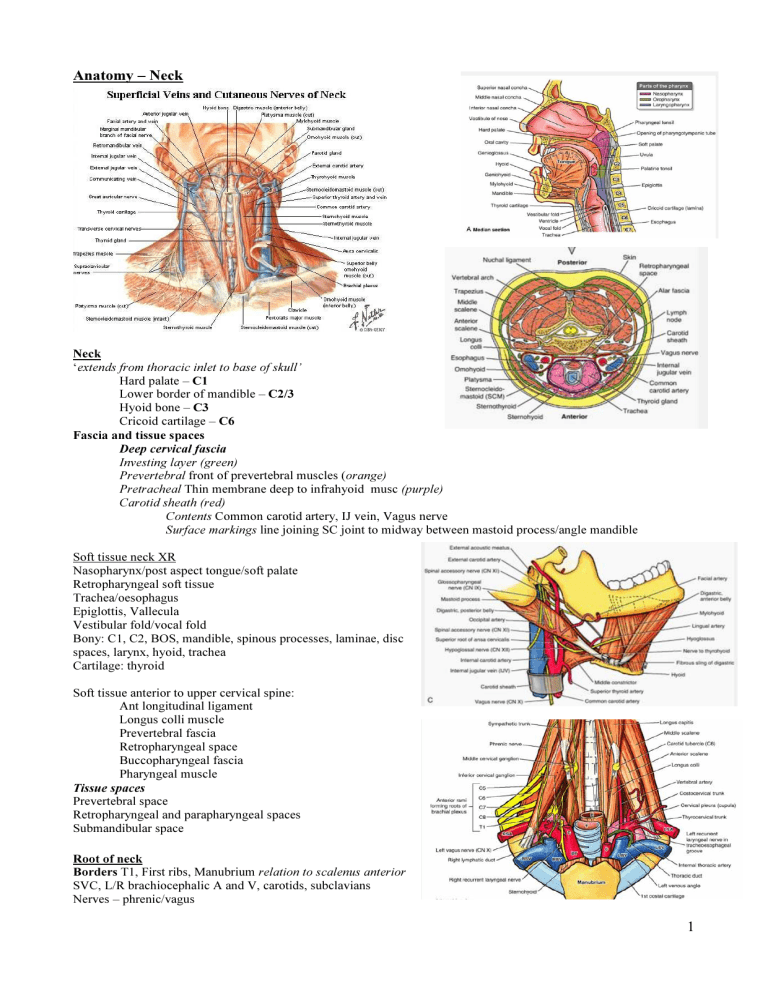
Anatomy – Neck
Neck
‘ extends from thoracic inlet to base of skull’
Hard palate – C1
Lower border of mandible – C2/3
Hyoid bone – C3
Cricoid cartilage – C6
Fascia and tissue spaces
Deep cervical fascia
Investing layer (green)
Prevertebral front of prevertebral muscles ( orange)
Pretracheal Thin membrane deep to infrahyoid musc (purple)
Carotid sheath (red)
Contents Common carotid artery, IJ vein, Vagus nerve
Surface markings line joining SC joint to midway between mastoid process/angle mandible
Soft tissue neck XR
Nasopharynx/post aspect tongue/soft palate
Retropharyngeal soft tissue
Trachea/oesophagus
Epiglottis, Vallecula
Vestibular fold/vocal fold
Bony: C1, C2, BOS, mandible, spinous processes, laminae, disc spaces, larynx, hyoid, trachea
Cartilage: thyroid
Soft tissue anterior to upper cervical spine:
Ant longitudinal ligament
Longus colli muscle
Prevertebral fascia
Retropharyngeal space
Buccopharyngeal fascia
Pharyngeal muscle
Tissue spaces
Prevertebral space
Retropharyngeal and parapharyngeal spaces
Submandibular space
Root of neck
Borders T1, First ribs, Manubrium relation to scalenus anterior
SVC, L/R brachiocephalic A and V, carotids, subclavians
Nerves – phrenic/vagus
1
Subclavian artery
Surface marking – line upwards 2cm above clavicle from SC joint to mid clavicle
From brachiocephalic R, aortic arch L – behind SC joints
Post to scal ant, crosses 1 st
rib, Ends lat border 1 st
rib → axillary
3 parts: med, behind, lat to scal ant
1 st
: 3 branch: vertebral, int thoracic, thyrocervical trunk
2 nd
: between scal ant/med 2 branch: supraclav, dorsal scap
3 rd
: No branches
Subclavian vein
From axill med to outer border 1 st rib → med/post to clavicle, sup to 1 st
rib
Lies ant to scal ant (subclavian A is behind this) groove 1 st
rib
→ brachioceph at med border scal ant joins IJV (thoracic duct L, lymphatic R)
Anterior to it: skin, pec major, clavipectoral fascia, pec min
Sternocleidomastoid
Origin Rounded tendon manubrium below sternal notch
Flat tendon from sup surface medial third clavicle
Insertion
Lateral surface mastoid process
Lateral half superior nuchal line of occipital bone
Crossed by External jugular vein
Deep to upper part lies cervical plexus
Deep to lower part lies carotid sheath
Nerve Spinal part of accessory nerve (C3,4)
Action tilts head to ipsilateral shoulder/rotates head opposite
Posterior triangle of the neck
Borders - SCM, trapezius, clavicle
Contents
3 trunks brachial plexus
Accessory nerve
Pulsation of subclavian artery
External jugular vein
Lymph nodes
Inferior belly of omohyoid
SCM
Ant dig
Carotid
Mandible
Digastric
Hyoid
Post dig
Submental
Anterior triangle of the neck
Omohyoid Midline
Muscular
Muscles of anterior triangle
Suprahyoid muscles
Mylohyoid (forms floor mouth)
Digastric – ant/post bellies
Stylohyoid, Geniohyoid
Infrahyoid muscles
Sternohyoid, Omohyoid, Thyrohyoid, Sternothyroid
Borders Mandible, SCM, Midline
Subdivided into
Carotid triangle
Borders SCM, post digastric, omohyoid
Contents Bifurcation common carotid and branches external carotid
Lingual, facial, sup thyroid veins
Hypoglossal, int/ext laryngeal n.
Jugulogastric lymph nodes
Digastric - Borders Mandible, ant/post digastric
Submental - Borders Ant belly digastric, hyoid, mid
Muscular - Borders SCM, omohyoid, midline
2
Cervical plexus
Anterior rami upper 4 cervical nerves
Branch - Phrenic nerve (C3/4/5) over scalene anterior, behind subclavian vein into mediastinum
Thyroid gland
Enclosed by capsule and pretracheal fascia, Ant neck C5-T1
Isthmus over 2nd, 3rd and 4th tracheal rings, Join lateral lobes
Blood supply Sup/inf thyroid arteries, Thyroidea ima artery; Sup/mid/inf thyroid veins
Lymph Mainly deep cervical nodes
Great vessels of the neck
Common carotid artery
Left aortic arch, Right brachiocephalic trunk, behind SC joint into common carotid and subclavian
Common carotid gives no branches
Lies in carotid sheath medial to internal jugular vein, with vagus nerve
Divides at upper border thyroid cartilage (upper border of C4) Carotid sinus , Carotid body
Surface marking – SC joint to upper border thyroid cartilage
External carotid artery
Main arterial supply neck & face
Origin upper border C4/thyroid cartilage
Med to ICA at commencement (later lat), ant to SCM, ascend ant to ICA
Surface marking – point bifurcation to in front of tragus of ear
BEFORE PAROTID
Some: Superior thyroid artery
Anaesthetists: Ascending pharyngeal artery
Like: Lingual artery
Fun: Facial artery
Others: Occipital artery
IN PAROTID
Prefer: Posterior auricular artery
S &: Superficial temporal artery (terminal)
M: Maxillary artery (terminal)
3 anterior, 2 behind, 1 medial
Ant: superior thyroid inferior, lingual above, facial above
Post: occipital inferior, posterior auricular above
Med: ascending pharyngeal
Internal carotid artery
4 parts – cervical, petrous, cavernous, cerebral
Main supply to brain and eye
Arises at bifurcation of common carotid artery (C3-4)
Lateral to external carotid at origin, Passes up poste and medial in carotid sheath
No branches in neck, Passes through carotid canal in petrous temporal bone
Surface marking – line from bifurcation to head of mandible
Internal jugular vein
Drains brain, neck & face
Thoracic duct opens into angle of union between L IJV & L subclavian vein, right lymphatic duct opens on right side
Emerges from jugular bulb at posterior part of jugular foramen
Lies on transverse process of atlas
Initially behind internal carotid artery in carotid sheath, then lateral
Covered by sternocleidomastoid in lower part
Join subclavian, form brachio behind sternal end clavicle
Surface marking – ear lobe to SC joint
Lymphatic drainage of head and neck
2 groups – superficial and deep:
Superficial: Occipital, Retro-auricular, Parotid, Buccal, Submandibular, Submental
These drain into: Anterior cervical, Superficial cervical
These drain into deep group which form a chain along the IJV in the carotid sheath and receive all lymph of head/neck
Retropharyngeal, Laryngeal, Paratracheal, Pretracheal
These form jugular lymph trunks which drain into thoracic duct or R lymphatic duct
3
Larynx
Function sphincter, phonation
Cartilages
Single Thyroid
Cricoid
Epiglottic
Median glossoepiglottic fold
Lateral pharyngoepiglottic fd
Valleculae (depression mucous membrane either side glossoepiglottic folds)
Paired Arytenoid, Corniculate , Cuneiform
Joints Cricothyroid, Cricoarytenoid joint
Ligaments and membranes
Extrinsic membranes
Thyrohyoid, Cricotracheal
Intrinsic membranes
Quadrangular membrane arytenoid to epiglottis; Mucous membrane covered, form aryepiglottic folds (vestibular)
Cricothyroid membrane
Ant cricothyroid ligament
Lat cricothyroid ligaments Free edge is vocal ligament (vocal cord)
Hypoepiglottic ligament
Thyroepiglottic ligament
Muscles
Adduction of cords and closing of glottis Transverse arytenoid, Oblique arytenoids, Lateral cricoarytenoid
Abduction of cords and opening of glottis Posterior cricoarytenoid
Shortens and relaxes cords Thyroarytenoid
Lengthens and tightens cords Cricothyroid
Nerve supply
Motor - Muscles
Extrinsic
Infrahyoid/suprahyoid/stylopharyngeus
CN X – recurrent laryngeal
Intrinsic
All muscles > recurrent laryngeal
Except cricothyroid > external laryngeal
Sensory - Mucous membranes
Above cords internal laryngeal, Below cords recurrent laryngeal
Pharynx
Nasopharynx (behind nasal, above soft palate), Oropharynx (behind oral cavity),
Laryngopharynx (behind larynx)
Rules of Nerve Supply for Muscle Groups
Pharynx - Pharyngeal plexus - Except stylopharyngeus (glossopharyngeal – IX)
Palate - Pharyngeal plexus - Except tensor veli palatini
Tongue - Hypoglossal (XII) - Except palatoglossus (pharyngeal plexus)
Facial expression and buccinator - Facial (VII) - Except levator palpebrae superioris (occulomotor)
Mastication - Mandibular division trigeminal (Vc) - Except buccinator (facial – VII)
Larynx - Recurrent laryngeal - Except cricothyroid (ext branch sup laryngeal – X)
4
