Report for Carotid Duplex Examinations
advertisement
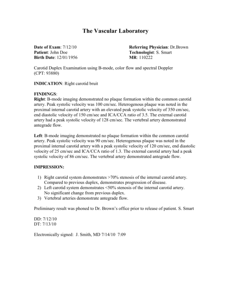
The Vascular Laboratory Date of Exam: 7/12/10 Patient: John Doe Birth Date: 12/01/1956 Referring Physician: Dr.Brown Technologist: S. Smart MR: 110222 Carotid Duplex Examination using B-mode, color flow and spectral Doppler (CPT: 93880) INDICATION: Right carotid bruit FINDINGS: Right: B-mode imaging demonstrated no plaque formation within the common carotid artery. Peak systolic velocity was 100 cm/sec. Heterogenous plaque was noted in the proximal internal carotid artery with an elevated peak systolic velocity of 350 cm/sec, end diastolic velocity of 150 cm/sec and ICA/CCA ratio of 3.5. The external carotid artery had a peak systolic velocity of 128 cm/sec. The vertebral artery demonstrated antegrade flow. Left: B-mode imaging demonstrated no plaque formation within the common carotid artery. Peak systolic velocity was 90 cm/sec. Heterogenous plaque was noted in the proximal internal carotid artery with a peak systolic velocity of 120 cm/sec, end diastolic velocity of 25 cm/sec and ICA/CCA ratio of 1.3. The external carotid artery had a peak systolic velocity of 86 cm/sec. The vertebral artery demonstrated antegrade flow. IMPRESSION: 1) Right carotid system demonstrates >70% stenosis of the internal carotid artery. Compared to previous duplex, demonstrates progression of disease. 2) Left carotid system demonstrates <50% stenosis of the internal carotid artery. No significant change from previous duplex. 3) Vertebral arteries demonstrate antegrade flow. Preliminary result was phoned to Dr. Brown’s office prior to release of patient. S. Smart DD: 7/12/10 DT: 7/13/10 Electronically signed: J. Smith, MD 7/14/10 7:09
