Enantioselective Michael Addition of Water
advertisement
DOI: 10.1002/chem.201405579
Full Paper
& Biocatalysis
Enantioselective Michael Addition of Water
Bi-Shuang Chen,[a] Verena Resch,[a, b] Linda G. Otten,[a] and Ulf Hanefeld*[a]
Abstract: The enantioselective Michael addition using water
as both nucleophile and solvent has to date proved beyond
the ability of synthetic chemists. Herein, the direct, enantioselective Michael addition of water in water to prepare important b-hydroxy carbonyl compounds using whole cells of
Introduction
The direct addition of water to C=C bonds is a highly attractive
transformation, yielding (chiral) alcohols.[1] However, the enantioselective addition of water to a,b-unsaturated carbonyl (Michael) acceptors still represents a chemically very challenging
reaction,[2] due to the poor nucleophilicity of water and its
small size, which make regio- and stereoinduction difficult.
Equally, the often unfavorable equilibrium of water-addition reactions remains to be solved. Although this reaction benefits
from its simplicity and excellent atom economy, no protocol
with broad applicability has to date been developed. Indirect
methods[3] using complex catalysts[4] or strong alternative nucleophiles[5] have been employed. Some of the described
methods require either cumbersome catalyst preparation or reductive/oxidative follow-up chemistry. Selective direct methods
have been reported by Roelfes and co-workers, applying DNAbased CuII catalysts[6] or the use of a protein as chiral ligand.[7]
However, they are limited to a,b-unsaturated 2-acyl imidazoles
as substrates and yield the corresponding alcohols in moderate enantiomeric purities. The only chemocatalytic process run
on industrial scale was the addition of water to acrolein.[1d]
Nevertheless, due to its poor selectivity and productivity, even
this seemingly straightforward reaction has been replaced by
a fermentative process.[1d, 8]
Rhodococcus strains is described. Good yields and excellent
enantioselectivities were achieved with this method. Deuterium labeling studies demonstrate that a Michael hydratase
catalyzes the water addition exclusively with anti-stereochemistry.
In contrast, enzymes such as fumarase, malease, citraconase,
aconitase, and enoyl-CoA hydratase have been successfully
used on industrial scale, and their excellent (enantio-) selectivities are highly valued.[1d, 9] Unfortunately, most hydratases are
part of the primary metabolism where perfect substrate specificity is required. This very high substrate selectivity severely
limits their practical applicability in organic synthesis.[2a] A
recent report on an asymmetric hydration of hydroxystyrenetype substrates catalyzed by phenolic acid decarboxylases
showed that a broader flexibility in the substrate spectrum for
hydratases is possible.[10] In order to broaden the biocatalytic
toolbox of hydratases, the work represented herein is dedicated to the search for a Michael hydratase with a more relaxed
substrate specificity.
In our search for a straightforward approach for the preparation of b-hydroxy carbonyl compounds via the direct Michael
addition of water, it was noted that whole cells of Rhodococcus
rhodochrous ATCC 17895 convert 3-methylfuran-2(5 H)-one 1 a
into (S)-3-hydroxy-3-methylfuranone 2 a; as briefly described in
1998.[11] Neither substrate 1 a nor product 2 a are part of the
primary metabolism indicating the involvement of a putative
Michael hydratase with possibly a broader substrate scope.
Since whole cells were used in this transformation, the hydratase activity needed to be critically evaluated.[11–14] Instead of
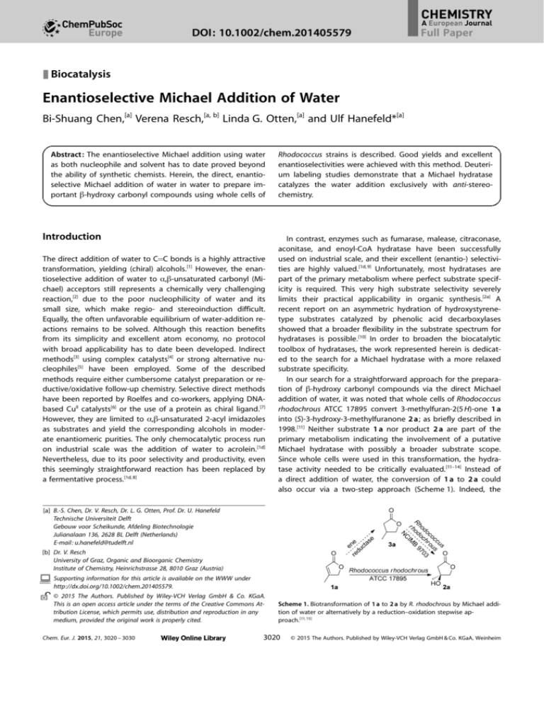
a direct addition of water, the conversion of 1 a to 2 a could
also occur via a two-step approach (Scheme 1). Indeed, the
[a] B.-S. Chen, Dr. V. Resch, Dr. L. G. Otten, Prof. Dr. U. Hanefeld
Technische Universiteit Delft
Gebouw voor Scheikunde, Afdeling Biotechnologie
Julianalaan 136, 2628 BL Delft (Netherlands)
E-mail: u.hanefeld@tudelft.nl
[b] Dr. V. Resch
University of Graz, Organic and Bioorganic Chemistry
Institute of Chemistry, Heinrichstrasse 28, 8010 Graz (Austria)
Supporting information for this article is available on the WWW under
http://dx.doi.org/10.1002/chem.201405579.
2015 The Authors. Published by Wiley-VCH Verlag GmbH & Co. KGaA.
This is an open access article under the terms of the Creative Commons Attribution License, which permits use, distribution and reproduction in any
medium, provided the original work is properly cited.
Chem. Eur. J. 2015, 21, 3020 – 3030
Scheme 1. Biotransformation of 1 a to 2 a by R. rhodochrous by Michael addition of water or alternatively by a reduction–oxidation stepwise approach.[11, 15]
3020
2015 The Authors. Published by Wiley-VCH Verlag GmbH & Co. KGaA, Weinheim
Full Paper
enantioselective hydroxylation of a range of THF and THP derivatives was reported for R. rhodochrous strains.[15] Therefore, it
is of high interest to probe whether the conversion of 1 a to
2 a is actually a Michael addition of water and how broadly it
is applicable.
Herein we report the results of screening several Rhodococcus strains as promising biocatalysts for the enantioselective
Michael addition of water to a variety of a,b-unsaturated carbonyl compounds.
Results and Discussion
Optimization
3 a as substrate under aerobic conditions (Table 1, control 1),
no conversion to 2 a was detected, indicating that no oxidation
occurs. In previous studies[2b, 14] we were able to show that
a chemically catalyzed addition reaction occurs when 2-cyclohexenone (1 h) is used as a substrate. Therefore, any undesired
background reaction needed to be ruled out. Heat-denatured
cell preparations in control experiments (Table 1, control 2)
clearly showed that there is no chemically catalyzed reaction
taking place; thus the reaction is effected by the active
enzyme.
Encouraged by the complete conversion after 17 h, we evaluated the rate of the reaction with 330 mg mL1 of wet cells.
This revealed an almost linear increase in product formation
during the first 6 h of the reaction and 2 a was formed in 75 %
yield (Figure 1 A). Complete conversion based on the consump-
To fully assess the potential of the putative Michael addition of
water, the previously reported conversion of 3-methylfuran2(5 H)-one (1 a)[11] was repeated and optimized. 1 a was synthesized using a modified literature procedure (see the Supporting Information, S3).[16] Whole cells of R. rhodochrous ATCC
17895 were used in two different concentrations (100 mg mL1
and 330 mg mL1 of wet cells; Table 1). The reaction with
100 mg mL1 cells gave a maximum conversion of 69 % after
17 h and, even after a prolonged reaction time (4 days), no further increase in conversion was observed. Furthermore, an ee
of 91 % was determined, which is in agreement with the previously reported study.[11] An increase of the cell concentration
to 330 mg mL1 of wet cells resulted in full conversion after
17 h, while ee values remained unchanged (90 %). When using
Table 1. Influence of the catalyst concentration on the conversion.
Catalyst
this
study
resting
cells
resting
cells
ref. [11] resting
cells
control 1 resting
cells[c]
control 2 denatured
cells[d]
Catalyst
conc. (wet
cells)
Substrate Conversion[a] Yield[a]
of 1 a [%]
of
2 a[b]
[%]
Figure 1. Time course (A), temperature profile at reaction time 6 h (B), pH
profile at reaction time 6 h (C) and Michaelis–Menten kinetics (D, based on
the yield of 2 a) of the putative Michael addition catalyzed using whole cells
of R. rhodochrous ATCC 17895. For reaction conditions, see the Experimental
Section. Conversion, yield, and ee values were determined by GC. Filled circles represent ee of 2 a. Filled triangles represent consumption of 1 a. Filled
squares represent yield of 2 a. Empty triangles represent consumption of 1 a
in blank reactions. Empty squares represent yield of 2 a in blank reactions (in
A and D, blank reaction was carried out with heat-denatured cells; in C,
blank reaction was carried out without the addition of cells).
ee[a]
of
2a
[%]
100 mg mL1
1a
69
57
91
330 mg mL1
1a
99
87
90
100 mg mL1
1a
55
55
95
330 mg mL1
3a
–
<3
n.d.[f]
330 mg mL1
1a
12[e]
<3
n.d.[f]
[a] Conversion, yield, and ee values were determined by GC; [b] absolute
configuration of 2 a has been established by converting 2 a into the corresponding methyl ester [methyl S-()-3,4-dihydroxy-3-methylbutanoate];[12, 13] [c] reaction with 3 a was carried out to rule out possible oxidation; [d] reaction with heat-denatured cells was carried out to ensure no
background reaction is taking place; [e] conversion is caused by the ring
opening of lactone 1 a, no water addition product (2 a) was detected;
[f] n.d. = not determined.
Chem. Eur. J. 2015, 21, 3020 – 3030
www.chemeurj.org
tion of 1 a was obtained after 9 h. No significant changes of
the product ee within the first 9 h were observed (from 99 %
to 95 %; see the Supporting Information, S11 for GC chromatographs). It should be noted that, since the desired Michael addition products (2 a) are highly soluble in water, the choice of
the organic solvent for extraction is crucial. For example, using
ethyl acetate or dichloromethane as the extraction solvent
only allowed recovery of 30 % of the product (data not
shown). In extraction studies, isoamyl alcohol gave the best
result for extraction of (S)-3-hydroxy-3-methylfuranone (2 a).
However, due to the similar polarity of isoamyl alcohol and
water-addition product 2 a, severe problems, such as separation issues during purification, arose. Therefore, for preparative-scale experiments, reaction mixtures were always continu-
3021
2015 The Authors. Published by Wiley-VCH Verlag GmbH & Co. KGaA, Weinheim
Full Paper
ously extracted overnight using a liquid–liquid extractor and
ethyl acetate as the organic solvent (see the Supporting Information, S9). This procedure had no influence on the ee values
of the product (data not shown).
The temperature profile of the reaction was evaluated as
well. Temperatures ranging from 18 8C to 48 8C were tested.
Conversions and ee values at different temperatures are summarized in Figure 1 B. When increasing the temperature above
28 8C a decrease in enzyme activity was observed. At 48 8C,
a yield of only 5 % was detected (an additional 12 % was
brought about by ring opening of lactone 1 a). Due to the low
amount of product at 48 8C, no reliable ee determination was
possible. Taking both the conversion and enantioselectivity
into account, the best results were achieved at 28 8C. These results are in agreement with the reported optimal growth temperature of 26 8C for R. rhodochrous.[17]
Since water serves not only as the reaction medium but also
as a substrate, the pH needs to be considered as a very important parameter. To quantify this effect, the reaction system was
tested at different pH values using potassium phosphate
buffer (pH 5.2–8.2) and citrate/phosphate buffer (pH 4.2) to
control the pH of the reaction medium. The results from this
study clearly show the dependence on pH. The conversion increased with increasing pH (Figure 1 C, filled triangles), as expected from our previous study,[2b] demonstrating that the hydration reaction is generally base-catalyzed. However, at neutral and slightly basic conditions (pH 7.2 and pH 8.2), significant ring opening of lactone 1 a took place (Figure 1 C, empty
triangles), which explains the rather poor product yield (Figure 1 C, filled squares). This effect can be explained by the
spontaneous hydrolysis of the lactone in basic aqueous
medium, which is an often observed phenomenon.[18] To confirm this, the blank reaction mixtures at pH 7.2 and 8.2 were
acidified with conc. HCl to pH 1. This leads to complete recovery of the substrate 1 a, validating our hypothesis. No desired
enantioselective Michael addition product 2 a was detected in
the blank reactions (Figure 1 C, empty squares) indicating that
chemical/base-catalyzed Michael addition does not occur
within the measured pH ranges. Therefore, the conversion in
the blank reactions (which is based on the decrease in amount
of substrate) is caused by the hydrolysis of 1 a. Moreover, product 2 a showed good stability under strongly acidic conditions
and only 10 % yield was lost overnight. Comparing the mass
balance and reaction rate, slightly acidic conditions (pH 6.2)
represented the optimal pH for this substrate.
Control experiments (Table 1 and Figure 1 A–C) confirmed
that the formation of 2 a is based on an enzymatic reaction
with high enantioselectivity and that no chemical background
reaction occurred. Therefore, the kinetic parameters Km, Vmax,
and Vmax/Km were determined with the optimized conditions.
The Michaelis–Menten Plot (Figure 1 D) allowed calculation of
the affinity constant Km as 1.7 102 m and Vmax as
6.9 nmol s1 g1 (wet cells), providing further support for one
enzymatic reaction, rather than a sequence of reactions
(Scheme 1).
To establish the distribution of the enzymatic activity over
different organisms, we proceeded with testing different closeChem. Eur. J. 2015, 21, 3020 – 3030
www.chemeurj.org
ly related Rhodococcus strains. The selection was based on
phylogenetic analysis (Table 2). The previously reported strain
R. rhodochrous ATCC 17895 was shown to be much more
closely related to R. erythropolis than to R. rhodochrous.[17] For
this reason, strains R. erythropolis DSM 43296, R. erythropolis
CCM 2595, R. erythropolis NBRC 100887, and R. erythropolis
DSM 43066 were evaluated (Table 2). Experiments for comparing the different organisms were carried out under conditions
optimized for R. rhodochrous ATCC 17895. Gratifyingly, in each
case, 3-methylfuran-2(5 H)-one (1 a) was converted into (S)-3hydroxy-3-methylfuranone (2 a) with good yields and excellent
enantioselectivities (see the Supporting Information, S12 for
GC chromatographs). Encouraged by these results, the less
closely related strain R. rhodochrous DSM 43241 was also
tested for water addition activity. Interestingly, the enantiomerically enriched water-addition product (S)-3-hydroxy-3-methylfuranone 2 a was also obtained in 75 % yield and with an
86 % ee, which was slightly lower than that with R. erythropolis
strains. All the results suggest that this promising hydratase activity is not limited to R. rhodochrous ATCC 17895 but may be
a general feature in several Rhodococcus strains. Taking the
conversion, enantioselectivity, and available genome sequence
into account, we decided to continue to use strain R. rhodochrous ATCC 17895 for all further studies.
Substrate scope and limitations
Since the very limited substrate scope of the known hydratases
is one of the challenging factors for their broad application,
we were interested in the scope of the Michael hydratase from
R. rhodochrous ATCC 17895. Neither substrate 1 a nor product
2 a are known to be part of primary metabolic pathways,
therefore the substrate scope of the hydratase from R. rhodochrous ATCC 17895 might be more relaxed than that for
other known hydratases. Hence we tested a set of different
substrates to evaluate the limitations of the enzyme (Table 3).
For a,b-unsaturated lactones (X = O; Table 3, entries 1–3) with
substituents in the b-positions, the reaction proceeded
smoothly in all cases to yield the corresponding hydration
products, whereas for R1 = H (X = O; Table 3, entries 4 and 5),
no water addition product was obtained. This result is surprising, as the tertiary alcohols obtained are sterically much more
demanding than the secondary alcohols, and suggests that
substituents in the b-position might play an important role for
proper orientation of the lactones in the enzyme’s active site.
However, the enzyme did not accept substrates with substituents in both b- and g-positions, such as 1 f (Table 3, entry 6),
which is probably due to its more bulky structure. Products 2 a
and 2 b are tertiary alcohols, representing a class of compounds that are difficult to prepare by chemical methods, to
date only accessible via this route.[11] The enantioselectivity
was measured using a chiral Ivadex 7/PS086 GC column and,
in parallel, the ee was confirmed by analysis of 1H and 19F NMR
spectra of their corresponding Mosher esters (see the Supporting Information, S4, S5, and S27–S29). In both cases, results
from 1H and 19F NMR spectra of the Mosher esters and chiral
GC analysis of the alcohols were comparable, showing excel-
3022
2015 The Authors. Published by Wiley-VCH Verlag GmbH & Co. KGaA, Weinheim
Full Paper
Table 2. Comparison of closely related Rhodococcus strains. Phylogenetic tree based on 16 rRNA
Entry[a]
Catalysts
Conversion[b] of 1 a [%]
Yield[b] of 2 a [%]
ee[b] of 2 a [%]
1
2
3
4
5
6
7
R. rhodochrous ATCC 17895
R. erythropolis DSM 43296
R. erythropolis CCM 2595
R. erythropolis NBRC100887
R. erythropolis DSM 43066
R. rhodochrous DSM 43241
90 8C heat-denatured cells of R. rhodochrous ATCC 17895
87
82
88
80
90
87
12
75
70
76
68
78
75
<3
95
93
95
93
95
86
n.d.
Genome sequence
+
+
+
[a] List of entries comparing activities using different organisms. [b] Conversion, yield, and ee values were determined by GC.
lent enantioselectivities. The absolute configuration of the
product was established by converting 2 a into the corresponding methyl ester [methyl S-()-3,4-dihydroxy-3-methylbutanoate].[12, 13]
Interestingly, the hydration of substrate 1 c (Table 3, entry 3)
gave access to the natural product mevalonolactone 2 c, the
salt form of which represents an intermediate in the pathway
leading to terpenoids.[19] Absolute configuration of (R)-2 c was
determined by comparison with previously reported optical rotation data.[20] Mevalonate is a product of acetate metabolism
and thus a key building block in secondary metabolism.[21] To
identify whether the putative Michael hydratase is a promiscuous enzyme of the mevalonate pathway, bioinformatics studies
were performed. We have sequenced and annotated the
genome of R. rhodochrous ATCC 17895 in a previous study.[17]
Looking for annotated hydratases in this genome only showed
known hydratases with their narrow substrate specificity, emphasizing that the hydratase of this study has not been described before. This enzyme could therefore be one of the
many unknown gene functions in the genome, or a promiscuous activity of a known enzyme. Screening through all three
sequenced Rhodococci genomes (Table 2, entry 1, 3 and 4), we
unexpectedly found that most of the typical enzymes from the
mevalonate pathway were missing. Instead the full deoxyxyluChem. Eur. J. 2015, 21, 3020 – 3030
www.chemeurj.org
lose phosphate (mevalonate-independent) pathway for terpenoid biosynthesis was present.
For a,b-unsaturated ketones (X = C), substrates without substituent in the b-position were surprisingly accepted by the
putative Michael hydratase (Table 3, entries 7–9) but no activity
towards the b-substituted 3-methylcyclohex-2-enone and
3-methylcyclopent-2-enone was found (Table 3, entries 10 and
11). This might lead to the conclusion that b-substituted
a,b-unsaturated ketones may be challenging for Michael addition of water using R. rhodochrous, although the opposite is
true for a,b-unsaturated lactones. The a,b-unsaturated ketones
1 g–i, were mostly reduced into ketones 3 g–i (75 %, 76 %, and
80 % yields, respectively), which explains the rather poor yield
of the water-addition reaction (Table 3, entries 7–9). Experiments
to rule out the reduction–oxidation as a possible reaction pathway were performed for these cyclic ketones (Scheme 2). Reaction using 1 h as substrate was performed under a nitrogen atmosphere to exclude air as a potential oxygen source. Even so,
22 % yield of 2 h was obtained with 65 % ee, ruling out the involvement of O2 as an active species in the reaction. Furthermore, when 3 h was used as a substrate directly under aerobic
conditions, no product 2 h was detected. These two control experiments demonstrate that the alcohol 2 h was the result of
the enantioselective Michael addition of water to 1 h. The com-
3023
2015 The Authors. Published by Wiley-VCH Verlag GmbH & Co. KGaA, Weinheim
Full Paper
Table 3. Substrate scope for the enantioselective Michael addition of water.
Entry Substrate Product Conversion[a] Yield[a] ee of EnantioEquilibrium
of 1 [%]
of 2
2
preference yield of 2
[%]
[%]
[%]
1
2
3[b]
4
5
6
7
8
9
10
11
12
13
14
15
1a
1b
1c
1d
1e
rac-1 f
1g
1h
1i
1j
1k
1l
1m
1n
1o
2a
2b
2c
2d
2e
2f
2g
2h
2i
2j
2k
2l
2m
2n
2o
87
80
75
12
32
12
93
98
95
<3
<3
42
<3
<3
<3
75
68
62
<3
<3
<3
18
22
15
<3
<3
40
<3
<3
<3
95[c,d]
94[d]
73[c]
n.d.
n.d.
n.d.
22[c]
65[c]
20[c]
n.d.
n.d.
48[c]
n.d.
n.d.
n.d.
S[e]
S
R[f]
n.d.
n.d.
n.d.
R[g]
R[g]
R[h]
n.d.
n.d.
R[f]
n.d.
n.d.
n.d.
> 97[i]
–[j]
–[j]
–[j]
–[j]
–[j]
–[j]
25[k]
–[j]
–[j]
–[j]
–[j]
–[j]
–[j]
–[j]
peting reduction reaction to 3 g, 3 h, and 3 i is most likely
due to an ene reductase also present in the Rhodococcus
cells.
To further probe whether the putative Michael hydratase also accepts acyclic a,b-unsaturated carbonyl compounds, methyl crotonate (1 l), crotonic acid (1 m),
(Z)-ethyl-4-hydroxy-3-methylbut-2-enoate (1 n), and benzylideneacetone (1 o) were subjected to the resting cell suspensions (Table 3, entries 12–15). Gratifyingly, the enzyme
readily accepted acyclic a,b-unsaturated ester (1 l), although no activity was observed for acyclic a,b-unsaturated carboxylic acid 1 m, ester 1 n, or ketone 1 o. Notably, in
many water-addition reactions to carbon–carbon double
bonds, the equilibrium can be on the side of the starting
material although the reaction is performed in water.[1d, 6c, 22] The unfavorable equilibrium might impede the
Michael addition of water; for example, the equilibrium
yield of 3-hydroxycyclohexanone (2 h) was determined to
be 25 % (Table 3, entry 8),[2b, 23] corresponding with the
measured yield of 22 %.
Finally we tested the scalability of the developed reaction system. Therefore the reaction was scaled to gram
scale using 1 a (2 g, 20 mmol, 200 g of wet cells) to give
2 a in 69 % isolated yield after column chromatography
and an ee of 90 % was determined.
Recyclability and enzyme investigation
One of the most important characteristics of a catalyst is
its operational stability and reusability over an extended
period of time, to ensure a practical application.[26] From
the viewpoint of process economics, the higher the
number of cycles that an enzyme remains stable, the
more efficiently a process can be run. Experiments were
[a] Conversion and yield were determined by GC; [b] reaction was performed at
performed to examine this recyclability of the whole cells
pH 5.2 to suppress ring open of lactone 1 c at pH 6.2; [c] ee was determined by
of strain R. rhodochrous ATCC 17895 for the Michael addiGC; [d] ee was determined by 1H and 19F NMR of the corresponding Mosher
tion of water to 3-methylfuran-2(5 H)-one (1 a). Based on
ester; [e] changing CIP priorities; [f] (R)-enantiomers commercially available;
the results summarized in Table 1, every reaction was car[g] absolute stereochemistry was determined by converting them into literature-known derivatives, following a procedure established earlier in our laboraried out in a 50 mL Erlenmeyer flask at 28 8C with 180 rpm
tory;[24] [h] absolute configuration was determined by comparison of the retenfor 23 h. At the end of the reaction, the cells were centrition times using the same GC column with a reported method;[25] [i] reverse refuged, washed twice with potassium phosphate buffer
action with 2 a as substrate was performed, analysis of this sample showed no
(100 mm, pH 6.2), and then reused for the next cycle.
dehydration and no decrease of the ee; [j] no literature values available; [k] see
references [2b, 23] .
Whole cells showed high activity and complete conversion
for 4 cycles (Figure 2). Only a slight decrease was observed
in cycle 5, whereas 27 % lower conversion was detected in
the cycle 6. However, even after 9 consecutive cycles, the
whole cells retained 20 % of the initial activity. Notably, no significant changes in enantioselectivities of the water-addition
reactions were detected during the 9 cycles (see the Supporting Information, S12 for GC chromatography).
To isolate the putative Michael hydratase for further investigation, we first broke the whole cells of R. rhodochrous ATCC
17895. The desired hydratase activity (yielding 2 a) was only
found in the cell pellets, rather than in the cell-free extract,
when 1 a was used as a substrate (Figure 3). Furthermore, no
significant difference was found between the initial rate of
Scheme 2. Control experiments to confirm that 2 h was formed by enzywhole cells and pelleted cell debris (Figure 3). Additionally,
matic water addition, rather than a reduction–oxidation sequence.
Chem. Eur. J. 2015, 21, 3020 – 3030
www.chemeurj.org
3024
2015 The Authors. Published by Wiley-VCH Verlag GmbH & Co. KGaA, Weinheim
Full Paper
95 % enantioselectivity was still obtained, ruling out that O2 is
involved as an active species in the reaction. The second approach included the use of substrate 3 a, which might be
formed by reduction of 1 a. Therefore 3 a was synthesized (see
the Supporting Information, S5) according to a standard procedure.[27a] When using 3 a with a resting cell suspension, no corresponding oxidation product was detected under aerobic
conditions (Scheme 3). These two control experiments demon-
Figure 2. Repeated water-addition reactions catalyzed by whole cells of
R. rhodochrous ATCC 17895. Conversion, yield, and ee values were determined by GC.
Scheme 3. Control experiments to confirm that 2 a was formed by enzymatic water addition, rather than a reduction–oxidation sequence.
Figure 3. Michael hydratase activities in different biocatalyst preparations
(whole cells, pelleted cell debris, and cell-free extract). Conversion was determined by GC.
with 1 h as a substrate, most of the ene reductase activity
(Table 3, entries 7–9) was retained in the cell-free extract,
whereas only minor activity was still detectable in the cell pellets. These results indicate that the putative Michael hydratase
is not a soluble protein but bound to either the membrane or
cell wall, whereas the reductase activity apparently resides
within another enzyme that is soluble. This natural immobilization of the putative Michael hydratase explains the high reusability of the whole cells (Figure 2).
Mechanistic studies
As mentioned above, Rhodococci have been shown to mediate
the hydroxylation of unactivated CH bonds on selected THF
and THP derivatives.[15] To clearly rule out the possibility of detecting a hydroxylation reaction instead of a hydration, we
turned our attention to the mechanism, including the stereochemical course of the reaction. The reaction was performed
under a nitrogen atmosphere to exclude air as a potential
oxygen source. Under these conditions, 87 % conversion with
Chem. Eur. J. 2015, 21, 3020 – 3030
www.chemeurj.org
strated that the enantiomerically enriched tertiary alcohol 2 a
was the result of the direct enantioselective Michael addition
of water to 1 a.
The stereochemical course of this water-addition reaction
was further evaluated by carrying out the biotransformation in
D2O using lyophilized cells as catalyst. The reaction in D2O was
found to be slower than that in H2O, which might be due to
activity loss caused by the lyophilization or an isotope effect.
However, upon elongation of the reaction time to 24 h, deuterium oxide-addition product 4 a was found at a conversion of
90 %. After extraction with ethyl acetate and column purification, compound 4 a, containing the optically active OD group,
was exchanged back into an OH group, which is an often-observed phenomenon.[28] NMR and GC-MS measurements
showed that the obtained compound (4 b) contained one deuterium at the a carbon (Figure 4 A). In the 1H NMR spectrum of
2 a, the geminal coupling constant between the two a protons
is 17.6 Hz, whereas the 1H NMR spectrum of 4 b showed only
one a proton, which indicates one deuterium at the a carbon.
Comparing with the singlet signal of 2 a in the 13C NMR spectrum, the triplet signal (coupling constant of 19.75 Hz) of 4 b
again indicates one deuterium at the a carbon. GC-MS spectra
also show 4 b to be one unit heavier than 2 a (for full spectra,
see the Supporting Information, S21–S23). A control experiment was performed by shaking pure 3-hydroxy-3-methylfuranone (2 a) in D2O. Analysis of this sample showed that no deuterium was incorporated at the a-position; hence, the 2H-labeled product 4 b must have resulted from enzymatic water
addition. This further supports a one-step hydration mechanism, via a Michael addition reaction.
According to the NMR measurements, the reaction of substrate 1 a and lyophilized R. rhodochrous ATCC 17895 cells in
D2O yielded monodeuterated 4 b as a sole diastereoisomer
3025
2015 The Authors. Published by Wiley-VCH Verlag GmbH & Co. KGaA, Weinheim
Full Paper
Figure 4. Diastereoselective Michael addition of water catalyzed by lyophilized cells of R. rhodochrous ATCC 17895.
Chem. Eur. J. 2015, 21, 3020 – 3030
www.chemeurj.org
3026
2015 The Authors. Published by Wiley-VCH Verlag GmbH & Co. KGaA, Weinheim
Full Paper
(Figure 4 A; for full spectra see the Supporting Information, S21
and S22). The observation brought us to investigate the diastereospecificity of the Michael addition of water, which until
now had been reported to show, depending on the enzyme,
either syn or anti preference.[29a] For example, in the case of
enoyl-CoA hydratase, selectivity towards syn-addition was observed,[29b] whereas enzymes belonging to the aspartase/fumarase superfamily, such as fumarase, aconitase, or enolase,
showed anti preference.[29c]
Nuclear Overhauser effect (NOE) experiments unfortunately
did not give conclusive results on the stereochemical course of
the water addition (see the Supporting Information, S32 and
S33). To further probe the stereoselectivity of the addition of
water, an anti E2 elimination of the deuterium oxide addition
product 4 b was performed.[30] The reaction was accomplished
with acetic anhydride/triethylamine in the presence of a catalytic amount of 4-dimethylaminopyridine (DMAP) and the corresponding product was measured directly by NMR and GC-MS
without further purification. The results showed 1 a as the only
elimination product, HDO being expelled during the elimination process (Figure 4 B). 1H NMR spectroscopy showed the appearance of signal for a proton at 5.91 ppm, indicating that
HDO was eliminated via an anti E2 elimination. The appearance of a singlet (which was visible as a triplet at 43.6 ppm
previously in 4 b) at 116.2 ppm for the a carbon in the 13C NMR
spectrum also proved the loss of molecular HDO. The D-NMR
spectrum shows only one peak at 2.60 ppm, which belongs to
the unconverted 4 b, but no peak at 5.91 ppm, again proving
that HDO was eliminated. This elimination was further confirmed by GC-MS analysis (for full spectra, see the Supporting
Information, S24 and S25). Since the E2 elimination always
occurs exclusively in anti fashion, and removed a-D and b-OH
groups, the enzymatic D2O addition must have proceeded
with exclusive anti stereochemistry. Our results are supported
by the findings of Mohrig et al.,[29a, d] who described that the
stereopreference of water addition depends on the position of
the abstracted proton: If the proton is in the a-position to the
carboxylate group, as in the case of our studies, anti-selectivity
is observed; abstracted protons that are in the a-position to
the carbonyl group of the thioester lead to syn-selectivity.[29a, b]
Conclusion
b-Hydroxy carbonyl compounds represent an important class
of compounds that is often found as a structural motif in natural products. Although the molecules themselves look rather
simple, their synthesis can be challenging. A straightforward
route for the preparation of chiral b-hydroxy carbonyl compounds was established, employing whole cells from several
Rhodococcus strains harboring a Michael hydratase. They catalyzed the enantioselective Michael addition of water in water
with good yields and excellent enantioselectivities. Compared
to the very narrow substrate scope of known hydratases, the
particularly intriguing feature and advantage of this new hydratase is its broad substrate range; a,b-unsaturated lactones
with substituents in b-position (1 a, 1 b and 1 c), a,b-unsaturated cyclic ketones with no substituent in b-position (1 g, 1 h
Chem. Eur. J. 2015, 21, 3020 – 3030
www.chemeurj.org
and 1 i), and an a,b-unsaturated ester (1 l). A series of control
experiments and deuterium labeling studies demonstrate that
the reaction is diasterospecific, with only the anti hydration
product formed. The biocatalytic reaction system was carefully
optimized for gram-scale synthesis, resulting in good conversions and excellent enantioselectivities. Under the optimized
conditions, whole cells could be reused for 4 cycles without
significant loss of activity while maintaining up to 90 % ee. Our
study suggests that this promising Michael hydratase is not
soluble but membrane-bound or cell wall-associated. In summary, whole cells from Rhodococcus strains are able to catalyze
the enantioselective Michael addition of water to several different substrates using water as both solvent and substrate
under mild conditions. This opens up an entirely new
approach to the synthesis of chiral b-hydroxy carbonyl
compounds.
Experimental Section
Material and Methods
All chemicals were purchased from Sigma–Aldrich (Schnelldorf,
Germany) and were used without further purification unless otherwise specified. The culture media components were obtained from
BD (Becton, Dickinson and Company, Germany).
1
H, 2H, 13C, and 19F NMR spectra were recorded with Bruker Advance 400 or Varian 300 (400 MHz, 61.4 MHz, 100 MHz and
376.33 MHz, respectively) instrument and were internally referenced to residual solvent signals. Data for 1H NMR are reported as
follows: chemical shift (d ppm), multiplicity (s = singlet, d = doublet,
t = triplet, q = quartet, m = multiplet), integration, coupling constant (Hz) and assignment. Data for 13C NMR and 19F NMR are reported in terms of chemical shift. Optical rotations were obtained
at 20 8C with a PerkinElmer 241 polarimeter (sodium D line).
Column chromatography was carried out with silica gel (0.060–
0.200 mm, pore diameter ca. 6 nm) and with mixtures of petroleum
ether (PE) and ethyl acetate (EtOAc) as solvents. Thin-layer chromatography (TLC) was performed on 0.20 mm silica gel 60-F plates.
Organic solutions were concentrated under reduced pressure with
a rotary evaporator.
Conversion of substrates and yield of products were quantified by
GC using calibration lines with dodecane as an internal standard
(specifications and temperature programs given in the Supporting
Information, S2) and the optical purity of the products [excepted
for 2 b] were determined using chiral GC (specifications and temperature programs given in the Supporting Information, S2). The
enantiomeric excess (ee) of 2 b was determined by 1H and 19F NMR
spectroscopy of the corresponding Mosher ester (see the Supporting Information, S4, S5, and S27–S29).
Microorganisms and culture conditions
Rhodococcus rhodochrous ATCC 17895 was purchased from ATCC
(American Type Culture Collection, Manassas, USA). Rhodococcus
erythropolis DSM 43296, Rhodococcus erythropolis DSM 43060, Rhodococcus erythropolis DSM 43066, and Rhodococcus rhodochrous
DSM 43241 were purchased from DSMZ (Germany). Rhodococcus
erythropolis PR4 NBRC 100887 was purchased from NBRC (Biological Resource Centre, Chiba, Japan). The organisms were maintained
on agar plates at 4 8C and these were subcultured at regular intervals. The medium used for cultivation[11] contained Solution A
3027
2015 The Authors. Published by Wiley-VCH Verlag GmbH & Co. KGaA, Weinheim
Full Paper
(980 mL) with potassium dihydrogen phosphate (0.4 g), dipotassium hydrogen phosphate (1.2 g), peptone (5 g), yeast extract (1 g),
glucose (15 g), final pH 7.2, sterilized at 110 8C in an autoclave; Solution B (10 mL) with magnesium sulphate (0.5 g), filter-sterilized;
Solution C (10 mL) with iron(II) sulphate (0.3 g), filter sterilized. Solutions were mixed before inoculation to make 1 L medium with
a buffer concentration of 3 mm. A loop of bacteria was used to inoculate 1 L medium in a 2 L Erlenmeyer flask. This culture was
shaken reciprocally at 28 8C for about 72 h to an optical density
(OD600) of around 6.3. The cells of Rhodococcus strains were harvested by centrifugation at 10 000 rpm and at 4 8C for 20 min. The
supernatant was removed and the cells were rinsed with potassium phosphate buffer (100 mm, pH 6.2) and centrifuged again. The
supernatant was discarded and the pellets were stored at 20 8C.
When needed, the wet pellets were freeze dried overnight and collected as lyophilized cells.
General biotransformation procedure for catalyst concentration study
Cells in the culture age of OD600 = 6.3 were harvested by centrifugation, washed twice with 100 mm potassium phosphate buffer
(pH 6.2). Around 100 mg mL1 or 330 mg mL1 of the cells were resuspended in the same buffer (15 mL) containing 33 mm 3-methylfuran-2(5 H)-one (1 a; 50 mg, 0.51 mmol). The resting cell reactions
were carried out in screw-capped Erlenmeyer flasks. Reactions
were shaken at 28 8C overnight (17 h). For the blank reaction the
setup was the same but heat-denatured cells (90 8C, 30 min) were
used. For workup, the cells were removed by centrifugation and
1 mL of the supernatant was saturated with NaCl followed by extraction with 2 0.5 mL of isoamyl alcohol (containing internal
standard) by shaking for 5 min. The combined organic layer was
dried over Na2SO4 and measured by GC for conversion, yield, and
ee (Table 1).
General biotransformation procedure for rate measurement
The reaction setup for rate determination was the same as for the
catalyst concentration study. Duplicate experiments were performed respectively in potassium phosphate buffer (100 mm,
90 mL, pH 6.2) containing 3-methylfuran-2(5 H)-one (1 a; 33 mm,
300 mg, 3.06 mmol) and resting cells (330 mg mL1). For the blank
reaction, the setup was the same but heat-denatured cells (90 8C,
30 min) were used. Reactions were allowed to proceed at 28 8C.
Every 1 hour, a 1.5 mL sample was taken from the reaction mixture.
Cells were removed by centrifugation and then 1 mL of the supernatant was saturated with NaCl followed by extraction with 2 0.5 mL of isoamyl alcohol (containing internal standard) by shaking
for 5 min. The combined organic layer was dried over Na2SO4 and
measured with GC for conversion, yield, and ee (Figure 1 A).
taining 1 a (50 mg, 0.51 mmol) and wet cells (330 mg mL1) at
given pH values (pH 5.2–8.2 were prepared as potassium phosphate buffers and pH 4.2 was prepared as citrate/phosphate buffer,
all at a buffer strength of 100 mm) at 28 8C for 6 h. For the blank
reactions the setup was the same but without the addition of
whole cells. For substrate recovery studies, experiments were performed by dissolving 3-methylfuran-2(5 H)-one (1 a; 6.5 mg,
0.07 mmol) in buffer (pH 7.2 or pH 8.2, 2 mL, 100 mm) and shaken
at 28 8C for 6 h (the same conditions as for the reaction), then
1 mL of the mixture was extracted directly while the remaining
1 mL was acidified with HCl to pH 1.0 before extraction. Workup
and analysis were as described in General biotransformation procedure for rate measurement (Figure 1 C).
Enzyme kinetic study
The reaction setup for the enzyme kinetic study was the same as
for the rate determination. Reactions were performed in potassium
phosphate buffer (100 mm, pH 6.2) at 28 8C for 2 h with various
substrate concentrations (1, 2, 4, 5, 8, 10, 15, 20, 25, 30, 35, 40, 50,
60, 70, 80, 90, 100 mm) of 1 a and with of wet cells (330 mg mL1).
For the blank reaction the setup was the same but heat-denatured
cells (90 8C, 30 min) were used. Workup and analysis were as described in General biotransformation procedure for rate measurement (Figure 1 D).
General procedure for organisms activity screening
The reaction setup for organisms activity screening was the same
as for the rate determination. Reactions were performed in 30 mL
of potassium phosphate buffer (100 mm, pH 6.2) containing 1 a
(100 mg, 1.02 mmol) with whole cells of given organisms at 28 8C
for 6 h. For the blank reaction the setup was the same but heat-denatured cells (90 8C, 30 min) were used. Workup and analysis were
as described in General biotransformation procedure for rate
measurement (Table 2).
General procedure for substrate screening
Reactions were carried out as described in the General bio-transformation procedure for rate measurement using the same concentration for each substrate. After extraction with isoamyl alcohol
(2 0.5 mL) samples were dried over Na2SO4 and crude samples
were analyzed by GC when product reference material was available or GC-MS (Varian FactorFour VF-1 ms column [25 m 0.25 mm 0.4 mm] and He as carrier gas) when product reference
material was not commercially available (Table 3).
General procedure for recyclability
Reaction temperature study
The reaction setup for the temperature study was the same as for
the rate determination. Reactions were performed in potassium
phosphate buffer (100 mm, 15 mL, pH 6.2) containing 1 a (50 mg,
0.51 mmol) and wet cells (330 mg mL1) at given temperatures for
6 h. Workup and analysis were as described above in General biotransformation procedure for rate measurement (Figure 1 B).
pH study
The reaction setup for the pH profile was the same as for the rate
determination. Reactions were performed in buffer (15 mL) conChem. Eur. J. 2015, 21, 3020 – 3030
www.chemeurj.org
Reactions were carried out with substrate 1 a (50 mg, 0.51 mmol)
in 15 mL of potassium phosphate buffer (100 mm, pH 6.2) and
330 mg mL1 of wet cells, shaken at 28 8C for 23 h. At the end of
the reaction, cells were centrifuged at 4000 rpm for 20 min to be
separated from the reaction mixture, then washed by potassium
phosphate buffer (100 mm, pH 6.2), and resuspended in 15 mL of
the same buffer containing the same substrates. The reaction mixture (1 mL of supernatant separated from cells) was saturated with
NaCl and then extracted with 2 0.5 mL of isoamyl alcohol (containing internal standard) by shaking for 5 min. The combined organic phase were dried over Na2SO4 and crude samples were analyzed by GC (Figure 2).
3028
2015 The Authors. Published by Wiley-VCH Verlag GmbH & Co. KGaA, Weinheim
Full Paper
Activities comparison using pelleted cell debris and cell free
extract
15 g of cells in the culture age of OD600 = 6.3 were harvested by
centrifugation, washed twice with 100 mm of potassium phosphate
buffer (pH 6.2) and resuspended in the same buffer (45 mL). The
cells were incubated first with lysozyme (1 mg mL1, 4 8C, 1 h) and
subsequently disrupted using a French press (2.05 kBar, 2 shots).
Cell-free extract and cell debris were separated by centrifugation
for 40 min at 10 000 rpm at 4 8C. Substrate 1 a (150 mg, 1.53 mmol)
was added to the supernatant (cell-free extract) and shaken at
28 8C (reaction A). Cell debris was resuspended in potassium phosphate buffer (45 mL, 100 mm, pH 6.2) containing the same concentration of substrates (reaction B). Every 1 hour, a 1.5 mL sample
was taken from both reaction A and B. For workup, the cells were
removed by centrifugation and 1 mL of the supernatant was saturated with NaCl followed by extraction with 2 0.5 mL of isoamyl
alcohol (containing internal standard) by shaking for 5 min. The
combined organic layer was dried over Na2SO4 and measured with
GC for conversion, yield and ee (Figure 3).
(S)-3-hydroxy-3-methylfuranone (2a); preparative scale)
For isolation and characterization of the Michael addition product,
the reaction was carried out on preparative scale. Pelleted cells
from 20 L medium were resuspended in potassium phosphate
buffer (600 mL, 100 mm, pH 6.2), and substrate 3-methylfuran2(5 H)-one (1 a; 2 g, 20.38 mmol) was added. Reaction was incubated at 28 8C and shaken at 180 rpm for 24 h. Then the cells were removed by centrifugation and the supernatant was saturated with
NaCl. Due to the high solubility of the resulting alcohols in water,
continuous extraction with ethyl acetate was performed overnight.
The extract was then concentrated under reduced pressure and
purified by flash column chromatography on silica gel (eluent: PE/
EtOAc 1:1) to yield 2 a (1.63 g, 14.06 mmol, 69 %) as a colorless oil;
[11]
[a]D + 53.92 (c 0.96 in CHCl3)];
½a20
D = + 46.6 (c 0.96 in CHCl3)
1
H NMR (400 MHz, CDCl3): d = 1.48 (s, 3 H), 2.57 (d, J = 17.6 Hz, 1 H),
2.63 (d, J = 17.6 Hz, 1 H), 3.71 (s, 1 H), 4.14 (d, J = 9.6 Hz, 1 H),
4.27 ppm (d, J = 9.6 Hz, 1 H); 13C NMR (100 MHz, CDCl3): d = 24.94,
43.06, 74.70, 79.82, 176.27 ppm (in accordance with literature[11]).
phy on silica gel (eluent: PE/EtOAc 1:1) yielding deuterium oxideaddition
product
(S)-[2-D]-3-hydroxy-3-methylfuranone
4b
(265 mg, 2.28 mmol, 67 %) as a colorless oil. 1H NMR (400 MHz,
CDCl3): d = 1.51 (s, 3 H), 2.64 (s, 1 H), 2.96 (s, 1 H), 4.15 (d, J = 9.6 Hz,
1 H), 4.28 ppm (d, J = 9.6 Hz, 1 H); 13C NMR (100 MHz, CDCl3): d =
24.96, 43.29 (t, 1JC,D = 19.8 Hz, CD), 74.61, 79.80, 176.49 ppm; m/z:
117 (M + , 2), 89 (5), 86 (4), 74 (3), 60 (9), 59 (100), 58 (20), 57 (4), 44
(36), 43 (87), 42 (10), 41 (4), 40 (7).
Dehydration of deuterium oxide addition product (4b)
To a solution of alcohol 4 b (30 mg, 0.26 mmol) in EtOAc (2 mL)
was slowly added acetic anhydride (35 mL) and triethylamine
(60 mL), followed by 4-dimethylaminopydridine (50 mL of 3 mg mL1
solution in EtOAc). The reaction was allowed to proceed for 30 min
at room temperature and was stopped by the addition of 0.5 mL
of water. The phases were separated and the organic layer was
dried over Na2SO4 and evaporated. The crude product was measured by NMR and GC-MS which showed the elimination product
was 3-methylfuran-2(5 H)-one (1 a). 1H NMR (400 MHz, CDCl3) d =
1.96 (s, 3 H), 4.00 (s, 2 H), 5.94 ppm (s, 1 H); 13C NMR (100 MHz,
CDCl3) d = 14.02, 73.86, 116.23, 166.32, 174.1 ppm (in accordance
with literature[27]); m/z: 98 [M + ] (26), 71 (16), 70 (62), 69 (100), 68
(13), 67 (3), 55 (3), 54 (3), 53 (6), 52 (3), 51 (3), 50 (6), 45(10), 44 (5),
43 (19), 42 (56), 41 (98), 40 (71).
Acknowledgements
The authors thank Dr. K. Djanashvili and Dr. J. Martinelli for
help with NMR measurements and analysis. We also thank M.
Gorseling and R. van Oosten for technical assistance and Prof.
S. de Vries for helpful discussions. A senior research fellowship
of China Scholarship Council–Delft University of Technology
Joint Program to B.S.C. is gratefully acknowledged. V.R. thanks
the Austrian Science Fund (FWF) for an “Erwin-Schroedinger”
Fellowship (J3292).
Keywords: biocatalysis · enantioselectivity · hydratases ·
Michael addition · water
(S)-3-hydroxy-3-ethylfuranone (2b; preparative scale)
For isolation and characterization of the Michael addition product,
the reaction was carried out on preparative scale. Using the biotransformation procedure described for 2 a, reaction of 3-ethylfuran-2(5 H)-one (1 b; 1 g, 8.92 mmol) gave 2 b (0.75 g, 5.80 mmol,
65 %) as a colorless oil; ½a20
(c 0.75 in CHCl3),[11]
D = + 49.6
[a]D = + 48.9 (c 0.72 in CHCl3)]; 1H NMR (400 MHz, CDCl3) d = 0.98 (t,
J = 7.6 Hz, 3 H), 1.72 (q, J = 7.4 Hz, 2 H), 2.49 (s, 1 H), 2.53 (s, 2 H),
4.13 (d, J = 9.6 Hz, 1 H), 4.21 (d, J = 9.6 Hz, 1 H); 13C NMR (100 MHz,
CDCl3) d = 8.70, 31.69, 42.36, 78.05, 79.26, 176.82 ppm (in accordance with literature[11]).
[2-D]-3-hydroxy-3-methylfuranone (4b)
Lyophilized cells (3 g) were resuspended in D2O (100 mL) containing 4 drops of potassium hydroxide solution (100 mm; final pD 6.5,
corresponds to pH 6.1). 1 a (330 mg, 3.40 mmol) was added. The
reaction mixture was shaken at 180 rpm, 28 8C for 24 h, then centrifuged and the supernatant was saturated with NaCl and continuously extracted with ethyl acetate (200 mL) overnight. The extract
was dried over Na2SO4 and evaporated under reduced pressure.
The crude product mixture was purified using flash chromatograChem. Eur. J. 2015, 21, 3020 – 3030
www.chemeurj.org
3029
[1] a) J. E. McMurry, Organic Chemistry, 8th ed., Cengage Learning, Melbourne, Australia, 2012; b) K. Schwetlick, Organikum, 23rd ed., WileyVCH, Weinheim, 2009; c) J. Clayden, N. Greeves, S. Warren, Organic
Chemistry, 2nd ed., Oxford University Press, Oxford, 2012; d) V. Resch, U.
Hanefeld, Catal. Sci. Technol. 2014, DOI: 10.1039/C4CY00692E.
[2] a) J. Jin, U. Hanefeld, Chem. Commun. 2011, 47, 2502 – 2510; b) V. Resch,
C. Seidler, B.-S. Chen, I. Degeling, U. Hanefeld, Eur. J. Org. Chem. 2013,
7697 – 7704.
[3] a) C. F. Nising, S. Brse, Chem. Soc. Rev. 2012, 41, 988 – 999; b) C. F.
Nising, S. Brse, Chem. Soc. Rev. 2008, 37, 1218 – 1228.
[4] a) X. Feng, J. Yun, Chem. Commun. 2009, 6577 – 6579; b) L. Xue, B. Jia, L.
Tang, X. F. Ji, M. Y. Huang, Y. Y. Jiang, Polym. Adv. Technol. 2004, 15, 346 –
349; c) S. Wang, Z. Zhang, C. Chi, G. Wu, J. Ren, Z. Wang, M. Huang, Y.
Jiang, React. Funct. Polym. 2008, 68, 424 – 430.
[5] a) C. D. Vanderwal, E. N. Jacobsen, J. Am. Chem. Soc. 2004, 126, 14724 –
14725; b) E. Hartmann, D. J. Vyas, M. Oestreich, Chem. Commun. 2011,
47, 7917 – 7932.
[6] a) A. J. Boersma, R. P. Megens, B. L. Feringa, G. Roelfes, Chem. Soc. Rev.
2010, 39, 2083 – 2092; b) R. P. Megens, G. Roelfes, Chem. Commun.
2012, 48, 6366 – 6368; c) A. J. Boersma, D. Coquiere, D. Geerdink, F.
Rosati, B. L. Feringa, G. Roelfes, Nat. Chem. 2010, 2, 991 – 995.
[7] J. Bos, A. Garca-Herraiz, G. Roelfes, Chem. Sci. 2013, 4, 3578 – 3582.
[8] E. Celinska, Biotechnol. Adv. 2010, 28, 519 – 530.
2015 The Authors. Published by Wiley-VCH Verlag GmbH & Co. KGaA, Weinheim
Full Paper
[9] G. Agnihotri, H. W. Liu, Bioorg. Med. Chem. 2003, 11, 9 – 20.
[10] a) C. Wuensch, J. Gross, G. Steinkellner, K. Gruber, S. M. Glueck, K. Faber,
Angew. Chem. Int. Ed. 2013, 52, 2293 – 2297; Angew. Chem. 2013, 125,
2349 – 2353; b) C. Wuensch, J. Gross, S. M. Glueck, K. Faber (Acib Gmbh,
Karl-Franzens Universitt Graz), WO186358, 2013.
[11] H. L. Holland, J.-X. Gu, Biotechnol. Lett. 1998, 20, 1125 – 1126. In this
study, the absolute stereochemistry was assigned as R, based on the
conversion of 2 a into the corresponding triol. However, the optical rotation {[a]D20 + 0.96 (c 5 in EtOH)} of this triol is very small and, therefore, impurities can easily lead to errors. Moreover, no experimental details and reaction conditions were given. Since our experiments with all
the other substrates gave the opposite orientation of the hydroxy
group, we converted 2 a into its literature-known methyl ester,[12] which
20
gave an optical rotation of ½a20
D = 9.3 (c 2.15 in CHCl3 ; Ref. [13]: ½aD =
+ 10.26 for the R-enantiomer). Therefore we reassigned the absolute
stereochemistry of 2 a to be S (for further details see the Supporting Information, S6 and S31).
[12] See the Supporting Information for experimental details.
[13] A. Kutner, M. Chodynski, S. J. Halkes, Synth. Commun. 1996, 26, 1175 –
1182.
[14] V. Resch, J. Jin, B.-S. Chen, U. Hanefeld, AMB Express 2014, 4, 30 – 37.
[15] E. O’Reilly, S. J. Aitken, G. Grogan, P. P. Kelly, N. J. Turner, S. L. Flitsch, Beilstein J. Org. Chem. 2012, 8, 496 – 500.
[16] A. Eisenfhr, P. S. Arora, G. Sengle, L. R. Takaoka, J. S. Nowick, M. Famulok, Bioorg. Med. Chem. 2003, 11, 235 – 249.
[17] B.-S. Chen, L. G. Otten, V. Resch, G. Muyzer, U. Hanefeld, Stand. Genomic.
Sci. 2013, 9, 175 – 184.
[18] S. Kara, D. Spickermann, J. H. Schrittwieser, A. Weckbecker, C. Leggewie,
I. W. C. E. Arends, F. Hollmann, ACS Catal. 2013, 3, 2436 – 2439.
[19] K. M. Byrne, S. K. Smith, J. G. Ondeyda, J. Am. Chem. Soc. 2002, 124,
7055 – 7060.
[20] T. Sugai, H. Kakeya, H. Ohta, Tetrahedron 1990, 46, 3463 – 3468.
[21] a) D. E. Wolf, C. H. Hoffman, P. E. Aldrich, H. R. Skeggs, L. D. Wright, K.
Folkers, J. Am. Chem. Soc. 1956, 78, 4499 – 4499; b) P. M. Dewick, Medici-
Chem. Eur. J. 2015, 21, 3020 – 3030
www.chemeurj.org
[22]
[23]
[24]
[25]
[26]
[27]
[28]
[29]
[30]
nal Natural Products: A Biosynthetic Approach, Wiley, Hoboken, 2nd ed.,
2001, pp. 167 – 175.
R. M. Bock, R. A. Alberty, J. Am. Chem. Soc. 1953, 75, 1921 – 1925.
M. Stiles, A. Longroy, Tetrahedron Lett. 1961, 2, 337 – 340.
a) S. K. Karmee, R. van Oosten, U. Hanefeld, Tetrahedron: Asymmetry
2011, 22, 1736 – 1739; b) B.-S. Chen, U. Hanefeld, J. Mol. Catal. B. Enzym.
2013, 85-86, 239 – 242.
O. Lifchits, M. Mahlau, C. M. Reisinger, A. Lee, C. Fares, I. Polyak, G. Gopakumar, W. Thiel, B. List, J. Am. Chem. Soc. 2013, 135, 6677 – 6693.
a) N. N. Rao, S. Ltz, K. Seelbach, A. Liese in Industrial Biotransformations,
2nd ed. (Eds.: A. Liese, K. Seelbach, C. Wandrey), Wiley-VCH, Weinheim,
2006, pp. 135 – 138; b) M. Schrewe, M. K. Julsing, B. Bhler, A. Schmid,
Chem. Soc. Rev. 2013, 42, 6346 – 6377.
a) V. Sharma, G. T. Kelly, C. M. H. Watanabe, Org. Lett. 2008, 10, 4815 –
4818; b) M. Egi, Y. Ota, Y. Nishimura, K. Shimizu, K. Azechi, S. Akai, Org.
Lett. 2013, 15, 4150 – 4153.
a) N. J. Patel, G. Britton, T. W. Goodwin, Biochim. Biophys. Acta. 1983,
760, 92 – 99; b) W. G. Niehaus, A. Kisic, A. Torkelson, D. J. Bednarczyk,
G. J. Schroepfer, J. Biol. Chem. 1970, 245, 3790 – 3797; c) D. H. Flint, Arch.
Biochem. Biophys. 1994, 311, 509 – 516; d) O. Gawron, T. P. Fondy, J. Am.
Chem. Soc. 1959, 81, 6333 – 6334.
a) J. R. Mohrig, Acc. Chem. Res. 2013, 46, 1407 – 1416; b) B. J. Bahnson,
V. E. Anderson, G. A. Petsko, Biochemistry 2002, 41, 2621 – 2629; c) H. M.
Holden, M. M. Benning, T. Haller, J. A. Gerlt, Acc. Chem. Res. 2001, 34,
145 – 157; d) J. R. Mohrig, K. A. Moerke, D. L. Cloutier, B. D. Lane, E. C.
Person, T. B. Onasch, Science 1995, 269, 527 – 529.
a) J.-M. Adam, J. Foricher, S. Hanlon, B. Lohri, G. Moine, R. Schmid, H.
Stahr, M. Weber, B. Wirz, U. Zutter, Org. Process Res. Dev. 2011, 15, 515 –
526; b) J. S. Dickschat, C. A. Citron, N. L. Brock, R. Riclea, H. Kuhz, Eur. J.
Org. Chem. 2011, 3339 – 3346.
Received: October 9, 2014
Published online on December 21, 2014
3030
2015 The Authors. Published by Wiley-VCH Verlag GmbH & Co. KGaA, Weinheim

