Petrifilm E. coli/Coliform Count Plate Interpretation Guide
advertisement
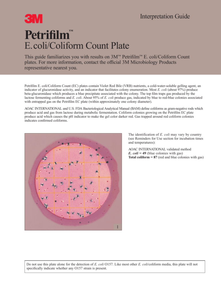
3 Petrifilm Interpretation Guide ™ E.coli/Coliform Count Plate This guide familiarizes you with results on 3M™ Petrifilm™ E. coli/Coliform Count plates. For more information, contact the official 3M Microbiology Products representative nearest you. Petrifilm E. coli/Coliform Count (EC) plates contain Violet Red Bile (VRB) nutrients, a cold-water-soluble gelling agent, an indicator of glucuronidase activity, and an indicator that facilitates colony enumeration. Most E. coli (about 97%) produce beta-glucuronidase which produces a blue precipitate associated with the colony. The top film traps gas produced by the lactose fermenting coliforms and E. coli. About 95% of E. coli produce gas, indicated by blue to red-blue colonies associated with entrapped gas on the Petrifilm EC plate (within approximately one colony diameter). AOAC INTERNATIONAL and U.S. FDA Bacteriological Analytical Manual (BAM) define coliforms as gram-negative rods which produce acid and gas from lactose during metabolic fermentation. Coliform colonies growing on the Petrifilm EC plate produce acid which causes the pH indicator to make the gel color darker red. Gas trapped around red coliform colonies indicates confirmed coliforms. The identification of E. coli may vary by country (see Reminders for Use section for incubation times and temperatures): AOAC INTERNATIONAL validated method E. coli = 49 (blue colonies with gas) Total coliform = 87 (red and blue colonies with gas) 1 Do not use this plate alone for the detection of E. coli O157. Like most other E. coli/coliform media, this plate will not specifically indicate whether any O157 strain is present. 3M Petrifilm ™ ™ E. coli/Coliform Count Plate 1 1 2 3 No growth = 0 E. coli count = 13 Notice the changes in gel color in figures 2 through 8. As the E. coli or coliform count increases, the color of the gel turns to dark red or purple-blue. Total coliform count = 28 Background bubbles are a characteristic of the gel and are not a result of E. coli or coliform growth. See square 1. Back Lighting 1 The counting range for the total population on Petrifilm EC plates is 15–150. Do not count colonies that appear on the foam barrier because they are removed from the selective influence of the medium. See circle 1. Front Lighting 2 4 5 E. coli count = 3 E. coli count = 17 Any blue in a colony (blue to red-blue) indicates the presence of E. coli. Front lighting will enhance the detection of blue precipitate formed by a colony. Estimated total coliform count = 150 Circle 1 shows a red-blue colony counted using back lighting. Circle 2 shows the same colony with front lighting. The blue precipitate is more evident in circle 2. The circular growth area is approximately 20 cm2. Estimates can be made on plates containing greater than 150 colonies by counting the number of colonies in one or more representative squares and determining the average number per square. Multiply the average number by 20 to determine the estimated count per plate. TNTC (Too Numerous to Count) To obtain a more accurate count, dilute the sample further. 6 7 Actual count ~ 106 Actual count ~ 108 Petrifilm EC plates with colonies that are TNTC have one or more of the following characteristics: many small colonies, many gas bubbles, and a deepening of the gel color from red to purple-blue. High concentrations of E. coli may cause the growth area to turn purple-blue. 8 9 Presumptive E. coli count ~ 8 Actual count ~ 108 Estimated total coliform count ~ 108 When high numbers of non-coliform organisms such as Pseudomonas are present on Petrifilm EC plates, the gel may turn yellow. When high levels of coliforms are present (>10 8), some strains of E. coli may produce less gas and blue colonies may be less definitive. Count all blue colonies without gas and/or blue zones as presumptive E. coli. Pick blue colonies without gas and confirm if necessary. Bubbles 1 3 2 10 11 Total coliform count = 3 Total coliform count = 78 Food particles are irregularly shaped and are not associated with gas bubbles. Bubble patterns may vary. Gas may disrupt the colony so that the colony “outlines” the bubble. See circles 1 and 2. Artifact bubbles may result from improper inoculation or from trapped air within the sample. They are irregularly shaped and are not associated with a colony. See circle 3. 3 1 4 2 5 8 6 9 7 10 12 Examples 1–10 show various bubble patterns associated with gas producing colonies. All should be enumerated. 3Petrifilm E. coli / Coliform Count Plates Reminders for Use For detailed WARNING, CAUTIONS, DISCLAIMER OF WARRANTIES / LIMITED REMEDY, LIMITATION OF 3M LIABILITY, STORAGE AND DISPOSAL information, and INSTRUCTIONS FOR USE see Product’s package insert. Storage Petrifilm Petrifilm Petrifilm 1 Store unopened packages at ≤8°C (≤46°F). Use before expiration date on package. In areas of high humidity where condensate may be an issue, it is best to allow packages to reach room temperature before opening. 2 To seal opened package, fold end over and tape shut. 3 Keep resealed package at ≤25°C (≤77°F) and <50%RH. Do not refrigerate opened packages. Use Petrifilm plates within one month after opening. 5 Add appropriate quantity of one of the following sterile diluents: Butterfield's phosphate buffer (IDF phosphate buffer, 0.0425 g/L of KH2PO4 adjusted to pH 7.2), 0.1% peptone water, peptone salt diluent (ISO method 6887), buffered peptone water (ISO method 6887-1), saline solution (0.85-0.90%), bisulfite-free letheen broth, or distilled water. 6 Blend or homogenize sample per current procedure. Sample Preparation 4 Prepare a 1:10 or greater dilution of food product. Weigh or pipette food product into an appropriate container such as a stomacher bag, dilution bottle, Whirl-Pak® bag, or other sterile container. Adjust pH of the diluted sample between 6.5 and 7.5. • for acid products, use 1N NaOH, • for alkaline products, use 1N HCI. Do not use buffers containing citrate, bisulfite, or thiosulfate; they can inhibit growth. Inoculation 7 Place Petrifilm plate on level surface. Lift top film. 8 With pipette perpendicular to Petrifilm plate, place 1 mL of sample onto center of bottom film. 9 Carefully ROLL top film down to avoid entrapping air bubbles. Do NOT let top film drop. RIDGE side FLAT side With FLAT side down, place spreader on top film over inoculum. 11 GENTLY apply pressure on spreader to distribute inoculum over circular area before gel is formed. Do not twist or slide the spreader. 12 Lift spreader. Wait a minimum of one minute for gel to solidify. 15 Colonies may be isolated for further identification. Lift top film and pick the colony from the gel. Interpretation Incubation <70°F 13 Incubate plates with clear side up in stacks of no more than 20. It may be necessary to humidify incubator to minimize moisture loss. 14 Petrifilm plates can be counted on a standard colony counter or other illuminated magnifier. Refer to the Interpretation Guide section when reading results. Incubation time and temperature varies by method. Most common approved methods: AOAC Official Method 991.14 for coliforms: incubate 24h ± 2h at 35°C ± 1°C for E.coli: incubate 48h ± 2h at 35°C ± 1°C AOAC Official Method 998.08 for E.coli (for meat, poultry and seafood): incubate 24h ± 2h at 35°C ± 1°C NMKL method (147.1993) for coliforms: incubate 24h ± 2h at 37°C for E.coli: incubate 48h ± 2h at 37°C Additional Comments 3 • Questions? U.S., Call 1-800-328-6553, Canada, call 1-800-265-1840 x6574 for technical service. • To order Petrifilm plates in the U.S., call 1-800-328-1671 • Latin America / Africa and Asia Pacific regions, call 1-651-733-7562. Microbiology Products 3M Canada 3M Europe 3M Center Bldg. 275-5W-05 St. Paul, MN 55144-1000 USA 1-800-228-3957 www.3M.com/microbiology Email:microbiology@mmm.com Post Office Box 5757 London, Ontario N6A4T1 Canada 1-800-563-2921 Laboratoires 3M Santé Boulevard de l’Oise 95029 Cergy Pontoise Cedex France 33 1 30 31 85 71 Recycled Paper 40% pre-consumer 10% post-consumer Petrifilm is a trademark of 3M. Whirl-Pak is a registered trademark of NASCO. Printed in U.S.A. © 3M 2001 70-2008-4574-4 (1211) DPI
