morphometric features and chromatin condensation abnormalities
advertisement
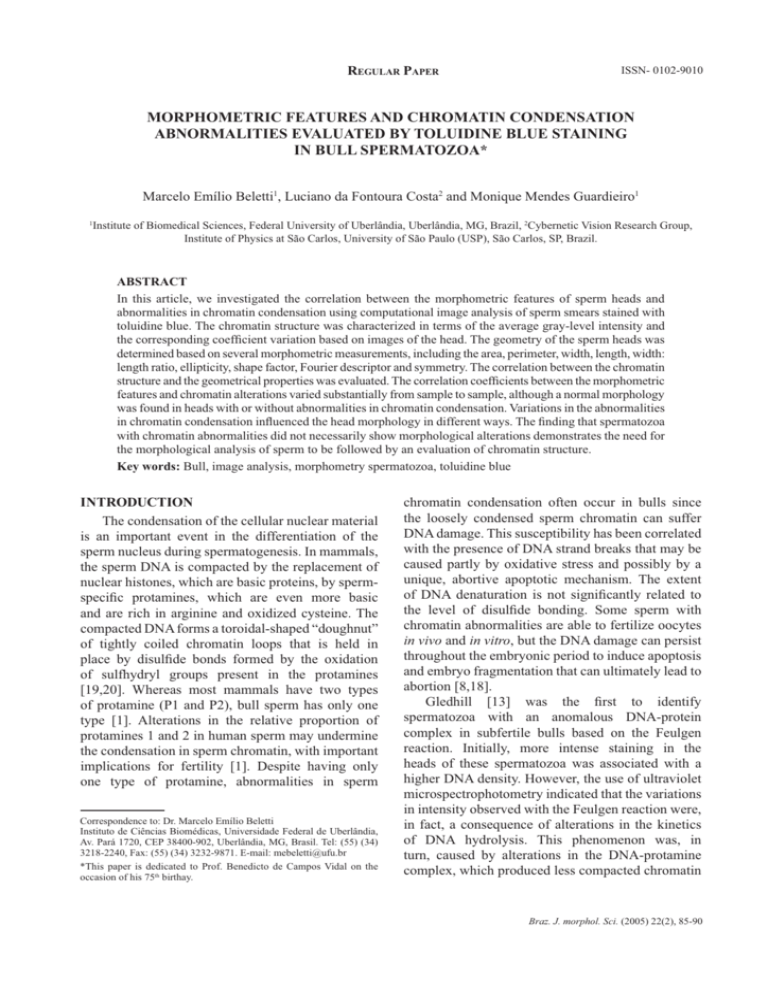
REGULAR PAPER ISSN- 0102-9010 MORPHOMETRIC FEATURES AND CHROMATIN CONDENSATION ABNORMALITIES EVALUATED BY TOLUIDINE BLUE STAINING IN BULL SPERMATOZOA* Marcelo Emílio Beletti1, Luciano da Fontoura Costa2 and Monique Mendes Guardieiro1 1 Institute of Biomedical Sciences, Federal University of Uberlândia, Uberlândia, MG, Brazil, 2Cybernetic Vision Research Group, Institute of Physics at São Carlos, University of São Paulo (USP), São Carlos, SP, Brazil. ABSTRACT In this article, we investigated the correlation between the morphometric features of sperm heads and abnormalities in chromatin condensation using computational image analysis of sperm smears stained with toluidine blue. The chromatin structure was characterized in terms of the average gray-level intensity and the corresponding coefficient variation based on images of the head. The geometry of the sperm heads was determined based on several morphometric measurements, including the area, perimeter, width, length, width: length ratio, ellipticity, shape factor, Fourier descriptor and symmetry. The correlation between the chromatin structure and the geometrical properties was evaluated. The correlation coefficients between the morphometric features and chromatin alterations varied substantially from sample to sample, although a normal morphology was found in heads with or without abnormalities in chromatin condensation. Variations in the abnormalities in chromatin condensation influenced the head morphology in different ways. The finding that spermatozoa with chromatin abnormalities did not necessarily show morphological alterations demonstrates the need for the morphological analysis of sperm to be followed by an evaluation of chromatin structure. Key words: Bull, image analysis, morphometry spermatozoa, toluidine blue INTRODUCTION The condensation of the cellular nuclear material is an important event in the differentiation of the sperm nucleus during spermatogenesis. In mammals, the sperm DNA is compacted by the replacement of nuclear histones, which are basic proteins, by spermspecific protamines, which are even more basic and are rich in arginine and oxidized cysteine. The compacted DNA forms a toroidal-shaped “doughnut” of tightly coiled chromatin loops that is held in place by disulfide bonds formed by the oxidation of sulfhydryl groups present in the protamines [19,20]. Whereas most mammals have two types of protamine (P1 and P2), bull sperm has only one type [1]. Alterations in the relative proportion of protamines 1 and 2 in human sperm may undermine the condensation in sperm chromatin, with important implications for fertility [1]. Despite having only one type of protamine, abnormalities in sperm Correspondence to: Dr. Marcelo Emílio Beletti Instituto de Ciências Biomédicas, Universidade Federal de Uberlândia, Av. Pará 1720, CEP 38400-902, Uberlândia, MG, Brasil. Tel: (55) (34) 3218-2240, Fax: (55) (34) 3232-9871. E-mail: mebeletti@ufu.br *This paper is dedicated to Prof. Benedicto de Campos Vidal on the occasion of his 75th birthay. chromatin condensation often occur in bulls since the loosely condensed sperm chromatin can suffer DNA damage. This susceptibility has been correlated with the presence of DNA strand breaks that may be caused partly by oxidative stress and possibly by a unique, abortive apoptotic mechanism. The extent of DNA denaturation is not significantly related to the level of disulfide bonding. Some sperm with chromatin abnormalities are able to fertilize oocytes in vivo and in vitro, but the DNA damage can persist throughout the embryonic period to induce apoptosis and embryo fragmentation that can ultimately lead to abortion [8,18]. Gledhill [13] was the first to identify spermatozoa with an anomalous DNA-protein complex in subfertile bulls based on the Feulgen reaction. Initially, more intense staining in the heads of these spermatozoa was associated with a higher DNA density. However, the use of ultraviolet microspectrophotometry indicated that the variations in intensity observed with the Feulgen reaction were, in fact, a consequence of alterations in the kinetics of DNA hydrolysis. This phenomenon was, in turn, caused by alterations in the DNA-protamine complex, which produced less compacted chromatin Braz. J. morphol. Sci. (2005) 22(2), 85-90 86 M. E. Beletti et al. that was more sensitive to hydrolysis. A number of methods are available for identifying alterations in the stability of sperm chromatin. Sperm chromatin structure analysis (SCSA), currently the most used of these methods, is based on a flow cytometric evaluation of the fluorescence of spermatozoa stained with acridine orange [6,9,10,15]. However, this approach has a shortcoming in that it does not allow the morphometric evaluation to be done concomitantly with the characterization of the sperm chromatin. Another method for chromatin evaluation was developed by Mello [14]. This method uses toluidine blue, a cationic dye that exhibits metachromasy, i.e. a color change to violet, induced by electron resonance between several stacked dye molecules. This property is useful for evaluating abnormalities in sperm chromatin condensation since when sperm smears are stained with toluidine blue at pH 4.0, the dye molecules bind to ionized phosphates in the DNA. The pH of 4.0 ensures that other possible anions binding sites are not ionized concurrently. In normal sperm chromatin, most of the phosphates are blocked by protamines and, consequently, few dye molecules bind to DNA; this results in staining that varies from green to light blue. Spermatozoa with less compacted chromatin have more binding sites for the dye molecules, resulting in staining that varies from dark blue to magenta. However, only the highest degrees of chromatin alteration are identified by this method. The sensitivity of this method can be increased by hydrolysis before staining. Normal sperm, which have highly compacted chromatin, are hardly affected by hydrolysis and consequently stain light blue. In contrast, sperm chromatin with a low degree of alteration would have their protamines partially extracted by hydrolysis, thereby allowing toluidine blue molecules to bind to DNA phosphates. Because mammalian sperm heads consist almost totally of chromatin, morphological abnormalities would be expected to follow alterations in chromatin structure [11,15,17]. Morphological defects in sperm can have severe implications for the hydrodynamic properties of these cells, including an effect on their normal rectilinear and uniform progression. Whereas human-based methods for assessing sperm nuclear shape involve a high degree of subjectivity in the visual analysis, computer-based methods for image processing and analysis are available that can provide a more objective evalu- Braz. J. morphol. Sci. (2005) 22(2), 85-90 ation of cell motility and sperm morphological abnormalities, in addition to greater sensitivity, accuracy, speed and reproducibility. Computational morphometric analysis of spermatozoa usually considers as basic measurements the area, perimeter, length and width, as well as features derived from the measurements, such as the width:length ratio, ellipticity, shape factor and others [12]. In addition to these basic measurements, the use of Fourier descriptors (usually considering from the fundamental to fifth harmonic) is also used [15,16]. Another approach is to use image analysis to characterize the sperm chromatin in smears stained with toluidine blue. Besides increasing the objectivity in sperm characterization, the latter approach also allows a morphometric analysis to be done concomitantly with the investigation of chromatin. Previous studies [11,15,17] reported significant correlations between chromatin anomalies and morphological alterations. However, in most cases, the investigations used SCSA to evaluate the chromatin, hence precluding the corresponding morphological evaluation. Ferrari et al. [11] used visual morphological analysis and cytophotometry of smears treated with the Feulgen reaction to identify correlations between a narrower neck or pear-shaped heads and abnormalities in chromatin condensation; similar correlations were not observed for other morphological alterations. The motive for using a customized computer-aided sperm analysis (CASA) system derived from the need for a comprehensive knowledge of the algorithms used and their properties and parameters, as well as the need to access several measurements not usually found in equivalent commercial systems [2]. The main objective of this work was to examine the correlation between a representative set of morphometric measurements and the occurence of abnormalities in chromatin condensation in bull sperm heads, based on a computational image analysis of sperm smears stained with toluidine blue. The use of computational methods allowed the same sperm heads to be examined for both of these analyses and increased the reliability of the resulting correlations. MATERIAL AND METHODS Ejaculates from five Simmental bulls of unknown fertility were used. The bulls were treated in accordance with established ethical guidelines. After collection with an artificial vagina, smears were obtained and fixed with 87 Morphometry and chromatin structure of bull sperm during image capture, all of which can influence the graylevel values. The coefficient of variation for the gray-level (in pixels) in each head was also considered as a parameter for identifying abnormalities in chromatin condensation that would otherwise have gone unnoticed had only the average values been used. Using this approach, heads with an intensely stained region (loosely compacted chromatin) bondered by a clearer region (rarefied DNA), that would produce a low average gray-level difference, could be properly identified. The area (A), perimeter (P), width (W), length (L), width:length ratio (W/l), ellipticity (E) and shape factor (SF) of all heads were determined using programs developed in SCILAB [2,4]. Fourier descriptors containing harmonic amplitudes from 0 to 2 (F0, F1, F2) were also considered since these features have traditionally been used for shape characterization and analysis [2,15,16]. Another estimated feature was the sperm head symmetry. The side symmetry (SS) is a measurement that identifies asymmetries along the principal sperm axis. Such asymmetries can have severe implications for the hydrodynamical properties of the sperm. The anterior-posterior symmetry (APS) measurement identifies asymmetries along the secondary sperm axis, and can reveal specific alterations such as a pyriform head and a narrower neck. All of these symmetries were calculated using the procedure described by Beletti and Costa [2], which involves flipping the object along its major (or minor) axis, filling in some voids created by the discrete representation of the object, and then identifying the area of overlap between the original and flipped areas [6]. The correlation coefficients (Pearson’s correlation) between each of the parameters measured and those characteristics of abnormalities in chromatin condensation (Dif, CV) were calculated. A value of p < 0.05 indicated significance. ethanol acetic acid (3:1, V/V) for 1 min and then in 70% ethanol for 3 min. The smears were hydrolyzed for 25 min in 4 mM HCl, washed in distilled water and air-dried. One droplet of 0.025% toluidine blue in McIlvaine buffer (sodium citrate-phosphate), pH 4.0, was placed over each smear and then coverslipped [5]. Fifty gray-level digital images of each slide were obtained randomly using an Olympus BX 40 microscope with a 100x objective (immersion) coupled to an OLY-200 Olympus camera that was connected to a PC microcomputer through a 3153 Data Translation card. In computing a gray-level, a grayscale or greyscale digital image was defined as an image in which the value of each pixel was a single sample or value. Displayed images of this sort typically consisted of shades of gray that varied from black at the weakest intensity to white at the strongest. The black color received a value of zero, the white color received a value 255 and the other shades of gray received values between zero and 255. The image analysis that evaluates the distribution and the values of pixels is referred to as texture analysis [7]. Using threshold-based image segmentation [7], 150 sperm heads were isolated for each smear. After head segmentation, which was done using software programs developed in the SCILAB environment, the average intensities of the gray-levels within the heads in each image were determined. The lower quartile for gray-level intensities was calculated and considered as the standard value. Subsequently, for each image, the difference between the standard value of the smear and the average value of each head analyzed was determined. This difference was transformed into a percentage (Dif%) of the gray-level intensities. The coefficient of variation (CV) of the graylevel intensity for each head was also calculated. Sperm heads with a percentage difference (Dif%) > 2.0 and/or a CV of gray-level intensity > 5.0 were considered to have anomalous chromatin, since in these cases it was generally possible to detect the differences by simple visual evaluation. This criterion was adopted to compensate for variations in smear thickness, in seminal liquid volume, and in the microscope and camera settings RESULTS Table 1 shows the measurements for all heads together, and for sperm heads with anomalous or Table 1. Measurements of all heads and of anomalous chromatin heads and normal heads separately. Head types Number A of heads (Pixels) P W L (Pixels) (Pixels) (Pixels) W/L E SF F0 F1 F2 SS APS CV Dif% All heads 750 2995.26 ±352.92 221.88 ±18.30 44.53 ±3.47 84.60 0.53 0.31 0.91 1438.55 236.55 116.47 0.97 0.90 2.82 ±5.98 ±0.04 ±0.04 ±0.10 ±258.16 ±77.14 ±52.81 ±0.01 ±0.03 ±1.27 1.3 ±1.8 Anomalous chromatin 115 2731.93 ±507.87 208.24 ±23.07 42.84 ±4.94 80.76 0.53 0.31 0.88 1302.63 227.64 112.04 0.97 0.89 4.30 ±8.63 ±0.06 ±0.05 ±0.08 ±344.50 ±71.98 ±47.26 ±0.02 ±0.04 ±2.24 4.0 ±3.0 Normal chromatin 635 3050.99 ±280.87 224.76 ±15.69 44.89 ±2.95 85.41 0.53 0.31 0.91 1467.31 238.44 117.40 0.97 0.90 2.51 0.76 ±4.89 ±0.04 ±0.03 ±0.10 ±225.98 ±78.12 ±53.90 ±0.01 ±0.03 ±0.58 ±0.52 The values are the mean ± SD of the number of heads indicated. A- area, APS- anterior-posterior symmetry, Dif(%)- percent gray-level differences, Eellipticity, F0- Fourier 0, F1- Fourier 1, F2- Fourier 2, L- length, P-perimeter, SF- shape factor, SS- side symmetry, CV- coefficient of variation (%) of the gray-level, W- width, W/L- width:length ratio. Braz. J. morphol. Sci. (2005) 22(2), 85-90 M. E. Beletti et al. 88 Table 2. Correlation coefficients between each measurement and the sperm chromatin features (Dif % and CV) in bull sperm heads with abnormal chromatin. Bull code Number of heads 1 Feature A P W L W/L E SF 22 CV Dif% -0.53* -0.56* -0.48* -0.51* -0.19 -0.19 -0.58* -0.67* 0.33 0.40* -0.33 -0.40* 0.11 0.14 2 33 CV Dif% -0.36* -0.39* -0.62* -0.64* -0.25 -0.27 -0.46* -0.52* 0.30 0.38* -0.30 -0.37* 3 35 CV Dif% -0.44* -0.24 -0.46* -0.42* -0.37* -0.11 -0.18 -0.26 -0.31 0.02 4 6 CV Dif% -0.53* -0.40* -0.52* -0.39* -0.49* -0.36* -0.52* -0.40* 5 19 CV Dif% -0.68* -0.45* -0.39* -0.60* -0.40* -0.01 -0.23 -0.46* F0 F1 F2 SS APS CV -0.52* -0.63* 0.19 0.11 -0.04 -0.06 -0.30 -0.23 -0.43* -0.35* 1.00 0.94* -0.54* -0.50* -0.41* -0.48* -0.26 -0.35* -0.27 -0.25 0.18 0.26 -0.09 -0.02 1.00 0.96* 0.33 0.01 -0.36* -0.38* 0.06* -0.15 0.41* 0.24 0.20 0.34 -0.15 -0.16 -0.66* -0.41* 1.00 0.70* 0.15 0.29 -0.16 -0.30 0.66* 0.55* -0.55* -0.42* -0.37* -0.23 -0.42* -0.28 -0.57* -0.45* -0.73* -0.62* 1.00 0.99* -0.14 0.20 0.17 -0.20 -0.03 -0.19 -0.12 -0.42* -0.24 -0.06 0.12 0.25 -0.37* -0.48* -0.04 0.06 1.00 0.79* The values are the mean ± SD of the number of heads indicated. A- area, APS- anterior-posterior symmetry, Dif(%)- percent gray-level differences, E-ellipticity, F0- Fourier 0, F1- Fourier 1, F2- Fourier 2, L- length, P- perimeter, SF- shape factor, SS- side symmetry, CV- coefficient of variation (%) of the gray-level, W- width, W/L- width:length ratio. *p < 0.05. Figure 1. Examples of sperm heads stained with toluidine blue, illustrating the various head types: normal morphology and abnormal chromatin (A, arrow), normal morphology and normal chromatin (B), abnormal morphology and normal chromatin (C), and abnormal morphology and abnormal chromatin (D, arrow). Braz. J. morphol. Sci. (2005) 22(2), 85-90 Morphometry and chromatin structure of bull sperm normal chromatin separately. The correlation coefficients between each of the parameters measured versus the Dif% or CV for each bull are shown in Table 2. Figure 1 shows bull sperm heads with a normal morphology and abnormal chromatin (A); a normal morphology and normal chromatin (B), an abnormal morphology and normal chromatin (C), and abnormal morphology and abnormal chromatin (D). Table 2 shows that significant correlations were obtained between various parameters and the graylevel differences (Dif%) and the coefficient of variation (CV). The negative correlation between the side symmetry (SS) and the chromatin features in bulls 4 and 5 indicated that in these animals the sperm heads that had more severe abnormalities in chromatin condensation were generally asymmetrical, whereas in the other bulls the abnormalities did not influence the SS. The correlations between abnormalities in chromatin condensation and the W/L and ellipticity showed opposite but similar absolute values. DISCUSSION Figure 1 shows a head with altered chromatin but an apparently normal morphology (A, arrow), and a head with modified morphology but apparently normal chromatin (C). This finding demonstrates that abnormalities in chromatin condensation can influence the head morphology in different ways, although chromatin abnormalities are not always followed by evident morphological alterations. Similarly, morphological alterations in the head were not always followed by abnormalities in chromatin condensation identifiable by the methodology used here. The significant correlation between the graylevel differences (Dif%) and the coefficients of variation (CV) showed that the abnormalities in chromatin condensation were generally heterogeneous (see Fig. 1A,D). Beletti et al. [3] used a computational approach to characterize alterations in bovine sperm chromatin and concluded that the chromatin abnormalities were generally heterogeneous and differentially affected the various regions of the sperm head. Human sperm chromatin has two types of protamine and an alteration in their relative proportion is considered an important cause of chromatin abnormalities. In contrast, only one type of protamine exists in bull sperm chromatin [1], which means that abnormalities in chromatin condensation in this species have other causes. The variations in 89 the correlations between the morphometrical and chromatin features seen in each bull indicated that different types of chromatin alterations occurred in bovine semen. The negative correlation between the side symmetry (SS) and chromatin features in bulls 4 and 5 demonstrates that for these bulls the sperm heads with chromatin condensation abnormalities are generally asymmetrical, and in the other bulls the chromatin condensation abnormalities do not influence the side symmetry. The finding that the W/L increased when the sperm head tended to be spherical explains its inverse correlation with ellipticity. The negative correlation between Dif% and the area and perimeter of sperm head showed that abnormalities in chromatin condensation were generally followed by a decrease in head size. This could be explained by the morphological changes caused by alterations in chromatin structure that would increase the height of spermatozoa without changing the area. The method used here did not allow us to measure the volume, so only changes in the image area (two dimensions) were considered in our analysis. The abnormalities in chromatin condensation that were accompanied by little or no increase in head size could be explained by the occurrence of only limited alterations in protamine or a structural protein (such as the matrix and annulus of the sperm nucleus) that would lead to imperfect DNA-protamine linking. Even normal protamine can have imperfect DNA binding that can facilitate the access of dye molecules to the phosphates of DNA. Ferrari et al. [11] examined the correlation between chromatin abnormalities and the morphological anomalies generally evaluated in routine spermiograms, and found a positive correlation between the former abnormalities and a narrower neck or pear-shaped heads; there was no correlation between the chromatin abnormalities and other morphological defects. However, these authors did not evaluate the influence of chromatin abnormalities on the morphometrical features generally used in computer evaluations of sperm. Other studies [15,17] have also examined the correlation between morphometrical features and chromatin imperfections using SCSA, but this methodology did not allow the simultaneous evaluation of the two groups of parameters in the same spermatozoon; different spermatozoa from the same sample had to be used to assess the correlation. In contrast, the computer-based evaluation described Braz. J. morphol. Sci. (2005) 22(2), 85-90 90 M. E. Beletti et al. here allowed a direct correlation between each morphometrical feature and the abnormalities in sperm chromatin condensation. The finding that spermatozoa with chromatin abnormalities did not necessarily have morphological alterations demonstrates the need for the morphological analysis of sperm to be followed by an evaluation of the chromatin. Characterization of the different types of abnormalities in chromatin condensation should provide useful information for future etiological and therapeutic studies. ACKNOWLEDGMENTS Luciano da F. Costa was supported by CNPq (308231/ 03-1) and FAPESP (99/12765-2), and Marcelo E. Beletti was supported by CNPq (150020/2002-3, 551879/2002-5, 350058/2003-2 and 304307/2004-1). REFERENCES 1. Balhorn R, Cosman M, Thornton K, Krishnan VV, Corzett M, Bench G, Kramer C, Lee IV J, Hud NV, Allen M, Prieto M, Meyer-Ilse W, Brown JT, Kirz J, Zhang X, Bradbury EM, Maki G, Braun RE, Breed W (1999) Protamine mediated condensation of DNA in mammalian sperm. In: The Male Gamete: from Basic Science to Clinical Applications (Gagnon C, ed). pp. 55-70. Cache River Press: Vienna. 2. Beletti ME, Costa LF (2003) A systematic approach to multispecies sperm morphometric characterization. Anal. Quant. Cytol. Histol. 25, 97-107. 3. Beletti ME, Costa LF, Viana MP (2004) A computational approach to characterization of bovine sperm chromatin alterations. Biotech. Histochem. 79, 17-23. 4. Beletti ME, Costa LF, Viana MP (2005) A comparison of morphometric characteristics of sperm from fertile Bos taurus and Bos indicus bulls in Brazil. Anim. Reprod. Sci. 85, 105-116. 5. Beletti ME, Mello MLS (2004) Comparison between the toluidine blue stain and the Feulgen reaction for evaluation of rabbit sperm chromatin condensation and their relationship with sperm morphology. Theriogenology 62, 398-402. 6. Bochenek M, Smorag Z, Pilch J (2001) Sperm chromatin structure assay of bulls qualified for artificial insemination. Theriogenology 56, 557-567. 7. Costa LF, Cesar Jr RM (2001) Shape acquisition and processing. In: Shape Analysis and Classification: Braz. J. morphol. Sci. (2005) 22(2), 85-90 Theory and Practice (Costa LF, Cesar Jr RM, eds). pp. 197-264. CRC Press: Boca Raton. 8. Ellington JE, Evenson DP, Fleming JE, Brisbois RS, Hiss GA, Broder SJ, Wright RW (1998) Coculture of human sperm with bovine oviduct epithelial cells decreases sperm chromatin structural changes seen during culture in media alone. Fertil. Steril. 69, 643-649. 9. Evenson DP, Darzynkiewicz Z, Melamed MR (1980) Relation of mammalian sperm chromatin heterogeneity to fertility. Science 210, 1131-1133. 10. Evenson DP, Larson KL, Jost LK (2002) The sperm chromatin structure assay (SCSA): clinical use for detecting sperm DNA fragmentation related to male infertility and comparisons with other techniques. J. Androl. 23, 25-43. 11. Ferrari MR, Spirito SE, Giuliano SM, Fernández HA (1998) Chromatin cytophotometric analysis of abnormal bovine espermatozoa. Andrologia 30, 85-89. 12. Garrett C, Baker HW (1995) A new fully automated system for the morphometric analysis of human sperm heads. Fertil. Steril. 63, 1306-1317. 13. Gledhill BL (1966) Studies on the DNA content, dry mass and optical even of morphologically normal and abnormal bull spermatozoa heads. Acta Vet. Scand. 7, 1-20. 14. Mello MLS (1982) Induced metachromasy in bull spermatozoa. Histochemistry 74, 387-392. 15. Ostermeier, GC, Sargeant GA, Yandell BS, Evenson DP, Parrish JJ (2001) Relationship of bull fertility to sperm nuclear shape. J. Androl. 22, 595-603. 16. Ostermeier, GC, Sargeant GA, Yandell BS, Parrish JJ (2001) Measurements of bovine sperm nuclear shape using Fourier harmonic amplitudes. J. Androl. 22, 584-594. 17. Sailer BL, Jost LK, Evenson, DP (1996) Bull sperm head morphometry related to abnormal chromatin structure and fertility. Cytometry 24, 167-173. 18. Twigg JP, Irvine DS, Aitken RJ (1998) Oxidative damage to DNA in human spermatozoa does not preclude pronucleus formation at intracytoplasmic sperm injection. Hum. Reprod. 13, 1864-1871. 19. Ward WS (1993) Deoxyribonucleic acid loop domain tertiary structure in mammalian spermatozoa. Biol. Reprod. 48, 1193-1201. 20. Ward WS, Coffey DS (1991) DNA packaging and organization in mammalian spermatozoa: comparison with somatic cells. Biol. Reprod. 44, 569-574. Received: January 26, 2005 Accepted: May 11, 2005
