LETTER
doi:10.1038/nature10379
In vitro centromere and kinetochore assembly on
defined chromatin templates
Annika Guse1, Christopher W. Carroll1, Ben Moree1, Colin J. Fuller1 & Aaron F. Straight1
1
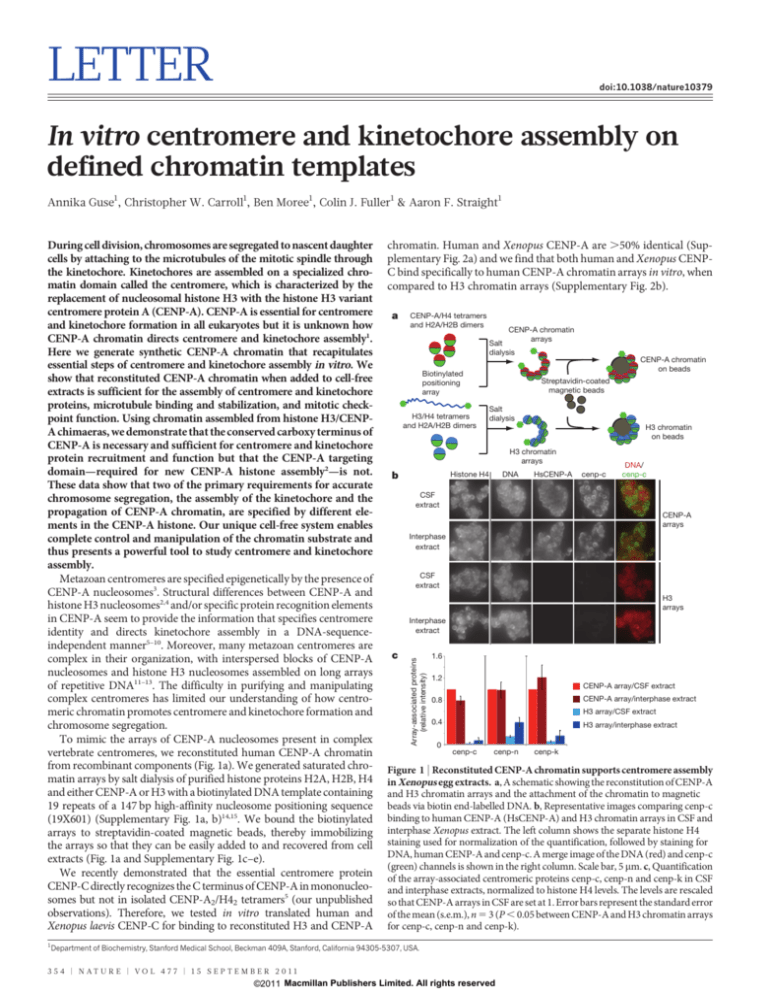
chromatin. Human and Xenopus CENP-A are .50% identical (Supplementary Fig. 2a) and we find that both human and Xenopus CENPC bind specifically to human CENP-A chromatin arrays in vitro, when
compared to H3 chromatin arrays (Supplementary Fig. 2b).
a
CENP-A/H4 tetramers
and H2A/H2B dimers
CENP-A chromatin
arrays
Salt
dialysis
CENP-A chromatin
on beads
Biotinylated
positioning
array
H3/H4 tetramers
and H2A/H2B dimers
Streptavidin-coated
magnetic beads
Salt
dialysis
H3 chromatin
on beads
H3 chromatin
arrays
b
DNA
Histone H4
HsCENP-A
cenp-c
DNA/
cenp-c
CSF
extract
CENP-A
arrays
Interphase
extract
CSF
extract
H3
arrays
Interphase
extract
c
Array-associated proteins
(relative intensity)
During cell division, chromosomes are segregated to nascent daughter
cells by attaching to the microtubules of the mitotic spindle through
the kinetochore. Kinetochores are assembled on a specialized chromatin domain called the centromere, which is characterized by the
replacement of nucleosomal histone H3 with the histone H3 variant
centromere protein A (CENP-A). CENP-A is essential for centromere
and kinetochore formation in all eukaryotes but it is unknown how
CENP-A chromatin directs centromere and kinetochore assembly1.
Here we generate synthetic CENP-A chromatin that recapitulates
essential steps of centromere and kinetochore assembly in vitro. We
show that reconstituted CENP-A chromatin when added to cell-free
extracts is sufficient for the assembly of centromere and kinetochore
proteins, microtubule binding and stabilization, and mitotic checkpoint function. Using chromatin assembled from histone H3/CENPA chimaeras, we demonstrate that the conserved carboxy terminus of
CENP-A is necessary and sufficient for centromere and kinetochore
protein recruitment and function but that the CENP-A targeting
domain—required for new CENP-A histone assembly2—is not.
These data show that two of the primary requirements for accurate
chromosome segregation, the assembly of the kinetochore and the
propagation of CENP-A chromatin, are specified by different elements in the CENP-A histone. Our unique cell-free system enables
complete control and manipulation of the chromatin substrate and
thus presents a powerful tool to study centromere and kinetochore
assembly.
Metazoan centromeres are specified epigenetically by the presence of
CENP-A nucleosomes3. Structural differences between CENP-A and
histone H3 nucleosomes2,4 and/or specific protein recognition elements
in CENP-A seem to provide the information that specifies centromere
identity and directs kinetochore assembly in a DNA-sequenceindependent manner5–10. Moreover, many metazoan centromeres are
complex in their organization, with interspersed blocks of CENP-A
nucleosomes and histone H3 nucleosomes assembled on long arrays
of repetitive DNA11–13. The difficulty in purifying and manipulating
complex centromeres has limited our understanding of how centromeric chromatin promotes centromere and kinetochore formation and
chromosome segregation.
To mimic the arrays of CENP-A nucleosomes present in complex
vertebrate centromeres, we reconstituted human CENP-A chromatin
from recombinant components (Fig. 1a). We generated saturated chromatin arrays by salt dialysis of purified histone proteins H2A, H2B, H4
and either CENP-A or H3 with a biotinylated DNA template containing
19 repeats of a 147 bp high-affinity nucleosome positioning sequence
(19X601) (Supplementary Fig. 1a, b)14,15. We bound the biotinylated
arrays to streptavidin-coated magnetic beads, thereby immobilizing
the arrays so that they can be easily added to and recovered from cell
extracts (Fig. 1a and Supplementary Fig. 1c–e).
We recently demonstrated that the essential centromere protein
CENP-C directly recognizes the C terminus of CENP-A in mononucleosomes but not in isolated CENP-A2/H42 tetramers5 (our unpublished
observations). Therefore, we tested in vitro translated human and
Xenopus laevis CENP-C for binding to reconstituted H3 and CENP-A
1.6
1.2
CENP-A array/CSF extract
0.8
CENP-A array/interphase extract
0.4
H3 array/interphase extract
H3 array/CSF extract
0
cenp-c
cenp-n
cenp-k
Figure 1 | Reconstituted CENP-A chromatin supports centromere assembly
in Xenopus egg extracts. a, A schematic showing the reconstitution of CENP-A
and H3 chromatin arrays and the attachment of the chromatin to magnetic
beads via biotin end-labelled DNA. b, Representative images comparing cenp-c
binding to human CENP-A (HsCENP-A) and H3 chromatin arrays in CSF and
interphase Xenopus extract. The left column shows the separate histone H4
staining used for normalization of the quantification, followed by staining for
DNA, human CENP-A and cenp-c. A merge image of the DNA (red) and cenp-c
(green) channels is shown in the right column. Scale bar, 5 mm. c, Quantification
of the array-associated centromeric proteins cenp-c, cenp-n and cenp-k in CSF
and interphase extracts, normalized to histone H4 levels. The levels are rescaled
so that CENP-A arrays in CSF are set at 1. Error bars represent the standard error
of the mean (s.e.m.), n 5 3 (P , 0.05 between CENP-A and H3 chromatin arrays
for cenp-c, cenp-n and cenp-k).
Department of Biochemistry, Stanford Medical School, Beckman 409A, Stanford, California 94305-5307, USA.
3 5 4 | N AT U R E | VO L 4 7 7 | 1 5 S E P T E M B E R 2 0 1 1
©2011 Macmillan Publishers Limited. All rights reserved
LETTER RESEARCH
a
0 min
Chromatin arrays
+ CSF extract
+ calcium
80 min
+1 vol. CSF extract
and nocodazole/DMSO
80 min
170 min
Fixation and
immunofluorescence
90 min
1.2
CENP-A
H3
1.0
0.8
0.6
0.4
0.2
0
– + – +
cenp-c
– + – +
ndc80
c
Extract
b
Array-associated proteins
(relative intensity)
Xenopus egg extract is a widely used cell-free system to study chromosome segregation16. Egg extracts are arrested in metaphase II of
meiosis by the activity of cytostatic factor (CSF) and the cell-cycle state
of the extract can be transitioned into interphase by adding calcium.
We developed a quantitative immunofluorescence assay to determine
whether centromere proteins bound to CENP-A chromatin arrays
when arrays were added to Xenopus egg extracts. CENP-N and
CENP-K are centromere proteins that are required for proper centromere and kinetochore assembly in somatic cells, and we have previously shown that CENP-N, similar to CENP-C, directly binds to the
CENP-A nucleosome6. We found that cenp-c, cenp-n and cenp-k
specifically associated with CENP-A arrays independent of the cellcycle stage of the extract (Fig. 1b, c and Supplementary Fig. 2c–f). The
centromere protein cenp-t that binds to either H3 nucleosomes or
DNA at centromeres did not selectively bind CENP-A chromatin
arrays (Supplementary Fig. 3a, b)17. Similarly, the inner centromere
protein incenp and polo-like kinase 1 (plk1) associated with both types
of chromatin arrays (Supplementary Fig. 3c). Xenopus incenp is targeted to chromatin through phosphorylation of both H2A and H3 and
thus may have affinity for both CENP-A and H3 chromatin18–20 and
plk1 associates with chromatin in Xenopus egg extract independent of
the kinetochore21. Furthermore, reconstituted chromatin segments are
unlikely to generate paired sister chromatids with inner centromeres
because naked DNA and linear DNA replicates inefficiently in these
egg extracts22. The specific recruitment of the centromere proteins
cenp-c, cenp-n and cenp-k, however, indicates that reconstituted
CENP-A chromatin arrays can support essential steps in the centromere assembly process in vitro.
Functional kinetochores assemble on sperm chromatin in metaphase Xenopus egg extract. At high sperm concentration, microtubule
depolymerization causes mitotic checkpoint activation, resulting in the
increased association of checkpoint proteins with kinetochores and
cell-cycle arrest23. We tested whether reconstituted CENP-A chromatin
arrays support kinetochore assembly and checkpoint protein binding
after microtubule depolymerization. We added CENP-A or H3 arrays
to CSF-arrested egg extracts and then cycled the extracts through interphase and back into mitosis, in the presence or absence of nocodazole,
as outlined in Fig. 2a and demonstrated in Supplementary Fig. 4a. The
constitutive centromere protein cenp-c and the microtubule-binding
kinetochore protein ndc80 bound to CENP-A arrays in the presence
or absence of nocodazole (Fig. 2b, c and Supplementary Fig. 4b). The
spindle assembly checkpoint proteins cenp-e, mad2, rod (also known
as kntc1) and zw10 associated with CENP-A chromatin at intermediate levels in the absence of nocodazole but upon microtubule
depolymerization their binding increased 2–4 fold (Fig. 2b). Western
blot analysis showed that cenp-c and ndc80 are precipitated with
CENP-A arrays independent of microtubule depolymerization.
Xenopus zw10 and rod are enriched on CENP-A arrays upon nocodazole treatment in metaphase, regardless of whether the extract has been
cycled through interphase (Fig. 2c). These results indicate that CENP-A
chromatin arrays respond to microtubule depolymerization by recruiting mitotic checkpoint proteins (Fig. 2b, c and Supplementary Fig. 4b).
Microtubule binding is a hallmark of kinetochore function and
decondensed sperm chromatin efficiently supports spindle formation
in egg extracts (Fig. 3a, left)24. However, chromatin assembled on naked
DNA induces spindle formation in Xenopus egg extracts independent
of kinetochores25. When we added CENP-A and H3 chromatin beads
into mitotic egg extract we observed microtubule polymerization
around the majority of CENP-A arrays but only around a subset of
H3 arrays (Fig. 3a, left). We quantified the amount of microtubule
polymer associated with each type of array and found significantly more
microtubules associated with CENP-A chromatin beads (Fig. 3b and
Supplementary Fig. 5a). This indicates that CENP-A chromatin preferentially stabilizes microtubules or promotes their polymerization.
We observed heterogeneous microtubule structures around the
CENP-A chromatin beads ranging from bipolar spindles to stabilized
– + – +
cenp-e
CSF extract
– + – +
mad2
– + – +
rod
– + – + NOC
zw10
Cycled extract
CENP-A H3 CENP-A H3
– + – + – + – + NOC
cenp-c
rod
zw10
ndc80
HsCENP-A
H4
Figure 2 | CENP-A chromatin specifically recruits kinetochore proteins as a
response to a mimic of kinetochore detachment from microtubules. a, A
schematic showing the experimental procedure. b, Quantification of
immunofluorescence analysis of cenp-c, ndc80, cenp-e, mad2, rod or zw10
recruitment to chromatin arrays with (1) and without (2) nocodazole (NOC).
The levels are rescaled so that CENP-A arrays with nocodazole are set at 1.
Error bars represent s.e.m., n 5 3 (P , 0.05 between (2) and (1) nocodazole
for cenp-e, mad2, rod and zw10 binding to CENP-A chromatin arrays).
c, Western blot analysis of cenp-c, ndc80, rod and zw10 recruitment to CENPA (HsCENP-A) and H3 chromatin arrays with and without nocodazole in CSF
and cycled egg extracts. H4 levels are shown as a loading control.
microtubules or microtubule bundles (Fig. 3a and Supplementary
Fig. 5a, b). A second property of functional kinetochores is that
kinetochore-associated microtubule bundles (k-fibres) are stable to cold
treatment, which depolymerizes non-kinetochore microtubules. We
asked whether kinetochores assembled on CENP-A chromatin could
stabilize microtubules to cold shock by incubating the microtubule
assembly reactions for 10 min at 4 uC. We found that kinetochores
assembled on CENP-A chromatin arrays stabilized microtubules to
cold shock similar to kinetochores assembled on native sperm chromatin whereas H3 chromatin arrays did not (Fig. 3a, c and Supplementary Fig. 5c). When we completely depolymerized microtubules
with nocodazole we observed mad2 recruitment to native sperm
centromeres and CENP-A chromatin beads but not H3 chromatin
beads (Fig. 3a, c and Supplementary Fig. 5c). These results indicate that
CENP-A chromatin arrays, similar to native sperm chromatin,
assemble functional kinetochores that promote microtubule binding,
k-fibre stabilization and spindle checkpoint function (Fig. 3a).
In cells, unattached kinetochores activate the mitotic checkpoint and
delay mitotic exit until all chromosomes are properly attached and
aligned26,27. We tested whether kinetochores assembled on CENP-A
chromatin arrays could generate a mitotic checkpoint response to
microtubule depolymerization and delay the cell cycle. We mixed
CENP-A and H3 chromatin with CSF extracts, cycled the reactions
through interphase and then cycled them back into mitosis in the
presence or absence of nocodazole (Fig. 2a). We then released the
extract from mitosis into interphase a second time and monitored
the kinetics of this transition by measuring the mitosis-specific phosphorylation of wee1 (phospho-wee1) (Fig. 3d). On release from mitosis,
phospho-wee1 levels rapidly declined and were undetectable after
30 min in control extracts containing CENP-A chromatin or H3 chromatin, as well as in extracts containing H3 chromatin in the presence of
1 5 S E P T E M B E R 2 0 1 1 | VO L 4 7 7 | N AT U R E | 3 5 5
©2011 Macmillan Publishers Limited. All rights reserved
RESEARCH LETTER
75
C ATD
114
133 140
C tail
CENP-A
LEEGLG
H3
ERA
CENP-A + H3C
ERA
H3 + CAC
Figure 3 | Kinetochores assembled on reconstituted CENP-A chromatin
bind microtubules and generate a mitotic checkpoint signal.
a, Representative images of microtubule polymerization induced by sperm or
reconstituted CENP-A and H3 chromatin. Microtubules (green) and mad2
(magenta) levels are shown. Scale bar, 10 mm. b, Quantification of tubulin and
DNA associated with CENP-A and H3 chromatin beads. Error bars represent
s.e.m., n 5 5. c, Quantification of tubulin and mad2 levels associated with
CENP-A and H3 chromatin beads after cold shock (4 uC) and nocodazole
(NOC) treatment. Error bars represent s.e.m., n 5 5. d, Western blot showing
phospho-wee1 (p-wee1) levels as an indicator of the cell-cycle stage and tubulin
levels as a loading control. Samples from different time points after release from
mitotic arrest are shown for CENP-A and H3 chromatin arrays, each incubated
with nocodazole (1) or with DMSO (2) as a control. e, Quantification of four
independent experiments showing the phospho-wee1 signal intensity (p-wee1
signal) over time (min). Error bars represent s.e.m., n 5 4.
nocodazole (Fig. 3d, e). In extracts containing CENP-A chromatin
and nocodazole, the phospho-wee1 signal increased until 20 min after
calcium addition and subsequently declined until 40 min after calcium
addition to a level only slightly lower than that before release (Fig. 3d, e).
In the presence of CENP-A chromatin and nocodazole, cyclin B levels
rapidly declined but then stabilized, similar to the response observed for
native sperm chromatin23. However, cyclin B was not stabilized in the
presence of H3 chromatin and nocodazole (Supplementary Fig. 5d, e).
We estimate that the number of CENP-A nucleosomes we are adding to
the egg extract exceeds the CENP-A nucleosome concentration required
to activate the checkpoint using sperm nuclei23. The lower efficiency of
reconstituted arrays for checkpoint signalling may be due to the comparatively short length of our reconstituted CENP-A chromatin to
native CENP-A chromatin or the lack of replicated sister chromatids
and inner centromeres important for tension-dependent checkpoint
activation. Despite these differences, our synthetic CENP-A chromatin
supports a mitotic checkpoint response that mimics the response of
native kinetochores to microtubule depolymerization.
The reconstituted chromatin system we have developed provides a
distinct experimental advantage over native metazoan centromeric
chromatin because the chromatin template can be easily manipulated
to dissect the roles of histone proteins in centromere function. A
central question in centromere function is how CENP-A chromatin
directs the assembly of the centromere and kinetochore. CENP-N
recognizes the CATD region of the CENP-A nucleosome while
CENP-C binds the C-terminal tail of CENP-A5,6. However, the relative
importance of these two recognition mechanisms in centromere and
kinetochore assembly is incompletely understood.
LEEGLG
H3 + CATD
d
ERA
70
60
50
40
30
20
10
0
800
600
400
200
CENP-A
b
H3
H3 + H3 +
HsCAC XlCAC
e
1.6
0.8
0.4
Tubulin
DNA
+ Calcium
Time after
release
1.2
0
0 min
40 min
CA
H3C
aaCENP-A
CAT
D
CAC
p-wee1
Tubulin
a
DNA
(integrated intensitiy)
Time after
0 min
10 min
20 min
30 min
40 min
release
CENP-A H3 CENP-A H3 CENP-A H3 CENP-A H3 CENP-A H3
– + – + – + – + – + – + – + – + – + – + NOC
H3
0
0
10
20
30
40
Time (min) after release from mitotic arrest
H3
4,000
P-A
8,000
H3
4 °C NOC 4 °C NOC
CENP-A arrays H3 arrays
12,000
H3C
0
16,000
CA
200
CENP-A control
CENP-A nocodazole
H3 control
H3 nocodazole
CEN
400
H3
arrays
CAT
D
CAC
600
20,000
CENP-A
arrays
H3
0
H3
200
P-A
Tubulin
mad2
p-wee1 signal
(integrated intensity)
e
800
d
400
DNA
Tubulin
mad2
Array-associated proteins
(integrated signal intensity)
c
600
H3
H3
arrays
Tubulin
DNA
800
Beads associated
with tubulin (%)
CENP-A
arrays
1,000
CEN
Sperm
We generated chromatin arrays containing chimaeric CENP-A/H3
proteins to ask how the CENP-A CATD domain and the CENP-A
C terminus influence centromere and kinetochore assembly (Fig. 4a).
We characterized the level of histone exchange and/or loss from the
arrays during incubation in extracts and found that the majority of
recombinant human CENP-A nucleosomes were stable during the
incubation, indicating low exchange and/or loss rates (Supplementary Fig. 6a, b). We detected a low level of phosphorylated histone
H3 on CENP-A chromatin arrays in CSF extract (11.7% 6 7% compared to H3 arrays) and in extract that had been cycled through interphase and back into mitosis (22% 6 13% compared to H3 arrays)
(Supplementary Fig. 6c, d). The chimaeric arrays containing CENP-A
with the histone H3 tail (CENP-A 1 H3C) exhibited similar levels of
exchange (Supplementary Fig. 6c, d). The Xenopus cenp-a present in
the extract did not appreciably exchange onto any of the arrays (detection limit ,5–10% exchange) (Supplementary Fig. 6c). The absence of
gross rearrangements or bulk histone exchange suggests that chromatin arrays can be used to dissect how individual domains of
CENP-A influence kinetochore assembly.
p-wee1
Tubulin
0
cenp-c
cenp-n
cenp-k
f
12,000
1.4
1.2
1.0
0.8
0.6
0.4
0.2
0 –+ –+ –+ –+ –+ –+ –+ –+ –+ –+ –+ –+ –+ –+ –+
NOC
ndc80
cenp-e
mad2
CENP-A
H3 + CAC
H3
H3 + CATD
CENP-A + H3C
10,000
c
p-wee1 signal
(integrated intensity)
b
Array-associated proteins
(relative intensity)
Nocodazole
Array-associated proteins
(integrated signal intensity)
4 °C
Array-associated proteins
(relative intensity)
Control
a
8,000
6,000
4,000
2,000
0
CENP-A
H3 + CAC
CENP-A + H3C
H3 + CATD
0
10
20
30
40
Time (min)
after release from mitotic arrest
Figure 4 | The CENP-A C terminus is required for centromere and
kinetochore assembly in Xenopus egg extract. a, A schematic showing the
different CENP-A/H3 chimaeras used in this study. The numbers at the top
represent the amino acid (aa) within human CENP-A. b, Quantification of
immunofluorescence analysis of cenp-c, cenp-k and cenp-n recruitment to
wild-type and chimaeric arrays. The relative amounts of each centromere
protein bound to the arrays are shown relative to CENP-A arrays set to 1. Error
bars represent s.e.m., n 5 3 (P # 0.05 for all proteins binding to CENP-A arrays
compared to chimaeric arrays except for the H3 arrays containing the CENP-A
C terminus). c, Quantification of immunofluorescence analysis of ndc80, cenp-e,
mad2 recruitment to chimaeric chromatin arrays with (1) and without (2)
nocodazole (NOC). Values are displayed relative to CENP-A arrays in the
presence of nocodazole set to 1. Error bars represent s.e.m., n 5 4. The
efficiencies of recruitment of kinetochore proteins to CENP-A and H3 1 CAC
arrays in nocodazole were not statistically distinguishable (P $ 0.26 for ndc80,
cenp-e and mad2). d, Quantification of microtubule binding to CENP-A, H3,
H3 1 human CAC (HsCAC) and H3 1 Xenopus CAC (XlCAC) chromatin
arrays represented as percentage of beads associated with tubulin levels above
threshold (dark grey bars, left y-axis). Average DNA levels on chromatin beads
are shown representing the levels of chromatin arrays bound to beads (light grey
bars, right y-axis). Error bars represent s.e.m., n 5 4 for CENP-A and H3 arrays,
n 5 5 for H3 1 human CAC arrays and n 5 2 for H3 1 Xenopus CAC arrays.
e, Western blot analysis shows phospho-wee1 (p-wee1) levels as an indicator of
the cell-cycle stage at 0 min and 40 min after mitotic exit. Tubulin levels are
shown as a loading control. f, Quantification of the phospho-wee1 signal
intensity over time. Error bars represent s.e.m., n 5 5.
3 5 6 | N AT U R E | VO L 4 7 7 | 1 5 S E P T E M B E R 2 0 1 1
©2011 Macmillan Publishers Limited. All rights reserved
LETTER RESEARCH
Using our in vitro centromere and kinetochore assembly assay, we
found that cenp-c bound with equal efficiency to chromatin arrays
assembled with either wild-type CENP-A or with chimaeras of histone
H3 with the CENP-A C-terminal six amino acids (H3 1 CAC) but not
CENP-A 1 H3C (Fig. 4b and Supplementary Fig. 7a, left). This
demonstrates that the CENP-A C terminus is necessary and sufficient
for recruiting cenp-c to CENP-A chromatin arrays in egg extracts, as it
is for CENP-A mononucleosome binding in vitro5.
Xenopus cenp-k depends on cenp-c for its association with sperm
centromeres28 and cenp-k also associated with the wild-type and
H3 1 CAC arrays (Fig. 4b and Supplementary Fig. 7a). Surprisingly,
we found that H3 1 CAC arrays recruited cenp-n as efficiently as
wild-type CENP-A arrays, even though these arrays lack the CATD
recognition element for CENP-N6. Xenopus cenp-n binding to either
CENP-A 1 H3C or H3 1 CATD arrays was no better than its binding
to H3 chromatin arrays, indicating that the CENP-A C terminus is
required for cenp-n association with CENP-A chromatin in Xenopus
egg extract (Fig. 4b and Supplementary Fig. 7a). The lack of Xenopus
cenp-n binding to H3 1 CATD and CENP-A 1 H3C chromatin
arrays is not due to species differences because Xenopus cenp-n binds
human CENP-A mononucleosomes in vitro in the absence of CENP-C
(Supplementary Fig. 7b). The association of cenp-n and cenp-k with
chromatin arrays was dependent on cenp-c, as cenp-c depletion from
the extract (Supplementary Fig. 8a) reduced the binding to background levels (Supplementary Fig. 8b, c). This was not due to depletion
of cenp-n or cenp-k by cenp-c, as we have previously shown that
complementation of cenp-c-depleted extracts restores cenp-k binding
and CENP-K is known to depend on CENP-N for its centromere
localization6,28,29,30. The dependence of CENP-N on CENP-C for its
localization to CENP-A arrays may reflect a role for CENP-C in altering the geometry of centromeric chromatin to promote access of
CENP-N to CENP-A nucleosomes, or it may reflect the assembly of
CENP-N into the larger CCAN complex recruited to the centromere
via CENP-C. Our results demonstrate that cenp-c recognition of the
CENP-A C terminus is necessary and sufficient for cenp-n and cenp-k
association with chromatin arrays in Xenopus egg extract.
We analysed the chromatin requirements for mitotic kinetochore
formation using the experimental strategy illustrated in Fig. 2a. The
kinetochore proteins ndc80, cenp-e, mad2, rod and zw10 are efficiently
recruited to wild-type and H3 1 CAC chromatin arrays, but not to
CENP-A 1 H3C or H3 1 CATD chromatin arrays (Fig. 4c, Supplementary Fig. 9a and Supplementary Fig. 10a). Similar to wild-type
CENP-A chromatin, only the checkpoint proteins cenp-e, mad2, zw10
and rod increased in their association with H3 1 CAC after microtubule depolymerization (Fig. 4c, Supplementary Fig. 9a and Supplementary Fig. 10a). As with wild-type CENP-A arrays, the
H3 1 CAC arrays showed increased associated microtubule polymer
indicating that the C terminus of CENP-A directs the formation of
microtubule binding or stabilization activity (Fig. 4d). Human and
Xenopus CENP-A differ by two amino acids in their C-terminal tail
(Supplementary Fig. 2a) and chimaeric nucleosome arrays containing
the Xenopus C-terminal tail of cenp-a fused to H3 (H3 1 Xenopus
CAC) were equally efficient in cenp-c recruitment and microtubule
binding as human H3 1 CAC arrays (Fig. 4d and Supplementary Fig. 10b);
indicating that the mode of interaction between CENP-C and CENP-A
is conserved.
We assayed the ability of chimaeric nucleosome arrays to promote
mitotic checkpoint arrest after microtubule depolymerization and
found that H3 1 CATD and CENP-A 1 H3C did not delay the exit
from mitosis but that H3 1 CAC did (Fig. 4e, f). The delay of mitotic
exit caused by H3 1 CAC arrays was less effective than that of CENP-A
chromatin arrays, indicating that regions of CENP-A in addition to the
C terminus increase the effectiveness of checkpoint signalling, possibly
by stabilizing CCAN and kinetochore protein interactions with
chromatin (Fig. 4e, f). Taken together, our data demonstrate that
the primary chromatin determinant for functional centromere and
kinetochore assembly is the C terminus of CENP-A and its recognition
by CENP-C.
Here we have shown that reconstituted CENP-A chromatin, in the
absence of native centromeric DNA, is necessary and sufficient for
centromere and kinetochore assembly. Our data imply that short
domains of CENP-A chromatin are sufficient for assembling core components of the centromere and kinetochore in the absence of higherorder organization of centromeric chromatin and interspersed domains
of H3 chromatin.
Using our in vitro system, we have directly assessed how domains of
CENP-A participate in centromere and kinetochore assembly, even
when the mutations we analyse would be expected to be lethal in vivo.
We find that the CENP-A C terminus is both necessary and sufficient for
the recruitment of centromere and kinetochore proteins, for microtubule
binding and for a checkpoint response to microtubule depolymerization.
We suggest that CENP-A performs two functions that can be separated
molecularly: (1) the CENP-A CATD provides a recognition mechanism
for targeting of CENP-A to centromeres to maintain centromeric chromatin2,6–8; and (2) the CENP-A C-terminal tail domain recruits the
conserved centromere protein CENP-C to promote centromere and
kinetochore assembly5. We envision the use of more complex chromatin
templates to understand the importance of higher-order chromatin
organization and regulatory modifications in centromere assembly
and function.
METHODS SUMMARY
Histone proteins and chimaeras were purified as described previously5,6,15 and
assembled onto a biotin end-labelled tandem array of 19 high-affinity nucleosome
positioning sequences (19X601) by salt dialysis14. Chromatin arrays were bound to
streptavidin-coated magnetic Dynabeads (Invitrogen). X. laevis extracts were prepared as previously described16 and centromere protein binding to chromatin
arrays was performed in freshly prepared CSF egg extract for 1 h with or without
calcium addition. Arrays were fixed in formaldehyde and stained for centromere
proteins by indirect immunofluorescence. Kinetochore and checkpoint protein
assembly was assayed by adding arrays to extracts released into interphase with
calcium for 80 min followed by re-addition of CSF extract in the presence or
absence of nocodazole (10 mg ml21) for another 90 min. To analyse microtubule
binding, chromatin arrays were incubated in CSF for 90 min. Reactions were
sedimented through a glycerol cushion onto a coverslip followed by tubulin immunofluorescence. Chromatin-array-dependent inhibition of mitotic exit was
assayed as described for kinetochore protein binding, but calcium was added a
second time to release extracts into interphase. The cell-cycle state was monitored
by western blotting using anti-phospho-wee1 antibody, provided by J. E. Ferrell.
Images were collected as 13 axial planes at 2 mm intervals on a Nikon Eclipse-80i
microscope using a 360, 1.4 NA PlanApo oil lens and a CoolSnapHQ CCD camera
(Photometrics) with MetaMorph software (MDS Analytical Technologies). Axial
stacks were maximum intensity projected and quantified using custom software.
For normalization of each experiment, a separate histone H4 staining was performed to quantify the exact array coupling efficiency.
Full Methods and any associated references are available in the online version of
the paper at www.nature.com/nature.
Received 18 November 2010; accepted 19 July 2011.
Published online 28 August 2011.
1.
2.
3.
4.
5.
6.
7.
8.
Cheeseman, I. M. & Desai, A. Molecular architecture of the kinetochoremicrotubule interface. Nature Rev. Mol. Cell Biol. 9, 33–46 (2008).
Black, B. E. et al. Structural determinants for generating centromeric chromatin.
Nature 430, 578–582 (2004).
Black, B. E. & Bassett, E. A. The histone variant CENP-A and centromere
specification. Curr. Opin. Cell Biol. 20, 91–100 (2008).
Sekulic, N., Bassett, E. A., Rogers, D. J. & Black, B. E. The structure of (CENP-A-H4)2
reveals physical features that mark centromeres. Nature 467, 347–351 (2010).
Carroll, C. W., Milks, K. J. & Straight, A. F. Dual recognition of CENP-A nucleosomes
is required for centromere assembly. J. Cell Biol. 189, 1143–1155 (2010).
Carroll, C. W., Silva, M. C. C., Godek, K. M., Jansen, L. E. T. & Straight, A. F. Centromere
assembly requires the direct recognition of CENP-A nucleosomes by CENP-N.
Nature Cell Biol. 11, 896–902 (2009).
Dunleavy, E. M. et al. HJURP is a cell-cycle-dependent maintenance and deposition
factor of CENP-A at centromeres. Cell 137, 485–497 (2009).
Foltz, D. R. et al. Centromere-specific assembly of CENP-A nucleosomes is
mediated by HJURP. Cell 137, 472–484 (2009).
1 5 S E P T E M B E R 2 0 1 1 | VO L 4 7 7 | N AT U R E | 3 5 7
©2011 Macmillan Publishers Limited. All rights reserved
RESEARCH LETTER
9.
10.
11.
12.
13.
14.
15.
16.
17.
18.
19.
20.
21.
22.
23.
24.
25.
Hu, H. et al. Structure of a CENP-A-histone H4 heterodimer in complex with
chaperone HJURP. Genes Dev. 25, 901–906 (2011).
Tachiwana, H. et al. Crystal structure of the human centromeric nucleosome
containing CENP-A. Nature. doi:10.1038/nature10258 (10 July, 2011).
Blower, M. D., Sullivan, B. A. & Karpen, G. H. Conserved organization of centromeric
chromatin in flies and humans. Dev. Cell 2, 319–330 (2002).
Ribeiro, S. et al. A super-resolution map of the vertebrate kinetochore. Proc. Natl
Acad. Sci. USA 107, 10484–10489 (2010).
Zinkowski, R. P., Meyne, J. & Brinkley, B. R. The centromere-kinetochore complex: a
repeat subunit model. J. Cell Biol. 113, 1091–1110 (1991).
Huynh, V., Robinson, P. & Rhodes, D. A method for the in vitro reconstitution of a
defined ‘‘30 nm’’ chromatin fibre containing stoichiometric amounts of the linker
histone. J. Mol. Biol. 345, 957–968 (2005).
Luger, K., Rechsteiner, T. J., Flaus, A. J., Waye, M. M. & Richmond, T. J.
Characterization of nucleosome core particles containing histone proteins made
in bacteria. J. Mol. Biol. 272, 301–311 (1997).
Desai, A., Murray, A., Mitchison, T. J. & Walczak, C. E. The use of Xenopus egg
extracts to study mitotic spindle assembly and function in vitro. Methods Cell Biol.
61, 385–412 (1999).
Hori, T. et al. CCAN makes multiple contacts with centromeric DNA to provide
distinct pathways to the outer kinetochore. Cell 135, 1039–1052 (2008).
Kawashima, S. A., Yamagishi, Y., Honda, T., Ishiguro, K.-i. & Watanabe, Y.
Phosphorylation of H2A by Bub1 prevents chromosomal instability through
localizing shugoshin. Science 327, 172–177 (2010).
Kelly, A. E. et al. Survivin reads phosphorylated histone H3 threonine 3 to activate
the mitotic kinase Aurora B. Science 330, 235–239 (2010).
Wang, F. et al. Histone H3 Thr-3 phosphorylation by Haspin positions Aurora B at
centromeres in mitosis. Science 330, 231–235 (2010).
Budde, P. P., Kumagai, A., Dunphy, W. G. & Heald, R. Regulation of Op18 during
spindle assembly in Xenopus egg extracts. J. Cell Biol. 153, 149–158 (2001).
Blow, J. J. & Laskey, R. A. Initiation of DNA replication in nuclei and purified DNA by
a cell-free extract of Xenopus eggs. Cell 47, 577–587 (1986).
Minshull, J., Sun, H., Tonks, N. K. & Murray, A. W. A MAP kinase-dependent spindle
assembly checkpoint in Xenopus egg extracts. Cell 79, 475–486 (1994).
Sawin, K. E. & Mitchison, T. J. Mitotic spindle assembly by two different pathways in
vitro. J. Cell Biol. 112, 925–940 (1991).
Heald, R. et al. Self-organization of microtubules into bipolar spindles around
artificial chromosomes in Xenopus egg extracts. Nature 382, 420–425 (1996).
26. Nicklas, R. B., Ward, S. C. & Gorbsky, G. J. Kinetochore chemistry is sensitive to
tension and may link mitotic forces to a cell cycle checkpoint. J. Cell Biol. 130,
929–939 (1995).
27. Rieder, C. L., Cole, R. W., Khodjakov, A. & Sluder, G. The checkpoint delaying
anaphase in response to chromosome monoorientation is mediated by an
inhibitory signal produced by unattached kinetochores. J. Cell Biol. 130, 941–948
(1995).
28. Milks, K. J., Moree, B. & Straight, A. F. Dissection of CENP-C-directed centromere
and kinetochore assembly. Mol. Biol. Cell 20, 4246–4255 (2009).
29. Foltz, D. R. et al. The human CENP-A centromeric nucleosome-associated
complex. Nature Cell Biol. 8, 458–469 (2006).
30. McClelland, S. E. et al. The CENP-A NAC/CAD kinetochore complex controls
chromosome congression and spindle bipolarity. EMBO J. 26, 5033–5047 (2007).
Supplementary Information is linked to the online version of the paper at
www.nature.com/nature.
Acknowledgements The authors would like to thank A.F.S. laboratory members for
support and comments, J. E. Ferrell, A. Murray, R.-H. Chen, G. Kops and P. T. Stukenberg
for providing antibodies. D. Rhodes, P. Robinson, K. Luger, J. Hansen, G. Narlikar and
J. Yang for providing reagents and advice. A.G. was supported by a postdoctoral
fellowship from the German Research Foundation (DFG). C.W.C. was supported by a
postdoctoral fellowship from the Helen Hay Whitney Foundation and the American
Heart Association (AHA). B.M. was supported by T32GM007276, C.J.F. was supported
by a Stanford Graduate Fellowship and this work was supported by National Institutes
of Health (NIH) R01GM074728 to A.F.S.
Author Contributions A.G. and A.F.S. designed the experiments and wrote the
manuscript. A.G. performed all the experiments. C.W.C. purified the CENP-A/H3
chimaeras and assembled arrays containing chimaeric proteins, analysed Xenopus
cenp-n binding to human CENP-A mononucleosomes and provided advice. B.M.
generated Xenopus centromere protein antibodies and C.J.F. designed and wrote the
image analysis software for quantitative analysis.
Author Information Reprints and permissions information is available at
www.nature.com/reprints. The authors declare no competing financial interests.
Readers are welcome to comment on the online version of this article at
www.nature.com/nature. Correspondence and requests for materials should be
addressed to A.F.S. (astraigh@stanford.edu).
3 5 8 | N AT U R E | VO L 4 7 7 | 1 5 S E P T E M B E R 2 0 1 1
©2011 Macmillan Publishers Limited. All rights reserved
LETTER RESEARCH
METHODS
Histone expression. CENP-A/H4 and H3/H4 wild-type and chimaeric tetramers, as
wellasH2AandH2Bdimerswereexpressedandpurifiedasdescribedpreviously5,6,15,31.
Preparation of biotinylated array DNA. A tandem array of 19 copies of the highaffinity nucleosome positioning sequence (19X601)14,32 was digested with EcoRI,
XbaI, DraI and HaeII (NEB) overnight to excise the 19-nucleosome positioning
sequence array and to digest the remaining backbone DNA to smaller DNA
fragments. The array DNA was then purified by PEG precipitation and dialysed
against 10 mM Tris-HCl pH 8.0, 0.25 mM EDTA as previously described14.
The array DNA was end labelled with biotin by end filling the EcoRI and XbaI
sites using Klenow DNA polymerase for 4 h at 37 uC in a reaction containing 35 mM
Biotin-14-dATP (Invitrogen), a-thio-dTTP and a-thio-dGTP (Chemcyte) and
dCTP. The labelled DNA was then purified using a PCR fragment purification kit
(Qiagen). The biotinylation efficiency was determined by adding FITC-streptavidin
(final concentration of 10 mg ml21) to 500 ng of purified array DNA and monitoring
the fraction of gel-shifted DNA after migration in a 0.7% agarose gel.
Chromatin array assembly. To assemble chromatin arrays, biotinylated DNA,
CENP-A/H4 or H3/H4 tetramers and H2A/H2B dimers were mixed at a stochiometry of 1:1:2.2 or 1:0.9:2.2, respectively, in high-salt buffer (10 mM Tris-HCl pH
7.5, 0.25 mM EDTA, 2 M NaCl) and then dialysed into low-salt buffer (10 mM
Tris-HCl pH 7.5, 0.25 mM EDTA, 2.5 mM NaCl) over 60–70 h at 4 uC. Final array
DNA concentration typically was 0.15 mg ml21 to 0.2 mg ml21.
To assess the efficiency of nucleosome assemblies, arrays were digested at room
temperature (approximately 22 uC) overnight with AvaI in a low-magnesium buffer
(50 mM potassium acetate, 20 mM Tris-acetate, 0.5 mM magnesium acetate, 1 mM
dithiothreitol, pH 7.9). Digested chromatin arrays were supplemented with glycerol
(20% final concentration) and separated on a native 5% acrylamide gel in 0.53 Tris/
Borate/EDTA buffer for 80 min at 10 mA. Gels were stained with EtBr (1 mg ml21)
to visualize DNA.
Coupling of biotinylated chromatin arrays to Dynabeads. Biotinylated chromatin arrays were coupled to prewashed streptavidin-coated magnetic Dynabeads
(Invitrogen) at a ratio of 10 mg DNA to 1 mg beads in 50 mM Tris-HCl pH 8.0,
75 mM NaCl, 0.25 mM EDTA, 2.5% polyvinyl alcohol (PVA) and 0.05% TritonX-100 for 1–2 h. The beads were then equilibrated in 75 mM Tris-HCl pH 8.0,
75 mM NaCl, 0.25 mM EDTA, 0.05% Triton-X-100 and either used directly or
stored at 4 uC for later use.
X. laevis egg extracts. X. laevis CSF extracts were prepared as previously
described16,33. To assess the binding of centromeric proteins to chromatin arrays in
CSF and interphase egg extracts, chromatin arrays were mixed with freshly prepared
CSF egg extract with or without CaCl2 (final concentration 0.6 mM) at a nucleosome
concentration of ,100 nM unless stated otherwise. The reactions were incubated for
1 h at 4 uC or at 16–20 uC in a water bath, the arrays were re-isolated from extracts by
exposure to a magnet and then washed three times in 13 CSF-XB buffer (10 mM
HEPES pH 7.7, 2 mM MgCl2, 0.1 mM CaCl2, 100 mM KCl, 5 mM EGTA, 50 mM
sucrose) supplemented with 0.05% Triton-X-100. Chromatin arrays were fixed in
CSF-XB buffer, 0.05% Triton-X-100, 2% formaldehyde for 5 min. After fixation, chromatin arrays were washed into antibody dilution buffer (20 mM Tris-HCl pH 7.5,
150 mM NaCl, 0.1% Triton-X-100, 2% BSA) and analysed by immunofluorescence.
Kinetochore and spindle checkpoint protein assembly were analysed by mixing
chromatin arrays with CSF extract and CaCl2 (final concentration 0.6 mM).
Reactions were incubated at 16–20 uC for 80 min to allow extracts to release into
interphase and mixed every 15 min. One volume of fresh CSF extract was added
together with nocodazole (or DMSO) at 10 mg ml21 and samples were held at 16–
20 uC for another 90 min. After 170 min total incubation time, samples for immunofluorescence analysis were washed and fixed as described above.
The cell-cycle state was verified by loading 2 ml extract of all relevant time points
onto SDS–PAGE, followed by western blotting using the anti-phospho-wee1
antibody34.
To assess the ability of chromatin arrays to inhibit mitotic exit, arrays were
mixed with CSF extract and CaCl2 (final concentration: 0.6 mM). The samples
were incubated for 80 min to induce the release into interphase. In the next step,
one volume of fresh CSF extract, supplemented with nocodazole/DMSO, was
added to cycle the extract back into a mitotic arrest. After 90 min, CaCl2 was added
again to release the extract from mitotic arrest. Western blot samples were taken at
all indicated time points and processed as described.
To analyse microtubule binding by CENP-A and H3 chromatin arrays, chromatin arrays were mixed with CSF extract and incubated for 90 min at 18–20 uC.
During incubation samples were mixed every 15 min. Reactions were fixed for
10 min in 2.5% formaldehyde, sedimented through a glycerol cushion onto coverslips and post-fixed for 5 min in ice-cold methanol followed by immunofluorescence analysis35. To assay for mad2 levels and microtubule stabilization, reactions
were either supplemented with nocodazole at a final concentration of 10 mg ml21
or shifted to 4 uC for 10 min after the 90 min incubation time.
Immunodepletion. Depletion of Xenopus cenp-c from Xenopus egg extracts was
performed as described previously28.
Cloning and antibody generation. The X. laevis cenp-n cDNA clone (GenBank
accessionnumberBC084956)waspurchased fromAmericanTypeCultureCollection.
PeptidesagainstXenopuscenp-n(acetyl-CPHKARNSFKITEKR-amide)weresynthesizedbyBio-Synthesisandpeptideantibodiesweregeneratedaspreviouslydescribed36.
Immunofluorescence. For immunofluorescence analysis, fixed chromatin arrays
were bound to poly-L-lysine-coated acid-washed coverslips. The following primary
antibodies were used for immunofluorescence staining and typically incubated at
4 uC overnight: anti-human CENP-A30 was directly coupled to Alexa 647
(Molecular Probes), anti-H4 (Abcam), anti-Xenopus cenp-c, anti-Xenopus cenpe, anti-Xenopus cenp-k and anti-Xenopus cenp-n and anti-tubulin (Dm1a; Sigma).
Rabbit antibodies were generated against the full-length Xenopus polo kinase
made in Sf9 cells and a GST fusion to the first 379 amino acids of Xenopus incenp
made in E. coli. The anti-mad2 antibody was provided by A. Murray (Harvard
University), and R.-H. Chen (Institute of Molecular Biology, Academia Sinica), the
anti-Xenopus zw10 and anti-Xenopus rod antibodies were provided by G. Kops
(University Medical Center Utrecht) and the anti-Xenopus ndc80 antibody was
provided by P. Todd Stukenberg (University of Virginia). Alexa-conjugated secondary antibodies were used at 1 mg ml21 (Molecular Probes). Propidium iodide at
1 mg ml21 or Hoechst at 10 mg ml21 was used to visualize DNA.
Microscopy and analysis. Images were collected on a Nikon Eclipse 80i microscope using a 360, 1.4 NA Plan Apo VC oil immersion lens, a Sedat Quad filter set
(Chroma Technology) using MetaMorph software (MDS Analytical Technologies)
and a charge-coupled device camera (CoolSnapHQ; Photometrics). Thirteen axial
planes at 2 mm intervals were acquired with an MFC-2000 Z-axis drive (Applied
Scientific Instrumentation). Axial stacks were maximum intensity projected and
then quantified using custom software (Matlab) to identify beads in each image and
to quantify the integrated intensity for each channel after background subtraction.
Briefly, the propidium iodide stained (DNA) channel was used to find beads. Bead
centroids were found by filtering the image using a structuring element that had a
peak at a 17 pixel radial distance from the structuring element centre, corresponding to the bright ring seen around the edges of the beads. A 35 pixel diameter circle
around the centroid of each bead identified was used as the region of interest for that
bead. After beads were identified, regions of interest were transferred automatically
to the remaining channels and the integrated signal intensity was calculated for each
bead in each channel, normalized to the area of the bead region (which was uniform
except in cases of partially overlapping beads), and background corrected using an
average of three bead-sized regions manually chosen to be away from any beads. For
each experiment, at least three images per coverslip were acquired and 20–300
beads were analysed per image. For the normalization of each experiment, a separate histone H4 staining was performed to quantify the exact coupling efficiency for
each type of chromatin array and for each experiment.
Immunofluorescence microscopy images of the microtubule binding assays that
were subjected to deconvolution were acquired with an Olympus IX70 microscope.
The microscope was outfitted with a Deltavision Core system (Applied Precision)
using an Olympus 360 1.4NA Plan Apo lens, a Sedat Quad filter set (Semrock) and a
CoolSnap HQ CCD Camera (Photometrics). The microscope was controlled via
softworx 4.1.0 software (Applied Precision) and images were deconvolved using
softworx v. 4.1.0 (Applied Precision). Microtubule quantification was performed
using a modification of the same software used for centromere protein quantification.
Immunoblotting. Western blot samples were separated by SDS–PAGE and transferred onto PVDF membrane (Bio-Rad) in CAPS transfer buffer (10 mM
3-(cyclohexylamino)-1-propanesulfonic acid, pH 11.3, 0.1% SDS and 20% methanol). The following primary antibodies were typically incubated overnight at
4 uC: anti-Xenopus cenp-c28, anti-tubulin (Dm1a, Sigma), anti-H4 (Abcam),
anti-phospho H3 (Ser10) (Millipore), anti-phospho-wee1. The anti-phospho-wee1
antibody was provided by J. E. Ferrell (Stanford University)34. For additional primary
antibodies, western blot samples were transferred onto PVDF membrane (Bio-Rad)
in 20 mM Tris-Base, 200 mM glycine. Alexa fluorophore conjugated anti-rabbit or
anti-mouse secondary antibodies (Molecular Probes) were used according to
manufacturer’s specification. Fluorescence was detected on a Typhoon 9400
Variable Mode Imager (Amersham Biosciences) and quantified using ImageJ
(http://rsb.info.nih.gov/ij/). Actin antibodies were provided by J. Theriot (Stanford
University) and anti-cyclin B was purchased from Santa Cruz Biotechnology.
In vitro binding of centromere proteins to chromatin arrays. Human and
Xenopus CENP-C were in vitro translated (IVT) in rabbit reticulocyte extracts
in the presence of 10 mCi ml21 [35S]methionine (Perkin Elmer) using the TnT
Quick-Coupled Transcription/Translation system (Promega) according to the
manufacturer’s instructions. For a binding reaction (60 ml total volume), 5 ml of
each IVT protein were mixed with chromatin arrays in bead buffer (75 mM TrisHCl pH 7.5, 50 mM NaCl, 0.25 mM EDTA, 0.05% Triton-X-100). The final
nucleosome concentration per reaction was 60 nM. Reactions were incubated at
©2011 Macmillan Publishers Limited. All rights reserved
RESEARCH LETTER
4 uC for 1 h. The beads were washed three times with bead buffer and resuspended
in 43 SDS loading buffer. Samples were separated on a SDS–PAGE, Coomassie
stained and after drying scanned using a phosphorimager (Typhoon 4200,
Amersham Biosciences) and quantified using ImageJ (http://rsb.info.nih.gov/ij/).
Statistical analysis. In each experiment, the relative levels of proteins associated
with the chromatin arrays were normalized to values for wild type CENP-A arrays
set to 1. For calculation of P values each data set was anchored at 1 and then log
transformed followed by calculation of P values using a Student’s t-test37.
31. Luger, K., Rechsteiner, T. & Richmond, T. Preparation of nucleosome core particle
from recombinant histones. Methods Enzymol. 304, 3–19 (1999).
32. Lowary, P. T. & Widom, J. New DNA sequence rules for high affinity binding to
histone octamer and sequence-directed nucleosome positioning. J. Mol. Biol. 276,
19–42 (1998).
33. Murray, A. W. Cell cycle extracts. Methods Cell Biol. 36, 581–605 (1991).
34. Kim, S., Song, E., Lee, K. & Ferrell, J. Jr. Multisite M-phase phosphorylation of
Xenopus Wee1A. Mol. Cell. Biol. 25, 10580–10590 (2005).
35. Hannak, E. & Heald, R. Investigating mitotic spindle assembly and function in vitro
using Xenopus laevis egg extracts. Nature Protocols 1, 2305–2314 (2006).
36. Field, C. M., Oegema, K., Zheng, Y., Mitchison, T. J. & Walczak, C. E. Purification of
cytoskeletal proteins using peptide antibodies. Methods Enzymol. 298, 525–541
(1998).
37. Osborne, J. W. Best Practices in Quantitative Methods (Sage Publications, 2008).
©2011 Macmillan Publishers Limited. All rights reserved


