Clinical Pearls for the Crashing Pediatric Patient
advertisement
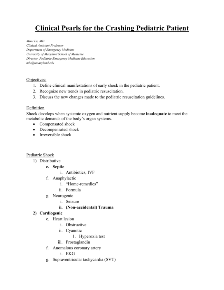
Clinical Pearls for the Crashing Pediatric Patient Mimi Lu, MD Clinical Assistant Professor Department of Emergency Medicine University of Maryland School of Medicine Director, Pediatric Emergency Medicine Education mlu@umaryland.edu Objectives: 1. Define clinical manifestations of early shock in the pediatric patient. 2. Recognize new trends in pediatric resuscitation. 3. Discuss the new changes made to the pediatric resuscitation guidelines. Definition Shock develops when systemic oxygen and nutrient supply become inadequate to meet the metabolic demands of the body’s organ systems. Compensated shock Decompensated shock Irreversible shock Pediatric Shock 1) Distributive e. Septic i. Antibiotics, IVF f. Anaphylactic i. “Home-remedies” ii. Formula g. Neurogenic i. Seizure ii. (Non-accidental) Trauma 2) Cardiogenic e. Heart lesion i. Obstructive ii. Cyanotic 1. Hyperoxia test iii. Prostaglandin f. Anomalous coronary artery i. EKG g. Supraventricular tachycardia (SVT) 3) Hypovolemic e. Blood loss i. 10 ml/kg PRBC f. Fluid loss i. 10-20 ml/kg normal saline bolus 4) Obstructive e. Malrotation with volvulus f. Diaphragmatic hernia g. Necrotizing enterocolitis 5) Endocrine/ metabolic e. Congenital adrenal hyperplasia (CAH) i. Low sodium, high potassium ii. Hydrocortisone f. Glucose!!! i. Rule of 50 g. Inborn error or metabolism i. Glucose ii. Lactate iii. Ammonia iv. Ketonuria T – trauma, non‐accidental trauma H – heart disease, hypovolemia, hypoxia E – endocrine (CAH, thyrotoxicosis) M – metabolic I – inborn errors of metabolism S – sepsis F – formula (dilution or concentration) I – intestinal catastrophies T – toxins S ‐ seizures Pediatric shock: early recognition and management a. Airway i. Cuffed tubes okay! ii. ETT size: (Age/4) + 4, decrease by ½ size if using cuffed Anatomy Infant Relatively large, intraoral Floppy, anterior, cephalad Inclined C3 level Small, narrowest point Small, short, collapsible Tongue Epiglottis Vocal cord angle Glottis Cricothyroid Membrane Trachea Adult Normal Firm Flat C5-C6 level Normal Large, stationary b. Breathing i. Measure RR over 30 seconds ii. Increase temp by 1˚C = increase RR by 2-5 Age adjusted rates Age RR Infant 30-60 Toddler 24-40 Preschooler 22-34 School-aged 18-30 < 30 Adolescent 12-16 < 15 < 60 c. Circulation v. (Age x 2) +90 = median 50% percentile for SBP vi. Early use of intraosseous access vii. NS bolus (20 ml/kg) EXCEPT neonates or congenital cardiac (10 ml/kg) viii. PRBC/ FFP 10 ml/kg (5 ml/kg in neonates) d. Dextrose!!! All ill-appearing infants are hypoglycemic until proved otherwise! i. Rule of 50-100 1. D10 5-10 ml/kg (age < 1 year) 2. D25 2-4 ml/kg (age 1 – 8 year) 3. D50 1-2 ml/kg (age > 8 year) e. Vital signs i. Always measure in kilograms!!! Newborns Brierley J, Carcillo JA, Choong K, et al. Clinical practice parameters for hemodynamic support of pediatric and neonatal septic shock: 2007 update from the American College of Critical Care Medicine. Crit Care Med. 2009 Feb;37(2):666-88. Infants and Children Brierley J, Carcillo JA, Choong K, et al. Clinical practice parameters for hemodynamic support of pediatric and neonatal septic shock: 2007 update from the American College of Critical Care Medicine. Crit Care Med. 2009 Feb;37(2):666-88. SIRS: Age-specific vital signs and laboratory values Age group Tachycardia Bradycardia Newborn Neonate Infant Toddler Child Adolescent >180 >180 >180 >140 >130 >110 <100 <100 <90 - Respiratory rate >50 >40 >34 >22 >18 >14 WBC x 103/mm3 >34 >19.5 or <5 >17.5 or <5 >15.5 or <6 >13.5 or <4.5 >11 or <4.5 Systolic BP mmHg <65 <75 <100 <94 <105 <117 Table 3. International pediatric sepsis consensus conference: definitions for sepsis and organ dysfunction in pediatrics. Goldstein B, Giroir B, Randolph A; International Consensus Conference on Pediatric Sepsis. Pediatr Crit Care Med. 2005 Jan;6(1):2-8. Therapies adults and children with septic shock Therapy Volume Children Adults Fluid resuscitation to CVP 12 Tight glycemic control Usually need more fluid, up to and over 60 ml/kg Early initiation of appropriate antibiotics within 1 hour First line peripheral epinephrine cold shock, transition to central when able. Central norepinephrine for warm shock Use for pulmonary hypertension Low CO, high SVR shock Unresolved ECMO Survival 80% neonates, 50% children Evolving H1N1 popularizing use Inhaled NO Neonates with RV failure No role Hydrocortisone Absolute adrenal insufficiency only; post ACTH cortisol level <18 µg/dL or baseline <5 µg/dL Use if continue on vasopressors regardless of adrenal status Antibiotics Inotropes and vasopressors Vasodilators Reprinted with permission from Marianne Gausche-Hill, MD Early initiation of appropriate antibiotics within 1 hour First line norepinephrine ± dobutamine Vasopressin for warm shock No role Harmful Pediatric Basic Life Support Change in sequence C-A-B Eliminated “look, listen and feel” to assess breathing after opening airway Lay rescuers: check for response of abnormal breathing (eliminate pulse check) Health care providers: allows 10 sec for pulse check o 30 compressions followed by 2 breaths (lone rescuer) o 15:2 if two providers “Push hard, push fast” at least 100 per minute, allowing recoil of chest o Infant chest compression 1.5 in (4 cm) o Children – 2 in (5 cm) Defibrillation o Manual defibrillator > AED with dose attenuator > AED without dose attenuator o 2-4 J/kg, followed by at least 4 J/kg, up to 10 J/kg Pediatric Advanced Life Support BVM recommended over ETI for out-of-hospital setting o Experienced providers may use LMAs Cricoid pressure NOT recommended Cuffed tubes okay! o Uncuffed: (age/4) + 4 = mm ID o Cuffed: (age/4) + 3.5 = mm ID Capnography recommended to confirm ETT placement and assess adequacy of CPR Avoid excessive ventilation (8-10 breaths per minute) Oxygen – increasing evidence for harm, especially in neonates o Avoid hyperoxemia o Start with 100% O2 then titrate to maintain SpO2 >94% Medications o Addition of procainamide as possible therapy for refractory SVT o Routine calcium administration NOT recommended unless clear indication o Etomidate may be used for RSI but NOT recommended in septic shock o Atropine may be added for symptomatic bradycardia but not cardio-pulmonary arrest Wide-complex tachycardia present of QRS width >0.09 sec Post-arrest Care o Therapeutic hypothermia may be considered in children with ROSC (large trial underway) Family presence during resuscitation is recommended New topics: o Specific guidance for cardiac arrest in infants with single-ventricle anatomy, Fontan or hemi-Fontan/ bidirectional Glenn physiology and pulmonary hypertension o Autopsies recommended for young victims of sudden cardiac arrest Neonatal resuscitation Maintain A-B-C sequence De-emphasis on peripartum suctioning ET suctioning for nonvigorous neonate in meconium-stained fluid. Vigorous newborns do not require ET suctioning of meconium regardless of how thick the meconium is. Determine degree of oxygenation by pulse ox on the right wrist or arm Term babies should be resuscitated with room air first, then supplemental oxygen if needed by blended oxygen-air delivery o If bradycardic after 90 sec of resuscitation, increased oxygen to 100% Start CPR for HR < 60 bpm despite assisted ventilation for 30 seconds. Preferred method is thumb-hand technique. Compression:ventilation ratio 3:1 (90 compressions with 30 ventilations per cycle) Infants >36 weeks with moderate to severe hypoxic-ischemic encephalopathy should be offered therapeutic hypothermia Sequence Compression rate (bpm) Depth Compression:ventilation ratio ‐ 1 rescuer ‐ 2 rescuers Pause for ventilation after intubation? Adults/ Adolescents Infants/ Children Neonates C-A-B 100 >2 inches C-A-B 100 1.5-2 inches A-B-C 90:30 events/min 1/3 AP diameter 30:2 30:2 No 30:2 15:2 No 3:1 3:1 Yes Key references: 1. 2. 3. 4. 5. 6. 7. Brierley J, Carcillo JA, Choong K, et al. Clinical practice parameters for hemodynamic support of pediatric and neonatal septic shock: 2007 update from the American College of Critical Care Medicine. Crit Care Med. 2009 Feb;37(2):666-88. Goldstein B, Giroir B, Randolph A, et al. International pediatric sepsis consensus conference: definitions for sepsis and organ dysfunction in pediatrics. International Consensus Conference on Pediatric Sepsis. Pediatr Crit Care Med. 2005 Jan;6(1):2-8. Arikan AA, Citak A. Pediatric shock: review. Signa Vitae. 2008; 3(1):13-23. Dellinger RP, Levy MM, Carlet JM, et al. Surviving Sepsis Campaign: international guidelines for management of severe sepsis and septic shock: 2008. Crit Care Med 2008;36:296-327. Kleinman ME, Chameides L, Schexnayder SM, et al. Part 14: pediatric advanced life support: 2010 American Heart Association Guidelines for Cardiopulmonary Resuscitation and Emergency Cardiovascular Care. Circulation. 2010 Nov 2;122(18 Suppl 3):S876-908. Berg MD, Schexnayder SM, Chameides L, et al. Part 13: pediatric basic life support: 2010 American Heart Association Guidelines for Cardiopulmonary Resuscitation and Emergency Cardiovascular Care. Circulation. 2010 Nov 2;122(18 Suppl 3):S862-75. Kattwinkel J, Perlman JM, Aziz K, et al. Part 15: neonatal resuscitation: 2010 American Heart Association Guidelines for Cardiopulmonary Resuscitation and Emergency Cardiovascular Care. Circulation. 2010 Nov 2;122(18 Suppl 3):S909-19.
