introduction to cognitive neuroscience i
advertisement
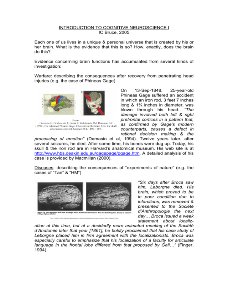
INTRODUCTION TO COGNITIVE NEUROSCIENCE I IC Bruce, 2005 Each one of us lives in a unique & personal universe that is created by his or her brain. What is the evidence that this is so? How, exactly, does the brain do this? Evidence concerning brain functions has accumulated from several kinds of investigation: Warfare: describing the consequences after recovery from penetrating head injuries (e.g. the case of Phineas Gage) On 13-Sep-1848, 25-year-old Phineas Gage suffered an accident in which an iron rod, 3 feet 7 inches long & 1¾ inches in diameter, was blown through his head. “The damage involved both left & right prefrontal cortices in a pattern that, as confirmed by Gage’s modern counterparts, causes a defect in rational decision making & the processing of emotion” (Damasio et al, 1994). Twelve years later, after several seizures, he died. After some time, his bones were dug up. Today, his skull & the iron rod are in Harvard’s anatomical museum. His web site is at http://www.hbs.deakin.edu.au/gagepage/pgage.htm. A detailed analysis of his case is provided by Macmillan (2000). Diseases: describing the consequences of “experiments of nature” (e.g. the cases of “Tan” & “HM”) “Six days after Broca saw him, Leborgne died. His brain, which proved to be in poor condition due to infarctions, was removed & presented to the Société d’Anthropologie the next day… Broca issued a weak statement about localization at this time, but at a decidedly more animated meeting of the Société d’Anatomie later that year [1861], he boldly proclaimed that his case study of Leborgne placed him in firm agreement with the localizationists. Broca was especially careful to emphasize that his localization of a faculty for articulate language in the frontal lobe differed from that proposed by Gall…” (Finger, 1994). Anatomy: analysis of the detailed structure of the brain (e.g. Brodmann’s “cytoarchitectonic” maps of the human cerebral cortex) “In the early part of the 20th century, Korbinian Brodmann divided the human cerebral cortex into 52 discrete areas on the basis of distinctive nerve cell structures & characteristic arrangements of cell layers. Brodmann’s scheme of the cortex is still widely used today & is continually updated… Several areas defined by Brodmann have been found to control specific brain functions. For instance, area 4, the motor cortex, is responsible for voluntary movement. Areas 1, 2, & 3 comprise the primary somatosensory cortex, which receives information on bodily sensation. Area 17 is the primary visual cortex, which receives signals from the eyes… Areas 41 & 42 comprise the primary auditory cortex…” (Kandel et al, 2000) Animal experiments: recording the activity of single neurons during specific behaviours (e.g. Eric Kandel’s work on the neural basis of learning & memory in the sea snail, Aplysia) “Responses of a single cell to the 14 facial stimuli… A neuronal response was evoked by all the animals with horns (including the schematic drawings), although small horns… were less effective.” (Kendrick & Baldwin, 1987) Correlations between cognitively significant stimuli, such as faces, & neural activity, reveal how such stimuli are processed & represented in the brain. Surgery: observing the consequences of direct brain stimulation during epilepsy surgery (e.g. Wilder Penfield’s maps of the functional organisation of the human cerebral cortex) “17 Patient was counting when stimulus was applied. Stimulation arrested speech completely. Patient added, “It raised my right arm.”… 19 Patient was counting. When stimulation was applied, she made an exclamation & then was silent. Her head turned to the right, & there was movement of the right arm… 22 The patient said, “Oh.” There was a movement of the body. The patient said afterward that her body seemed to arise, but she did not seem to be doing it. She said she experienced this at the beginning of her attacks.” (Penfield & Welch, 1954) Imaging: direct observation of changes in the normal human brain during the execution of specific tasks (e.g. PET, fMRI, MEG) In this functional magnetic resonance imaging (fMRI) study, areas that responded to real motion but not while motion was being imagined are shown in red, and areas that were active during motion imagery but not to real motion are shown in green. Areas that were active in both conditions appear as orange & yellow (Thompson & Kosslyn, 2000). So, how does the brain create our internal universe? All brains, from the relatively simple ones that operate jellyfish, to complex ones like those of primates, operate via networks of specialized cells: neurons. Any neuron is composed of three parts: a cell body (soma) that contains the genetic material & metabolic machinery to keep the neuron alive; a set of branching processes (dendrites) that constitute the input surface of the neuron – information arriving from other neurons arrives on the dendrites; and a single process (the axon) that constitutes the output of the neuron – conveying the results of the neuron’s calculation to other neurons. Neurons represent information in terms of brief (~1msec) electrical events (action potentials). For example, this temperature-sensitive neuron in the skin represents temperature as a frequency code. Higher frequency indicates lower temperature. The upper trace shows the baseline skin temperature of 34°C, followed by a cooling step of 110°. Warming the skin at the end of the step silences this cold-sensitive neuron (Kandel et al, 2000). Neurons are linked with one another at synapses, where the electrical events (action potentials) are converted to chemical events, a package of chemical being released by each action potential. This chemical then diffuses across the space (synapse) and affects the dendrites of the neurons with which the upstream neuron’s axon terminals are connected. While more than 100 of such chemicals have been identified, there are only two kinds of synapse. Some synapses are excitatory: An action potential in one (pink) neuron releases a chemical that turns on the second (blue) neuron. This second neuron is therefore more likely to transmit an action potential to other neurons. Some synapses are inhibitory: An action potential in one (pink) neuron releases a chemical that turns off the second (blue) neuron. So the second neuron is less likely to send action potentials to other neurons. Here, we have a dynamic network whose elements represent information as electrical events & this information is manipulated by the processes of excitation & inhibition. At this level of consideration, the human brain works the same way as any other brain. However, the level of “complexity” in the connectivity of the human brain is extraordinary. Each cubic millimetre of your cerebral cortex contains about 90,000 neurons, whose dendritic processes added together would extend about 400 meters, & whose axons would sum to about 3.4 kilometres. In this cubic millimetre, the number of connections from axons to dendrites (synapses) is about 700,000,000. The thickness of the cerebral cortex varies between 2 & 4 mm, & its surface area is about 250,000 mm2. What is the total number of synapses in your cerebral cortex? Since each synapse can be “on” or “off” (to excite or inhibit) from millisecond to millisecond, how many different states can your brain be in over a period of 1 second? I don’t know how to measure “connectional complexity”, but I think that between 50,000 & 100,000 years ago, the human brain may have crossed some kind of threshold of complexity that allowed new properties & therefore new functions to emerge. If this is true, then a machine of sufficient connectional complexity should be able to do what the human brain does. REFERENCES Damasio H, Grabowski T, Frank R, Galaburda AM, Damasio AR (1994) The return of Phineas Gage: Clues about the brain from the skull of a famous patient. Science 264, 1102-1105. Finger S (1994) Origins of Neuroscience: A History of Explorations into Brain Function. Oxford University Press. Kandel ER, Schwartz JH, Jessell TM (2000) Principles of Neural Science, 4th Edn, McGraw-Hill, New York. Kendrick KM, Baldwin BA (1987) Cells in temporal cortex of conscious sheep can respond preferentially to the sight of faces. Science 236, 448-450. Macmillan M (2000) An Odd Kind Of Fame: Stories Of Phineas Gage. MIT Press, Cambridge. Penfield W, Welch K (1954) The supplementary motor area of the cerebral cortex. A clinical & experimental study. Archives of Neurology & Psychiatry 66, 298-317. Thompson WL, Kosslyn SM (2000) Neural systems activated during visual mental imagery: A review & meta-analyses. In: Toga W, Mazziotta JC (Eds) Brain Mapping: The Systems. Academic Press, San Diego, pp 535-560. RECOMMENDED READING Calvin WH, Ojemann GA (1994) Conversations with Neil’s Brain: The Neural Nature of Thought & Language. Addison-Wesley Publ Co. You can read or download it free at: http://faculty.washington.edu/wcalvin/bk7/bk7.htm. Carter R (1999) Mapping The Mind. Seven Dials Press, London. The Hidden Mind. Scientific American Special Edition, August 2002. Downloadable from www.sciam.com (for a small fee). Society for Neuroscience information at: http://apu.sfn.org/content/Publications/BrainBriefings/index.html Recently, the world’s top science magazines “Nature” & “Science” have each published special editions on cognitive neuroscience. They can be found among the “e-journals” in the HKU library system. The references are: Nature, Volume 431, Number 7010, 14 Oct 2004 (“Story of a Neuron”). Science, Volume 306, Number 5695, 15 Oct 2004 (“Cognition & Behaviour”).
