Analysis of powder crystal structures of organic crystals using a high
advertisement

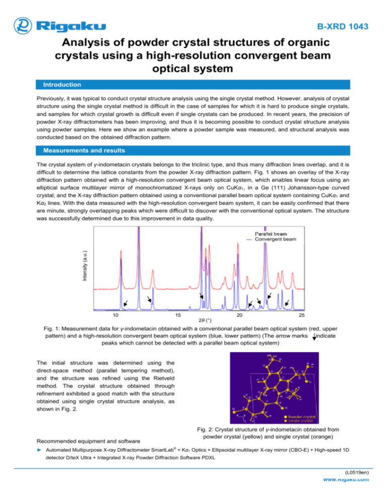
B-XRD 1043 Analysis of powder crystal structures of organic crystals using a high-resolution convergent beam optical system Introduction Previously, it was typical to conduct crystal structure analysis using the single crystal method. However, analysis of crystal structure using the single crystal method is difficult in the case of samples for which it is hard to produce single crystals, and samples for which crystal growth is difficult even if single crystals can be produced. In recent years, the precision of powder X-ray diffractometers has been improving, and thus it is becoming possible to conduct crystal structure analysis using powder samples. Here we show an example where a powder sample was measured, and structural analysis was conducted based on the obtained diffraction pattern. Measurements and results The crystal system of γ-indometacin crystals belongs to the triclinic type, and thus many diffraction lines overlap, and it is difficult to determine the lattice constants from the powder X-ray diffraction pattern. Fig. 1 shows an overlay of the X-ray diffraction pattern obtained with a high-resolution convergent beam optical system, which enables linear focus using an elliptical surface multilayer mirror of monochromatized X-rays only on CuKα1, in a Ge (111) Johansson-type curved crystal; and the X-ray diffraction pattern obtained using a conventional parallel beam optical system containing CuKα1 and Kα2 lines. With the data measured with the high-resolution convergent beam system, it can be easily confirmed that there are minute, strongly overlapping peaks which were difficult to discover with the conventional optical system. The structure was successfully determined due to this improvement in data quality. Intensity (a.u.) ― Parallel beam ― Convergent beam 10 15 2θ (°) 20 25 Fig. 1: Measurement data for γ-indometacin obtained with a conventional parallel beam optical system (red, upper pattern) and a high-resolution convergent beam optical system (blue, lower pattern) (The arrow marks indicate peaks which cannot be detected with a parallel beam optical system) The initial structure was determined using the direct-space method (parallel tempering method), and the structure was refined using the Rietveld method. The crystal structure obtained through refinement exhibited a good match with the structure obtained using single crystal structure analysis, as shown in Fig. 2. Fig. 2: Crystal structure of γ-indometacin obtained from powder crystal (yellow) and single crystal (orange) Recommended equipment and software ® ► Automated Multipurpose X-ray Diffractometer SmartLab + Kα1 Optics + Ellipsoidal multilayer X-ray mirror (CBO-E) + High-speed 1D detector D/teX Ultra + Integrated X-ray Powder Diffraction Software PDXL (L0519en)


