CELL INJURY
advertisement
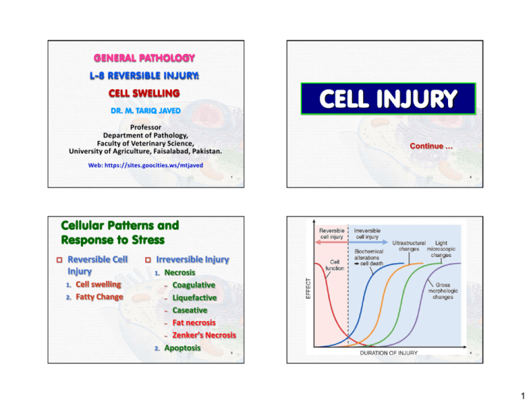
GENERAL PATHOLOGY CELL INJURY CELL SWELLING DR. M. TARIQ JAVED Professor Department of Pathology, Faculty of Veterinary Science, University of Agriculture, Faisalabad, Pakistan. Continue … Web: https://sites.goocities.ws/mtjaved 1 2 Cellular Patterns and Response to Stress Reversible Cell Injury 1. 2. Irreversible Injury 1. Cell swelling Fatty Change 2. Necrosis – Coagulative – Liquefactive – Caseative – Fat necrosis – Zenker’s Necrosis Apoptosis 3 4 1 CELL SWELLING REVERSIBLE INJURY Degeneration Necrosis = = Degeneration Reversible injury Irreversible injury Degeneration Cloudy swelling Cell Swelling Hydropic degeneration Fatty Change Fatty Infiltration Fatty Degeneration Degeneration -OSIS (hepatosis, nephrosis) (cellular oedema) Early and almost universal manifestation of injury. Failure in maintaining ionic and fluid homeostasis (Na ATPase). Difficult to appreciate under the light microscope. 5 6 CELL SWELLING Microscopically, Cells appear enlarged and compresses adjacent structures, CELL SWELLING Microscopically, Extensive water logging e.g., swelling of hepatocytes -- obliteration of sinusoid and swelling of renal tubular cells -obliteration of tubular lumen. In mildly swollen cells -- cloudy appearance of cytoplasm “cloudy swelling” (Rudolph Virchow, in 1860) 7 distension of organelles, “vacuolar degeneration”. distension of the cell, central areas cleared of proteins, shrunken nuclei pushed to the periphery -- “hydropic degeneration”. The vacuoles need to be differentiated from fat droplets or non-membrane-bound areas as in glycogen storage disease. If majority of cells appear swollen and many are actually dead (devoid of nucleus), the injury is more serious 8 2 CELL SWELLING CELL SWELLING The ultrastructural changes of reversible injury: Plasma membrane alterations, such as blebbing, blunting, and loss of microvilli Mitochondrial changes, including swelling and the appearance of small amorphous densities Dilation of the ER, with detachment of ribosomes; intra-cytoplasmic myelin figures may be present Nuclear alterations, with disaggregation of granular and fibrillar elements Normal kidney tubules Early (reversible) injury Necrosis (irreversible injury) 9 10 CELL SWELLING CELL SWELLING HYDROPIC DEGENERATION 11 12 3 CELL SWELLING CELL SWELLING Cloudy swelling Kidney HYDROPIC DEGENERATION 13 14 CELL SWELLING CELL SWELLING Cell Swelling Kidney Grossly pallor colouration, increased turgor, increase in weight rounding of edges water oozes out of cut surface the cut surface bulges out. they sink in water In capsulated organs, the capsule appears stretched. 15 16 4
