Cerebral Cortex November 2005;15:1666--1675
doi:10.1093/cercor/bhi043
Advance Access publication February 9, 2005
The Functional Anatomy of
Sleep-dependent Visual Skill Learning
Matthew P. Walker1, Robert Stickgold2, Ferenc A. Jolesz3 and
Seung-Schik Yoo3
1
Sleep and Neuroimaging Laboratory, Center for Sleep and
Cognition, Department of Psychiatry, Beth Israel Deaconess
Medical Center, Harvard Medical School, Boston, MA 02115,
USA, 2Center for Sleep and Cognition, Department of
Psychiatry, Beth Israel Deaconess Medical Center, Harvard
Medical School, Boston, MA 02115, USA and 3Department of
Radiology, Brigham and Women’s Hospital, Harvard Medical
School, Boston, MA 02115, USA
Learning of procedural skills develops gradually, with performance
improving significantly with practice. But improvement on some
tasks, including a visual texture discrimination task, continues in the
absence of further practice, expressly during periods of sleep and not
across equivalent waking episodes. Here we report that the brain
activation revealed significantly different patterns of performancerelated functional activity following a night of sleep relative to 1 h
post-training without intervening sleep. When task activation
patterns after a night of sleep were compared with activation
patterns without intervening sleep (1 h post-training), significant
regions of increased signal intensity were observed in the primary
visual cortex, the occipital temporal junction, the medial temporal
lobe and the inferior parietal lobe. In contrast, a region of decreased
signal intensity was found in the right temporal pole. Corroborating
these condition differences, correlations between behavioural performance and brain activation revealed significantly different patterns of performance-related functional activity following a night of
sleep relative to those without intervening sleep. Together, these
data provide evidence of overnight bi-directional changes in functional anatomy, differences that may form the neural basis of sleepdependent learning expressed on this task.
Keywords: memory, plasticity, sleep, visual skill learning
Introduction
The process of learning procedural skills is a gradual one. While
practice has long been known to trigger improvement during
training, recent studies demonstrate that performance on
certain tasks continues to improve in the absence of any further
practice exclusively during sleep, and not across equivalent
periods of time awake (for reviews, see Smith, 1995; Maquet,
2001; Stickgold et al., 2001; Walker, 2004).
Evidence of sleep-dependent learning has now been established across a wide range of visual (Karni et al., 1994; Gais et al.,
2000; Stickgold et al., 2000a,b), auditory (Atienza et al., 2004;
Gaab et al., 2004) and motor skills (Smith and MacNeill, 1994;
Walker et al., 2002, 2003a,b; Fischer et al., 2002; Korman et al.,
2003; Robertson et al., 2004). Within the visual domain, several
behavioural studies using a visual texture-discrimination task
(TDT) have identified a delayed, sleep-dependent learning
component which develops following initial training. Karni
et al. (1994) have established that learning of the TDT, which
does not benefit from 4--12 h of wake following acquisition
(Stickgold et al., 2000b), improves significantly following a night
of sleep. Furthermore, they demonstrated that selective disruption of rapid eye movement (REM) sleep, but not non-REM
(NREM) sleep, results in a loss of these performance gains
(Karni et al., 1994). Gais et al. (2000) have selectively deprived
subjects of early sleep (normally dominated by slow-wave sleep;
Ó The Author 2005. Published by Oxford University Press. All rights reserved.
For permissions, please e-mail: journals.permissions@oupjournals.org
SWS) or late-night sleep (normally dominated by REM and stage-2
NREM) following learning of the TDT, concluding that consolidation of this task is initiated by SWS-related processes,
while REM sleep then promotes additional enhancement.
Using the same task, Stickgold et al. (2000b) have shown that
these enhancements are specifically sleep-dependent, with the
overnight improvements correlating positively with the amount
of both early night SWS and late night REM sleep. It was also
demonstrated that these delayed performance benefits are
absolutely dependent on the first night of sleep following
training (Stickgold et al., 2000a). In addition, they have also
shown that short daytime sleep episodes can restore performance decrements caused by repeated practice on this task
(Mednick et al., 2002), and that 60--90 min sleep epochs,
containing both REM and SWS, can produce performance
enhancements equivalent to a normal night of sleep (Mednick
et al., 2003). Yet while the sleep-dependent nature of this TDT
has been extensively investigated, the underlying neural correlates of sleep-dependent learning remain largely unexplored.
Using neuroimaging techniques, Maquet et al. (2003) have
specifically investigated the effects of sleep deprivation on
memory consolidation and functional brain anatomy using
a visuo-motor task. Subjects were trained on the task and then
retested during an fMRI scan 72 h later. Half of the subjects
underwent total sleep-deprivation across the first night after
training, followed by two recovery nights of sleep, while the
remaining subjects slept all three nights. At the later 72 h retest,
subjects in the sleep deprivation condition demonstrated
a significantly altered pattern of brain activation relative to
control subjects. Moreover, these differences in functional brain
activation were also accompanied by significant learning impairments in the sleep-deprived group. Nevertheless, no study has
yet investigated the beneficial effect of a night of sleep on skill
learning and brain plasticity without employing sleep deprivation techniques.
In a seminal study, Schwartz et al. (2002) have recently
examined changes in brain activity using the TDT, comparing
task-dependent activation without training (‘untrained’), relative to performance following training (‘trained’) at a later 24 h
retest, which included a night of sleep. When the two
conditions were compared, significantly greater signal intensity
was evident in the primary visual cortex in the trained condition
relative to the untrained condition, indicative of a low-level
plastic change associated with task learning. However, these
findings could not determine whether such changes occurred
during training, across post-training wake, or across a subsequent night of sleep.
Regarding the brain basis of visual perceptual discrimination,
the highly specific characteristics of this form of learning (Sagi
and Tanne, 1994; Karni and Bertini, 1997) have lead to the
hypothesis that orientation discrimination should be reflected in
neural changes early in the visual cortex (Karni and Sagi, 1991;
Karni, 1996; Gilbert et al., 2001). While some experimental
evidence has confirmed these predictions (Gilbert et al., 2001),
other reports have not been successful in identifying substantial
differences in either the selectivity or responsivity of neurons in
V1 or V2 (Crist et al., 2001; Schoups et al., 2001; Ghose et al.,
2002). While the reasons for this discrepancy remain unclear, it
has led to the possibility that visual perceptual learning may be
mediated by changes downstream of these early visual processing regions. For example, a recent study by Yang and Maunsell
(2004) has demonstrated that orientation discrimination is
associated with plasticity in later stages of the visual cortex,
specifically area V4, indicating that perceptual learning can cause
overt changes in the tuning of neurons outside of V1. Therefore,
visual perceptual learning, during both training and potentially
following sleep, may be related to distinct neural changes
throughout several visual processing regions.
Using the sleep-dependent TDT, we now report a functional
magnetic resonance imaging (fMRI) study demonstrating
changes in human regional brain activity exclusively across
a night of sleep, relative to a condition without intervening
sleep but following equivalent amounts of training. Learning of
this task is both retinotopically and monocularly specific (Karni
and Sagi, 1991). That is to say, learning to discriminate these
texture targets is specific to the portion of the visual field in
which subjects are trained, and the learning achieved with
the trained eye does not transfer to the untrained eye. Based on
the visual specificity of this TDT, we were therefore able to
investigate, in the same subjects, during the same scanning
session, at the same circadian time and after equivalent amounts
of training, how functional brain activity and learning seen
during retesting differed following a night of sleep, compared
with retesting without intervening sleep.
Based on previous neurophysiological and functional imaging
studies of perceptual learning, we hypothesized that intervening sleep, and the associated learning benefit, would result in
significantly greater signal intensity within the corresponding
primary visual cortex. Moreover, considering that orientation
discrimination learning may also trigger corresponding neural
changes downstream of the primary visual cortex, we also
expected additional regions of increased activation in later
visual processing areas following a night of sleep.
Materials and Methods
The study was approved by the local human studies committee and
participants provided written informed consent.
Participants
Subjects (n = 18; 10 females, 8 males, mean age ± SD = 25.7 ± 2.9) had no
prior history of drug or alcohol abuse, neurological, psychiatric or sleep
disorders, and agreed to be drug, alcohol and caffeine free for 24 h prior
to and during the study period. All subjects maintained a standard sleep
schedule for 1 week prior to the study. On the intervening night of sleep
during the experimental phase, subjects obtained an average 8.3 ± 0.78 h
sleep, as measured by sleep-log diaries.
Experimental Protocol
All training and retesting on the TDT was performed monocularly.
Subjects were trained monocularly on one eye at 9 P.m. on day 1. The
following morning, at 9 a.m., subjects were trained monocularly using
the other eye. One hour later, subjects were then retested monocularly
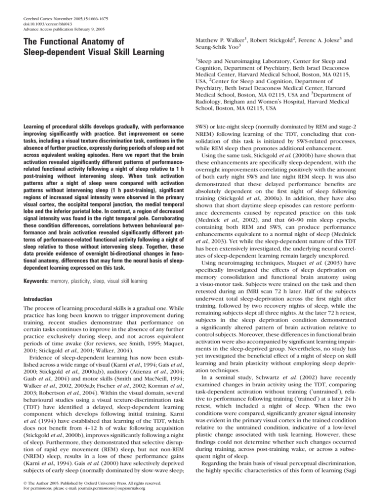
using each eye separately, during the fMRI scan (Fig. 1a). Retesting of
the eye with sleep and without sleep was counterbalanced with regard
to first and second retests. Assignment of the condition of sleep or
without sleep to either the left or right eye, and the dominant or nondominant eye, was also counterbalanced across subjects. In both the
training sessions and at the subsequent fMRI retest sessions, target
stimuli were presented in the lower right quadrant of the visual field.
Thus, each eye received identical amounts of training, but differed at
retest based on the presence or absence of intervening sleep between
training and the fMRI retest session (assigned as the ‘SLEEP’ or
‘WITHOUT-SLEEP’ conditions respectively; Fig. 1a).
TDT Task
The TDT was composed of a series of trials, organized into successive
blocks. Each 3 s trial consisted of 500 ms of fixation followed by a target
screen presented for 17 ms (Fig. 1b). Each target screen containing
either a ‘T’ or ‘L’ at the fixation point and a horizontal or vertical array of
three diagonal bars in the lower-right quadrant of the visual field. The
background was always a field of horizontal bars. After a blank interstimulus interval (ISI) screen of varying length (80--500 ms), a mask
was presented for 17 ms followed by a blank screen (1966--2386 ms)
producing a constant total trial length of 3 s. During the final blank
screen, subjects indicated with a button press which letter was
presented at the fixation point, and then whether the three diagonal
line segments in the lower-right quadrant were arranged in a horizontal
or vertical array. The central fixation cross was then redisplayed as the
next trial began. Monocularity was achieved using a custom cotton eye
patch worn during training, taped to the subject’s forehead, completely
occluding vision from the patched eye.
Training
Monocular training consisted of 10 blocks, with decreasing ISIs (increasing
difficulty) of 500, 400, 300, 250, 200, 160, 140, 120, 100 and 80 ms.
Each block began with 24 s of central fixation, followed by five sets of
eight TDT trials (24 s per set) separated by 24 s period of central
fixation, and ending with a final 24 s of fixation. During training, subjects
sat at 35--40 cm from a 15$ CRT screen. The target diagonal array was
2.58° in width and was located 58° from the fixation point.
fMRI Retest
Monocular retesting of each eye during the fMRI session involved a single
block (40 trials as described above) of the TDT at an ISI of 250 ms.
This ISI was identified in pilot testing as providing a manageable difficulty level across conditions, while still remaining challenging enough
to identify subtle differences. During retesting inside the MRI scanner,
subjects monocularly viewed the stimuli using a mirror box inside the
scanner. Stimuli (448 square resolution) were projected by an SVGA
resolution LCD projector on a central screen located 270 cm from the
subject, at a projected image size of 82 cm, resulting in a viewing angle
of 17°. Monocularity during the MRI retest was again achieved using the
same custom cotton eye patch as worn during training, taped to the
subjects forehead above the eye. Between each scan session, an
experimenter gently flipped up one eye patch and placed down the
other eye patch (from behind the subjects head in the magnet bore), to
occlude the opposite eye, preventing the need for subject head
movement or change of position. The shift in the center-of-view due
to monocularity was small, <1°, and was also compensated by adding the
crosshair for the visual fixation, preventing any effect on hemispheric
stimulation by shifting eye patch.
Measurement of Skill Level
Behavioural performance during both training and retesting was
measured as the discrimination accuracy within each ISI block. For
the training sessions, target discrimination accuracy was plotted against
ISI, and the detection threshold defined as the interpolated ISI which
produced 80% accuracy. The threshold value (ms) thus reflects the time
required for cortical discrimination of the visual target from its
background of horizontal bars. For the fMRI retest sessions, containing
one block of trials per eye, a single accuracy percentage was calculated.
The number of correct discriminations made during the single-block
retest of each eye in the scanner similarly reflects cortical processing
efficiency at the set ISI.
Cerebral Cortex November 2005, V 15 N 11 1667
Figure 1. Texture discrimination task and experimental protocol. (a) Subjects training monocularly in the lower right visual quadrant on day 1 (9 p.m.). After an intervening night of
sleep, subjects trained monocularly in the same visual quadrant using the other eye (9 a.m.). Training of the left eye versus right eyes was counterbalanced across subjects and
conditions, as was eye dominance. Each eye was then retested monocularly on one single block (ISI 5 250 ms) in the visual quadrant during fMRI scanning. Retesting as either the
first or second scanning session was also counterbalanced across each eye condition. (b) Each 3 s trial consisted of a series of stimulus screens, including a target stimulus containing
either a ‘T’ or ‘L’ letter at the fixation point, and a horizontal or vertical array of three diagonal bars in the lower-right quadrant of the visual field. During the blank response screen,
subjects indicated with a button press which letter was presented at the fixation point, and then whether the three diagonal line segments in the lower-right quadrant were arranged
in a horizontal or vertical array. Monocular training consisted of 10 blocks containing 40 trials, with decreasing ISIs: 500, 400, 300, 250, 200, 160, 140, 120, 100 and 80 ms.
MRI Scanning
MRI data were acquired with a 1.5 T GE Signa system (GE Medical
Systems, WI). Structural anatomical images were acquired using a 3DSpoiled Gradient Recalled (SPGR) sequence, covering the whole brain
volume with 1.5 mm sagittal slices (TE/TR = 6/35 ms, flip angle = 75°, field
of view = 24 cm, matrix = 256 3 256). Functional MRI images were
acquired using a gradient echo-planar T2*-sequence sensitive to the
blood-oxygenation level-dependent (BOLD) contrast. Functional image
volumes consisted of 28 oblique-axial slices (thickness = 5 mm, matrix =
64 3 64, TR/TE = 2500/50 ms, flip angle = 90°) covering the whole brain
volume.
fMRI Data Analysis
fMRI data were analyzed using the SPM99 software package (www.fil.
ion.ucl.ac.uk/spm/spm99.html). Each set of axial images for each
subject were realigned to the first functional image (following removal
of the first four initial functional images used to achieve steady-state
equilibrium), co-registered with the corresponding T1-weighted data
1668 Anatomy of Sleep-dependent Visual Learning
d
Walker et al.
set, spatially normalized to the SPM99 template, and smoothed with an
isotropic Gaussian kernel (6 mm full-width at half-maximum).
Condition and subject effects were estimated using a general linear
model (Friston et al., 1995). The effects of global differences in scan
intensity were removed by scaling each scan in proportion to its global
intensity, and low-frequency drifts were removed using a temporal highpass filter with the default cutoff of 110 s.
Individual contrast images were first produced by comparing taskdependent activation in the SLEEP condition relative to the WITHOUTSLEEP condition for each subject separately (therefore controlling for
within-subject variance). This condition contrast was then entered into
a second level, group analysis using a random-effects model. A onesample t-test was performed across the set of individual contrast images
to identify brain areas in which cerebral activity differed between the
SLEEP and WITHOUT-SLEEP conditions in each direction (SLEEP >
WITHOUT-SLEEP and SLEEP < WITHOUT-SLEEP), controlling betweensubject variances. To control for type I error through multiple
comparisons, regions of significant difference were identified using
a corrected joint expected probability distribution of extent (P < 0.05)
and height (P < 0.001).
In addition to the group subtractions, within each condition we
explored the relationship between behavioural performance (percentage correct responses for ‘horizontal’ and ‘vertical’ discrimination;
accuracy) and brain activation across subjects using a correlation
analysis. The behavioural retest accuracy score was therefore used as
a specified covariant in the design matrix, and correlated with brain
activation using the regression tool in SPM99 (Jansma et al., 2000;
Peigneux et al., 2003; Pleger et al., 2003; Madden et al., 2004). Regions
of significant correlation were again identified using a corrected joint
expected probability distribution of extent (P < 0.05) and height (P <
0.001) to control for type I error through multiple comparisons.
Results
Behavioural
During the initial training sessions, no significant difference in
threshold was observed between the two eyes [eye-SLEEP 110.5
ms versus eye-WITHOUT-SLEEP 107.56 ms; paired t-test; t (17) =
0.77, P = 0.45)], confirming equal performance by both eyes and
an absence of learning transfer between eyes (Fig. 2). Thus, both
eyes experienced identical amounts of training and both eyes
appeared to benefit equally from the initial training session.
During the fMRI retest session, significantly better visual
discrimination was expressed by the eye in the SLEEP condition
compared with the eye in the WITHOUT-SLEEP condition [96.8
versus 92.7% respectively, paired t-test; t (17) = 2.93, P = 0.009)].
Thus, the night of intervening sleep produced a significant
learning benefit, consistent with previous reports of overnight
sleep-dependent enhancement of TDT performance (Karni
et al., 1994; Gais et al., 2000; Stickgold et al., 2000a,b).
fMRI Group Comparisons
Group analysis of retest brain activation maps demonstrated
a region of significantly greater activation in the left primary visual
cortex (V1) in the SLEEP condition, relative to the WITHOUTSLEEP condition (SLEEP > WITHOUT-SLEEP; Fig. 3) — an area that
corresponds to the trained visual field and also supports the
achievement of successful monocularity. This finding of increased
activation in V1 is consistent with previous fMRI findings using the
TDT (Karni et al., 1995; Schwartz et al., 2002) and is consonant
with the suggested low-level specificity of the task. However,
there were additional lateralized regions of significantly increased
activity beyond the primary visual cortex following sleep. These
included portions of the ventral visual processing stream — the
left temporal--occipital junction and the left posterior temporal
lobe, as well as the left inferior parietal region of the dorsal visual
processing stream (Ungerleider and Mishkin, 1982; Ungerleider
and Haxby, 1994; Yantis and Serences, 2003).
In the opposite group contrast (SLEEP < WITHOUT-SLEEP),
there was a region of decreased signal intensity in the right
temporal pole (Fig. 3). Anatomical coordinates for all these
regions of difference, and the corresponding z-scores, are provided in Table 1.
Together, these data indicated clear differences in activation
between the two conditions, suggesting an influence of sleep on
task related function brain activation. However, since behavioural skill level was significantly higher in the SLEEP condition,
these data alone were not able to dissociate the possibility that
better performance skill, rather than a specific sleep-dependent
influence per se, formed the basis of these differences.
To address this issue, a correlation analysis was carried out
between behavioural performance and functional activation
patterns within each of the SLEEP and WITHOUT-SLEEP
conditions separately. Since there was a range of performance
values across subjects within each condition (i.e. both high and
low level performance scores), this offered the ability to
investigate whether superior accuracy in the SLEEP condition
correlated with the same or different brain regions, relative to
superior accuracy in the WITHOUT-SLEEP condition. If it were
the case that sleep, rather than skill level, determined differences in brain activation between the two conditions, then
better performance would be expected to correlate with
different brain regions in each of these conditions.
Performance Correlations
When behavioural task performance within each condition was
correlated with functional brain activation, clear differences in
the patterns of both positive and negative performance-related
brain activity emerged.
In the SLEEP condition, positive correlations with task
performance were identified bilaterally in the posterior temporal lobes, largely inferior, as well as a region of the right
Figure 2. Monocular training curves and initial discrimination thresholds. (a) Training curves for performance in the SLEEP and WITHOUT-SLEEP conditions across the 10 blocks of
decreasing ISI (increasing difficulty, resulting in decreasing performance accuracy). (b) The detection thresholds, defined as the interpolated ISI which produced 80% accuracy, for
the SLEEP and WITHOUT-SLEEP conditions.
Cerebral Cortex November 2005, V 15 N 11 1669
Figure 3. fMRI group (condition) effects. Increased fMRI signal activity (in red) and decreased signal activity (in blue) during task retest following training with sleep, relative to
training without sleep. Corresponding coronal sections (A--E) in ICBM152 (International Consortium for Brain Mapping) space are provided, with averaged BOLD signal change (in %
with respect to the baseline signal) for each condition (gray: trained & SLEEP; white: trained & WITHOUT-SLEEP).
Table 1
Anatomical coordinates for significant clusters of activation for the main group (condition)
comparisons
Region (Brodmann’s area)
x
y
SLEEP [ WITHOUT-SLEEP
Primary visual area (striate cortex; BA 17)
Occipital--temporal junction (BA 39)
Inferior parietal lobe (BA 7)
Inferior/middle temporal lobe (BA 21)
ÿ2
ÿ42
ÿ20
ÿ58
ÿ90
ÿ74
ÿ56
ÿ56
42
14
WITHOUT-SLEEP [ SLEEP
Temporal pole (BA 38)
z
Cluster
size (mm3)
Peak
z-score
10
22
38
2
280
1048
656
288
3.82
4.85
3.91
3.86
ÿ20
312
4.31
Taken as a whole, these results demonstrated that skill level in
the SLEEP condition (ranging from low to high) correlated with
a different pattern of brain activation compared with skill level
in the WITHOUT-SLEEP condition (also ranging from low to
high). It does not therefore appear that skill level alone
determined differences in brain activation between the two
conditions. If this were true, better performance should have
correlated with the same brain regions in both of the conditions. Instead, better performance in each group correlated
with quite different patterns of brain activation, indicating the
existence of distinct performance-related networks with or
without-sleep, rather than skill level per se.
The x--y--z coordinates are given in ICBM152 space. The Brodmann’s area location is identified
according to the atlas of Talairach and Tournoux (1988).
Discussion
medial frontal lobe [Brodmann’s area (BA) 6/8] (Fig. 4a). In
addition to these positive correlations, significant negative
correlations were identified in the left medial dorsal and
pulvinar nuclei of the thalamus (Fig. 4a).
In contrast to the diffuse regions of correlation in the SLEEP
condition, there was only one region of positive correlation
with behavioural performance in the WITHOUT-SLEEP condition, restricted to the right posterior medial temporal lobe (Fig.
4b). Furthermore, no negative correlations in any brain regions
were found in the WITHOUT-SLEEP condition.
Anatomical coordinates for these regions of correlation
in both the SLEEP and WITHOUT-SLEEP conditions, together
with the corresponding z-scores and r-values, are provided in
Table 2.
1670 Anatomy of Sleep-dependent Visual Learning
d
Walker et al.
By comparing brain activity with and without a night of sleep
following training on a visual TDT, we have identified regionally
specific increases and decreases in functional activation, associated with sleep-dependent learning. Moreover, when behavioural performance measures were correlated with brain
activation, significantly different patterns of covariance were
evident between the two conditions; indicative of a unique
performance-related brain network, post-sleep.
It should be noted that these differences cannot be explain
either by (i) variations in alertness due to differences in
circadian test times, since both conditions were measured at
the same time of day in the same scanning session; or (ii)
differences in training, since the two eyes, although trained
separately, underwent identical training regimens. Furthermore, each eye attaining near identical training thresholds,
Figure 4. Correlational analyses between behavioural task performance and functional brain activity within each group (condition). Positive correlations between task performance
and increased fMRI signal activity (in red) and negative correlations (in blue) within each of the experimental conditions of (a) SLEEP and (b) WITHOUT-SLEEP. Corresponding axial
and sagital sections in ICBM152 space are provided, together with correlational plots (positive in red, negative in blue) between peak signal intensity in each region and the
associated behavioural task performance score.
Cerebral Cortex November 2005, V 15 N 11 1671
Table 2
Anatomical coordinates for regions of activity correlating significantly with behavioural
performance score within each condition
Region (Brodmann’s area)
x
y
z
Cluster
size (mm3)
Peak
z-score
SLEEP
Positive correlations
Middle temporal gyrus (BA 21)
Middle temporal gyrus (BA 21)
Precentral gyrus (BA 6)
Negative correlations
Thalamus, medial dorsal nucleus
Thalamus, Pulvinar
ÿ42
51
57
ÿ33
ÿ41
ÿ2
3
ÿ1
31
1032
520
472
4.10
3.77
3.74
0.814
0.773
0.770
ÿ4
ÿ6
ÿ15
ÿ29
14
11
728
496
3.84
3.75
ÿ0.783
ÿ0.771
53
ÿ46
8
744
4.37
0.842
WITHOUT-SLEEP
Positive correlations
Middle temporal gyrus (BA 21)
r-value
The x--y--z coordinates are given in ICBM152 space. The Brodmann’s area location is identified
according to the atlas of Talairach and Tournoux (1988).
suggesting that the initial practice-dependent improvements
were similar.
We cannot, however, rule out the possibility that differences
observed between the two conditions are simply a factor of time
rather than sleep, since the two conditions differed not only in
the presence or absence of intervening sleep, but also in the
amount of total intervening time between training and retest —
an important limitation of this study. According to Karni et al.
(1994), a small percentage of subjects (1--2 of 11 subjects) did
express some performance gains without sleep. Nevertheless,
to verify that an intervening time period awake did not confer
any performance improvement on this modified version of the
task, an additional group of subjects (n = 10; 8 females, 2 males,
mean age 23.6 ± 1.7) were trained monocularly (10 blocks) in
the morning (9 a.m. ± 30 min) and 12h later, after waking, were
retested monocularly (10 blocks) without scanning, offering
a behavioural wake control. Consonant with previous studies,
no significant improvement was expressed at the later retest
without sleep [training threshold = 116.3 ms versus retest
threshold = 114.2; t (9) = 0.37, P = 0.71)], indicating that the
behavioural characteristics of this modified task are similar to
those of previous task versions (Karni et al., 1994; Stickgold
et al., 2000a,b; Mednick et al., 2002, 2003) and that an
intervening time period awake did not confer any learning
enhancements, in contrast to improvements expressed
overnight.
This evidence, together with data from prior investigations
demonstrating that performance on this task does not improve
across waking episodes of 60 min, 90 min, 3 h, 6 h, 9 h or even
12 h (Karni et al., 1994; Stickgold et al., 2000a,b; Mednick et al.,
2002, 2003), leads us to consider that differences in functional
activation patterns between each condition are mostly likely
explained by intervening overnight sleep. Nevertheless, it will
be vital for future studies to clarify that similar neural changes to
those identified overnight do not occur across equivalent time
periods awake, truly confirming the sleep-dependency of such
brain plasticity.
Group Difference between Conditions
The retinotopic and monocular specificity of the TDT task
suggests that at least part of the underlying neural substrates of
this form of learning reside early in the visual processing
pathway (Karni and Sagi, 1991; Karni, 1996; Gilbert et al.,
1672 Anatomy of Sleep-dependent Visual Learning
d
Walker et al.
2001; but see Crist et al., 2001; Schoups et al., 2001; Ghose et al.,
2002). Neuroimaging studies that have compared brain activation between trained and untrained conditions using this task
have demonstrated increased activation in the primary visual
cortex (V1) (Karni et al., 1995; Schwartz et al., 2002), findings
that are in accordance with the known cell selectivity for
stimulus orientation in this region (Hubel and Wiesel, 1959).
However, since learning on this task continues to develop
across sleep, independent of further practice, the plastic
changes associated with overnight improvement may rely on
a different functional anatomy to those changes which mediate
practice-dependent learning. This possibility is supported by
evidence of neural changes in later stages of the visual system
associated with orientation discrimination learning, indicating
that a more distributed system can support perceptual learning,
beyond V1 (Yang and Maunsell, 2004). It is also of note that
increased proficiency of visual object recognition has been
related to changes in downstream regions of the visual
processing pathway (Tanaka et al., 1991; Sakai and Miyashita,
1994; Sakai et al., 1994; Zohary et al., 1994), although these
tasks have usually involved visual stimuli of a more complex
nature. Thus, additional overnight improvement leading to
enhanced visual discrimination accuracy may not only be
evident in low-level processing regions of the primary visual
regions (as may be the case during training), but may extend
beyond this region (Gilbert et al., 2001).
Our findings describe a region of increased activation within
the primary visual cortex following a night of sleep, relative to
without sleep, supporting previous neuroimaging data comparing untrained to trained performance using the TDT (Karni
et al., 1995; Schwartz et al., 2002). In this sense, the overnight
changes in functional brain activation (and the associated
improved task performance) reported here appear, in part, to
be a continuance of the neural changes associated with initial
practice-dependent learning. However, our data also demonstrate further changes that extended beyond V1, into later visual
processing areas, including the inferior temporal and inferior
parietal regions. One interpretation of these findings is that
overnight, sleep-dependent learning produces orientationspecific changes exclusively in V1; plastic changes that then
drive increased responsivity in associated downstream regions
involved in object and spatial recognition, but which do not
themselves undergo a plastic change. An alternative interpretation is that the entire collection of cortical regions which show
greater activation represent a genuine large-scale, systems-level,
plastic change that takes place throughout several posterior
visual processing areas, as well as in V1. In this respect, sleep
would be considered to trigger a modification in the locus of the
memory representation beyond the primary visual cortex
(Gilbert et al., 2001). It is also possible that regions of increased
activation in later visual processing areas, particularly in the
parietal lobe, reflect an overnight adjustment of top-down
attentive modulation on the primary visual cortex, thus improving object recognition (Kastner and Ungerleider, 2000). This is
especially pertinent considering the fact that perceptual learning appears to require a degree of conscious attentive involvement (Hochstein and Ahissar, 2002). However, based on
the temporal resolution capabilities of fMRI, the current study is
not able to differentiate between these possibilities.
Nevertheless, these data do argue for a potential dissociation
between initial training-dependent plastic changes, relative to
the subsequent delayed, overnight changes, which may recruit
greater involvement of later visual processing regions (Karni
et al., 1995; Schwartz et al., 2002). This dissociation is
supported by behavioural models which propose unique stages
of memory processing during initial practice and across the
subsequent brain states of wake and sleep (for review, see
Walker, 2004), and recent molecular studies describing the
temporal stage progression of plasticity-associated zif-268 gene
expression across intervals of wake, slow-wave sleep and REM
sleep (Ribeiro et al., 2002).
In association with the increased activations in the SLEEP
condition, there was also a region of decreased signal in the
right temporal pole, an area considered to be a higher order
visual structure (Nakamura and Kubota, 1996), potentially
representing a reduced visual memory load. Interestingly,
Lane et al., have reported greater temporal pole activation
specifically related to the processing of visual stimuli with
strong emotional meaning (Lane et al., 1999). In this sense, it
would not be surprising if processing demands related to the
emotional burden of the task decreased during retesting
following sleep due to greater ease of task performance
(because of the sleep-dependent learning benefit), thereby
reducing activity in the temporal pole.
Performance Correlations
While the main group contrasts revealed significant differences
between the sleep and WITHOUT-SLEEP conditions, an alternative explanation was that superior skill level in the SLEEP
condition, rather than the intervening sleep itself, was the cause
of these differences. That is to say, differences in activation were
more learning related rather than sleep related. To explore this
possibility further, behavioural performance was correlated
with brain activation within each group separately.
Contrary to a simple learning-related explanation, behavioural performance in the SLEEP condition, which ranged from
high to low across individual subjects, correlated with a very
different pattern of brain activity relative to performance in the
WITHOUT-SLEEP condition, which also ranged from high to
low across subjects. Specifically, in the SLEEP condition,
performance scores correlated positively with brain activity in
bilateral anterior temporal lobe regions, as well as in the right
prefrontal area (BA6/8), while negative correlations were
evident throughout the thalamus. In contrast, only a unilateral
positive correlation in the right temporal lobe was observed in
the WITHOUT-SLEEP condition. This would suggest that skill
level across subjects does not correlate with the same brain
regions in each condition, but instead, distinct performancerelated brain networks had developed in the two conditions,
determined by the presence or absence of intervening sleep.
It is interesting to note that processing of complex stimuli
along the ventral stream, particularly in the inferior temporal
(IT) regions, appears to play a crucial role in object recognition
(Tanaka, 1996), and that the tuning properties of these cells can
be modified by practice (Logothetis et al., 1994; Booth and
Rolls, 1998; Kobatake et al., 1998). Neuroimaging studies in
humans have largely confirmed these findings, reporting a similar human homologue in the temporal lobe (Malach et al., 1995;
Grill-Spector et al., 2001), and Grill-Spector et al. (2001) have
termed this human ventral processing area the lateral occipital
complex (LOC). Moreover, Grill-Spector et al. (2000) have
found that training on a visual object recognition task improves
behavioural performance considerably, and that the degree of
LOC activation shows a strong positive correlation with in-
creased recognition ability. Thus the LOC appears to play
a functional role in object familiarity and hence enhanced visual
stimulus recognition.
Our finding that bilateral temporal lobe regions (which
conform to the LOC) correlated with better task performance
in the SLEEP condition may therefore be a reflection of
improved visual stimulus recognition post-sleep, although it
should be noted that these previous studies have utilized much
more complex visual stimuli. While the characteristics and
neural underpinnings of these different forms of visual learning
(basic orientation discrimination and complex object identification) are somewhat different, it may indicate that sleepdependent learning offers a greater degree of stimulus familiarity and recognition; with those subjects who benefit most from
the sleep-dependent process (characterized by better performance at retest), demonstrating proportionally more intense
and diffuse (bilateral) LOC activation.
Finally, there was also a region of positive correlation in the
SLEEP condition within BA6, located in the area of the frontaleye fields (FEF) (Paus, 1996). Considering that the FEF have
been consistently associated with visual selection, even in the
absence of eye movements (Muggleton et al., 2003; O’Shea
et al., 2004), we speculate that sleep-dependent learning is
accompanied by a proportional overnight increase within this
functional region, allowing the ability for improved visual target
discrimination (Muggleton et al., 2003; O’Shea et al., 2004).
In parallel with these changes, a significant negative correlation developed in the SLEEP condition in the left medial dorsal
and pulvinar nuclei of the thalamus — a region involved in visual
attention (Grieve et al., 2000). In this respect, the improved
recognition abilities of the LOC following sleep might allow for
reduce attentional demands and thus decreased thalamic involvement, consonant with the notion that sleep-dependent
learning promotes task automation (Atienza et al., 2004;
Kuriyama et al., 2004).
In summary, we have demonstrated overnight, bi-directional
changes in functional brain activity during retesting on a visual
skill task which lead to specific increases in activity within both
the dorsal and ventral visual processing streams following sleep,
together with a region of reduced activity in the temporal pole
post-sleep. Furthermore, task performance after a night of sleep
correlated with a significantly different pattern of functional
brain activation relative to activation without intervening sleep.
These findings provide evidence of overnight changes in brain
activation and may represent the neural substrate of sleepdependent visual skill learning.
Notes
The authors wish to thank Dr Sophie Schwartz for her guidance and
advice in experimental design, and helpful comments regarding the
findings. This work was supported in part by grants from the National
Institutes of Health (MH 48,832; MH 65,292; MH 67,754 and MH
06,9935), and by a grant from the Korean Ministry of Science and
Technology (to Y.S.-S., 2004-55-02038).
Address correspondence to Seung-Schik Yoo, Department of Radiology, Brigham and Women’s Hospital, Harvard Medical School, 75 Francis
Street, Boston, MA 02115, USA. Email: yoo@bwh.harvard.edu.
References
Atienza M, Cantero JL, Stickgold R (2004) Posttraining sleep enhances
automaticity in perceptual discrimination. J Cogn Neurosci 16:53--64.
Cerebral Cortex November 2005, V 15 N 11 1673
Booth MC, Rolls ET (1998) View-invariant representations of familiar
objects by neurons in the inferior temporal visual cortex. Cereb
Cortex 8:510--523.
Crist RE, Li W, Gilbert CD (2001) Learning to see: experience and
attention in primary visual cortex. Nat Neurosci 4:519--525.
Fischer S, Hallschmid M, Elsner AL, Born J (2002) Sleep forms memory
for finger skills. Proc Natl Acad Sci USA 99:11987--11991.
Friston KJ, Holmes AP, Poline JB, Grasby PJ, Williams SC, Frackowiak RS,
Turner R (1995) Analysis of fMRI time-series revisited. Neuroimage
2:45--53.
Gaab N, Paetzold M, Becker M, Walker MP, Schlaug G (2004) The
influence of sleep on auditory learning — a behavioral study.
Neuroreport 15:731--734.
Gais S, Plihal W, Wagner U, Born J (2000) Early sleep triggers memory for
early visual discrimination skills. Nat Neurosci 3:1335--1339.
Ghose GM, Yang T, Maunsell JH (2002) Physiological correlates of
perceptual learning in monkey V1 and V2. J Neurophysiol
87:1867--1888.
Gilbert CD, Sigman M, Crist RE (2001) The neural basis of perceptual
learning. Neuron 31:681--697.
Grieve KL, Acuna C, Cudeiro J (2000) The primate pulvinar nuclei:
vision and action. Trends Neurosci 23:35--39.
Grill-Spector K, Kushnir T, Hendler T, Malach R (2000) The dynamics of
object-selective activation correlate with recognition performance
in humans. Nat Neurosci 3:837--843.
Grill-Spector K, Kourtzi Z, Kanwisher N (2001) The lateral occipital
complex and its role in object recognition. Vision Res 41:1409--1422.
Hochstein S, Ahissar M (2002) View from the top: hierarchies and
reverse hierarchies in the visual system. Neuron 36:791--804.
Hubel DH, Wiesel TN (1959) Receptive fields of single neurones in the
cat’s striate cortex. J Physiol 148:574--591.
Jansma JM, Ramsey NF, Coppola R, Kahn RS (2000) Specific versus
nonspecific brain activity in a parametric N-back task. Neuroimage
12:688--697.
Karni A (1996) The acquisition of perceptual and motor skills:
a memory system in the adult human cortex. Brain Res Cogn Brain
Res 5:39--48.
Karni A, Bertini G (1997) Learning perceptual skills: behavioral probes
into adult cortical plasticity. Curr Opin Neurobiol 7:530--535.
Karni A, Sagi D (1991) Where practice makes perfect in texture
discrimination: evidence for primary visual cortex plasticity. Proc
Natl Acad Sci USA 88:4966--4970.
Karni A, Tanne D, Rubenstein BS, Askenasy JJ, Sagi D (1994) Dependence
on REM sleep of overnight improvement of a perceptual skill.
Science 265:679--682.
Karni A, Weisberg J, Lalonde F, Ungerleider LG (1995) Slow changes in
primary and secondary visual cortex associated with perceptual skill
learning: an fMRI study. Neuroimage S543.
Kastner S, Ungerleider LG (2000) Mechanisms of visual attention in the
human cortex. Annu Rev Neurosci 23:315--341.
Kobatake E, Wang G, Tanaka K (1998) Effects of shape-discrimination
training on the selectivity of inferotemporal cells in adult monkeys.
J Neurophysiol 80:324--330.
Korman M, Raz N, Flash T, Karni A (2003) Multiple shifts in the
representation of a motor sequence during the acquisition of skilled
performance. Proc Natl Acad Sci USA 100:12492--12497.
Kuriyama K, Stickgold R, Walker MP (2004) Sleep-dependent learning
and motor skill complexity. Learn Mem (in press).
Lane RD, Chua PM, Dolan RJ (1999) Common effects of emotional
valence, arousal and attention on neural activation during visual
processing of pictures. Neuropsychologia 37:989--997.
Logothetis NK, Pauls J, Bulthoff HH, Poggio T (1994) View-dependent
object recognition by monkeys. Curr Biol 4:401--414.
Madden DJ, Whiting WL, Provenzale JM, Huettel SA (2004) Age-related
changes in neural activity during visual target detection measured by
fMRI specific versus nonspecific brain activity in a parametric N-back
task. Cereb Cortex 14:143--155.
Malach R, Reppas JB, Benson RR, Kwong KK, Jiang H, Kennedy WA,
Ledden PJ, Brady TJ, Rosen BR, Tootell RB (1995) Object-related
activity revealed by functional magnetic resonance imaging in
human occipital cortex. Proc Natl Acad Sci USA 92:8135--8139.
1674 Anatomy of Sleep-dependent Visual Learning
d
Walker et al.
Maquet P (2001) The role of sleep in learning and memory. Science
294:1048--1052.
Maquet P, Schwartz S, Passingham R, Frith C (2003) Sleep-related
consolidation of a visuomotor skill: brain mechanisms as assessed by
functional magnetic resonance imaging. J Neurosci 23:1432--1440.
Mednick SC et al. (2002) The restorative effect of naps on perceptual
deterioration. Nat Neurosci 28:28.
Mednick S, Nakayama K, Stickgold R (2003) Sleep-dependent learning:
a nap is as good as a night. Nat Neurosci 6:697--698.
Muggleton NG, Juan CH, Cowey A, Walsh V (2003) Human frontal eye
fields and visual search. J Neurophysiol 89:3340--3343.
Nakamura K, Kubota K (1996) The primate temporal pole: its putative role in object recognition and memory. Behav Brain Res 77:
53--77.
O’Shea J, Muggleton NG, Cowey A, Walsh V (2004) Timing of target
discrimination in human frontal eye fields. J Cogn Neurosci 16:
1060--1067.
Paus T. (1996) Location and function of the human frontal eye-field:
a selective review. Neuropsychologia 34:475--483.
Peigneux P, Laureys S, Fuchs S, Destrebecqz A, collette F, Delbeuck X,
Phillips C, Aerts J, Del Fiore G, Degueldre C, Luxen A, Cleeremans A,
Maquet P (2003) Learned material content and acquisition level
modulate cerebral reactivation during posttraining rapid-eyemovements sleep. Neuroimage 20:125--134.
Pleger B, Foerster AF, Ragert P, Dinse HR, Schwenkreis P, Malin JP,
Nicolas V, Tegenthoff M (2003) Functional imaging of perceptual
learning in human primary and secondary somatosensory cortex.
Neuron 40:643--653.
Ribeiro S, Mello CV, Velho T, Gardner TJ, Jarvis ED, Pavlides C
(2002) Induction of hippocampal long-term potentiation during
waking leads to increased extrahippocampal zif-268 expression
during ensuing rapid-eye-movement sleep. J Neurosci 22:
10914--10923.
Robertson EM, Pascual-Leone A, Press DZ (2004) Awareness modifies
the skill-learning benefits of sleep. Curr Biol 14:208--212.
Sagi D, Tanne D (1994) Perceptual learning: learning to see. Curr Opin
Neurobiol 4:195--199.
Sakai K, Miyashita Y (1994) Neuronal tuning to learned complex forms
in vision. Neuroreport 5:829--832.
Sakai K, Naya Y, Miyashita Y (1994) Neuronal tuning and associative
mechanisms in form representation. Learn Mem 1:83--105.
Schoups A, Vogels R, Qian N, Orban G (2001) Practising orientation
identification improves orientation coding in V1 neurons. Nature
412:549--553.
Schwartz S, Maquet P, Frith C (2002) Neural correlates of perceptual
learning: a functional MRI study of visual texture discrimination.
Proc Natl Acad Sci USA 99:17137--17142.
Smith C (1995) Sleep states and memory processes. Behav Brain Res
69:137--145.
Smith C, MacNeill C (1994) Impaired motor memory for a pursuit rotor
task following Stage 2 sleep loss in college students. J Sleep Res
3:206--213.
Stickgold R, James L, Hobson JA (2000a) Visual discrimination learning
requires sleep after training. Nat Neurosci 3:1237--1238.
Stickgold R, Whidbee D, Schirmer B, Patel V, Hobson JA (2000b) Visual
discrimination task improvement: a multi-step process occurring
during sleep. J Cogn Neurosci 12:246--254.
Stickgold R, Hobson JA, Fosse R, Fosse M (2001) Sleep, learning, and
dreams: off-line memory reprocessing. Science 294:1052--1057.
Tanaka K (1996) Inferotemporal cortex and object vision. Annu Rev
Neurosci, 19:109--139.
Tanaka K, Saito H, Fukada Y, Moriya M (1991) Coding visual images of
objects in the inferotemporal cortex of the macaque monkey.
J Neurophysiol 66:170--189.
Ungerleider LG, Haxby JV (1994) ‘What’ and ‘where’ in the human brain.
Curr Opin Neurobiol 4:157--165.
Ungerleider LG, Mishkin M (1982) Two cortical visual systems. In:
Analysis of visual behavior (Ingle I, Goodale MA, Mansfield R, eds),
pp 549--586. Cambridge, MA: MIT Press.
Walker MP (2004) A refined model of sleep and the time course of
memory formation. Behav Brain Sci (in press).
Walker MP, Brakefield T, Morgan A, Hobson JA, Stickgold R (2002)
Practice with sleep makes perfect: sleep dependent motor skill
learning. Neuron 35:205--211.
Walker MP, Brakefield T, Allan Hobson J, Stickgold R (2003a) Dissociable
stages of human memory consolidation and reconsolidation. Nature
425:616--620.
Walker MP, Brakefield T, Seidman J, Morgan A, Hobson JA, Stickgold R
(2003b) Sleep and the time course of motor skill learning. Learn
Mem 10:275--284.
Yang T, Maunsell JH (2004) The effect of perceptual learning on
neuronal responses in monkey visual area V4. J Neurosci 24:
1617--1626.
Yantis S, Serences JT (2003) Cortical mechanisms of space-based
and object-based attentional control. Curr Opin Neurobiol 13:
187--193.
Zohary E, Celebrini S, Britten KH, Newsome WT (1994) Neuronal
plasticity that underlies improvement in perceptual performance.
Science 263:1289--1292.
Cerebral Cortex November 2005, V 15 N 11 1675


