PDF - Thinking Writing - Queen Mary University of London
advertisement
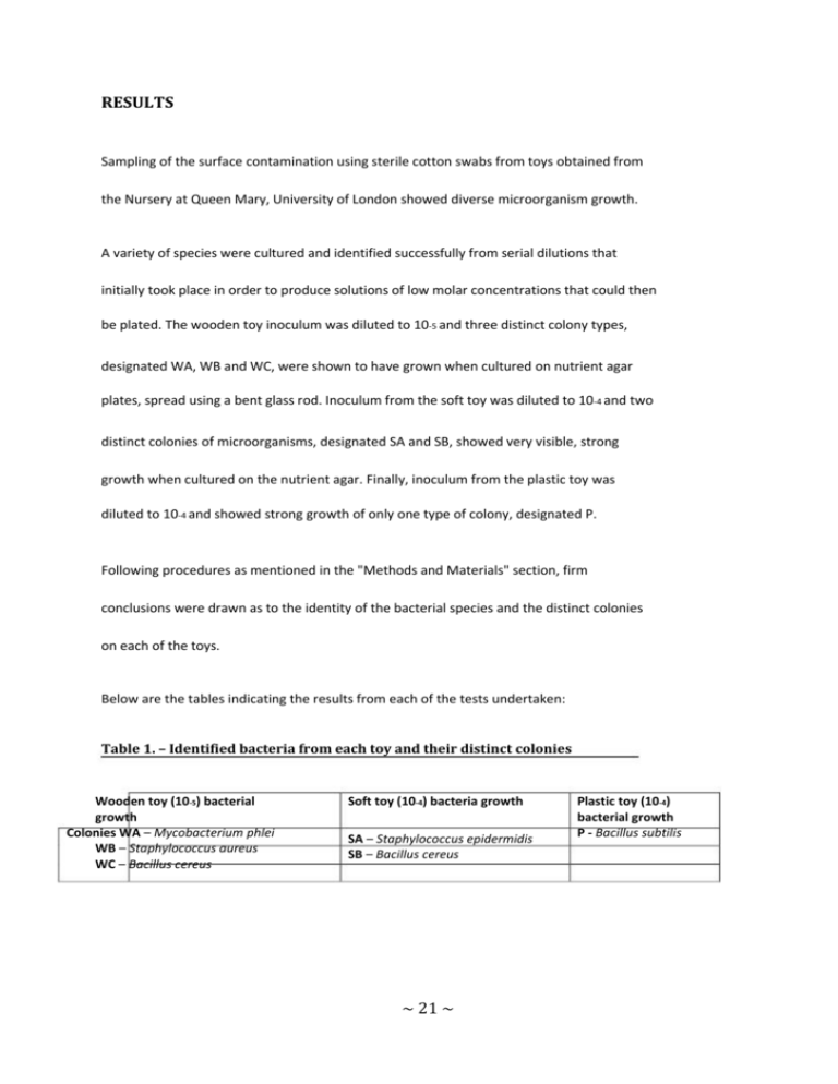
RESULTS Sampling of the surface contamination using sterile cotton swabs from toys obtained from the Nursery at Queen Mary, University of London showed diverse microorganism growth. A variety of species were cultured and identified successfully from serial dilutions that initially took place in order to produce solutions of low molar concentrations that could then be plated. The wooden toy inoculum was diluted to 10-5 and three distinct colony types, designated WA, WB and WC, were shown to have grown when cultured on nutrient agar plates, spread using a bent glass rod. Inoculum from the soft toy was diluted to 10-4 and two distinct colonies of microorganisms, designated SA and SB, showed very visible, strong growth when cultured on the nutrient agar. Finally, inoculum from the plastic toy was diluted to 10-4 and showed strong growth of only one type of colony, designated P. Following procedures as mentioned in the "Methods and Materials" section, firm conclusions were drawn as to the identity of the bacterial species and the distinct colonies on each of the toys. Below are the tables indicating the results from each of the tests undertaken: Table 1. – Identified bacteria from each toy and their distinct colonies Wooden toy (10-5) bacterial growth Colonies WA – Mycobacterium phlei WB – Staphylococcus aureus WC – Bacillus cereus Soft toy (10-4) bacteria growth SA – Staphylococcus epidermidis SB – Bacillus cereus ~ 21 ~ Plastic toy (10-4) bacterial growth P - Bacillus subtilis Table 2. – Results of Gram-staining procedure Gram Stain Gram positive organismsGram negative organisms WB – observed as coccus WC – observed as rods, possible spores SA – observed as coccus SB – observed as rods P – observed as rods, clear sporulation WA was inconclusive WA – Mycobacterium phlei, WB – Staphylococcus aureus, WC – Bacillus cereus SA – Staphylococcus epidermidis, SB – Bacillus cereus, P - Bacillus subtilis Table 3. – Results of Catalase test Catalase test Catalase positive WB WC SA SB P Catalase negative WA WA – Mycobacterium phlei, WB – Staphylococcus aureus, WC – Bacillus cereus SA – Staphylococcus epidermidis, SB – Bacillus cereus, P - Bacillus subtilis ~ 22 ~ Table 4. – Results of Dryspot Staphytect Plus for the identification of Staphylococcus aureus Dryspot Staphytect Plus Positive result WB – agglutination Negative result WC SA SB P WA WA – Mycobacterium phlei, WB – Staphylococcus aureus, WC – Bacillus cereus SA – Staphylococcus epidermidis, SB – Bacillus cereus, P - Bacillus subtilis The visible agglutination implied that this unknown was likely to be Staphylococcus aureus. No visible agglutination from SA (Gram-positive coccus) implied that the unknown was possibly Staphylococcus epidermidis. Table 5. – Results for the growth on Mannitol Salt agar to distinguish between Staphylococcus. Mannitol Salt agar Growth WB – pale yellow colonies observed SA – pink/red colonies observed No growth WC SB P WA WA – Mycobacterium phlei, WB – Staphylococcus aureus, WC – Bacillus cereus SA – Staphylococcus epidermidis, SB – Bacillus cereus, P - Bacillus subtilis Growth on the highly selective mannitol salt agar led to confirmation of unknown WB as Staphylococcus aureus. Unknown SA was confirmed as Staphylococcus epidermidis due to characteristic growth. ~ 23 ~ Table 6. - Results for the growth on Bacillus cereus agar to distinguish between Bacillus Bacillus cereus selective agar Positive resultNegative result SB – pink colonies observedSA WC – pink colonies observedWA P – yellow colonies with slight precipitateWB observed WA – Mycobacterium phlei, WB – Staphylococcus aureus, WC – Bacillus cereus SA – Staphylococcus epidermidis, SB – Bacillus cereus, P - Bacillus subtilis Visible pink/red colonies confirmed the already suspected presence of Bacillus cereus following catalase testing and also viewing the unknowns SB and WC from under the light microscope. Yellow colonies are characteristic of Bacillus subtilis. Table 7. - Results of spore staining and acid fast staining to identify possible Mycobacterium phlei A spore stain was undertaken with unknown colony WA to distinguish it from possible Bacillus cereus. Under the lens of the light microscope it was difficult to differentiate whether the unknown was rod-shaped or cocci-shaped. Following a negative test result, the acid-fast stain was then undertaken to determine whether the unknown was indeed Mycobacterium. Spore stain Acid-fast stain Unknown colony WA Negative result Positive result ~ 24 ~ As shown, Bacillus spp. were found to be common. Bacillus endospores are often found to disperse rapidly through the air and in soils and other environments are an ever-present microorganism (Nicholson, 2002) (Vilain et al,2006) so their abundant presence here was not startling. This is predominantly because the toys that are played with are often moved from the inside and the outside of the nursery premises, by children who are running around. Therefore, if dropped on the ground or more precisely in areas of grass and soil, the toys are able to accumulate such bacteria and these bacteria can then be transferred onto other toys as well as children from inside the nursery. In addition to this, Staphylococcus spp. were also identified as well as Mycobacterium phlei. Bacillus cereus is a prevalent, gram-positive, rod-shaped bacterium that resides in soil. Most commonly, Bacillus cereus is known to be harmful to humans due to their ability to cause illnesses arising from improper preparation of food. Cases of food-borne illnesses predominantly arise due to the production of bacterial endospores produced by the microorganism and their survival (Turnbull, 1996). These endospores, and the general bacterial growth, may then lead to the production of enterotoxins and the onset of nausea, diarrhea and vomiting in humans (Kotiranta et al, 2000). In the case of children within a healthcare environment, the transmission and possible onset of disease is therefore of obvious concern. As acknowledged, the spores of B. cereus can be found widely in the nature around us, including samples of dust, dirt and water amongst others. Normal contamination levels are generally known to be <100/g (Hobbs and Gilbert, 1974) however such levels are unlikely to be found within a carefully and often sanitized child-care environment. ~ 25 ~ Bacillus subtilis, a gram positive rod, is commonly found in plant undergrowth and soil as well, as such, it is often known as the grass Bacillus (Madigan and Martinko, 2005). This particular species of Bacillus is also known to be non-pathogenic in humans. Much like Bacillus cereus, they are acknowledged to contaminate food products but are nowhere near as potentially harmful as Bacillus cereus and very seldom result in food-borne illnesses prior to ingestion of the contaminated foodstuffs (Ryan and Ray, 2004). Therefore, incidence of the bacteria, particularly in a child care environment can be regarded as harmless. Staphylococcus spp. were also cultured and identified. This is a genus of Gram-positive coccus that are said to form in "grape-like" structures when viewed under a light microscope following staining (Ryan and Ray, 2004). It is well understood that most Staphylococcus are harmless and are commonly found to reside on the skin and mucous membranes of humans very comfortably (Kluytmans et al, 1997). It is interesting to note that Staphylococci are microorganisms that also universally dwell in soil and dirt. Staphylococcus aureus in particular is a well known specie of the genus and resides in the nose and on the skin of healthy human beings (Kluytmans et al, 1997). Research has shown that seemingly almost 20% of the human populations are carriers of Staphylococcus aureus (Kluytmans et al, 1997). Stapylococcus epidermidis detected on the surface of the soft toy is also found as part of our skin flora and therefore it is understood to be, for the most part, harmless. There are exceptional circumstances however such as in humans that are immuno-compromised (Salyers and Whitt, 2002). Furthermore, due to various methods of contamination, it is known to be probably the most common microorganism species found when performing tests within a laboratory (Queck and Otto, 2008). ~ 26 ~ Mycobacterium phlei was the unknown identified last during the research undertaken. Being Mycobacterium it was initially difficult to classify as simple staining, such as that of the gram stain, does not provide good, conclusive results as to whether this particular unknown was indeed gram positive or negative. The reason because of this is due to the presence of the long chain fatty acid, mycolic acid, within the cells walls of bacterium of the particular the genus (Vance et al, 1973). Mycobacterium phlei, much like Bacillus subtilis is also found principally on grass and more importantly is known to be non-pathogenic in humans. From the results gathered and obtained from laboratory experimentation it is evident that amongst almost all the unknowns identified there is no immediate threat to the children of the child-care environment. Bacillus cereus poses the most cause for concern as it is a well defined pathogen in humans causing only a small minority of all food-borne illnesses (Kotiranta et al, 2000). However, it should also be understood that the bacteria that were found to be present on the surface of the toys are ever-present whether they remain a part of our external environment or even our internal environment making up much of our normal human flora. ~ 27 28 ~
