Idosi Publications
advertisement
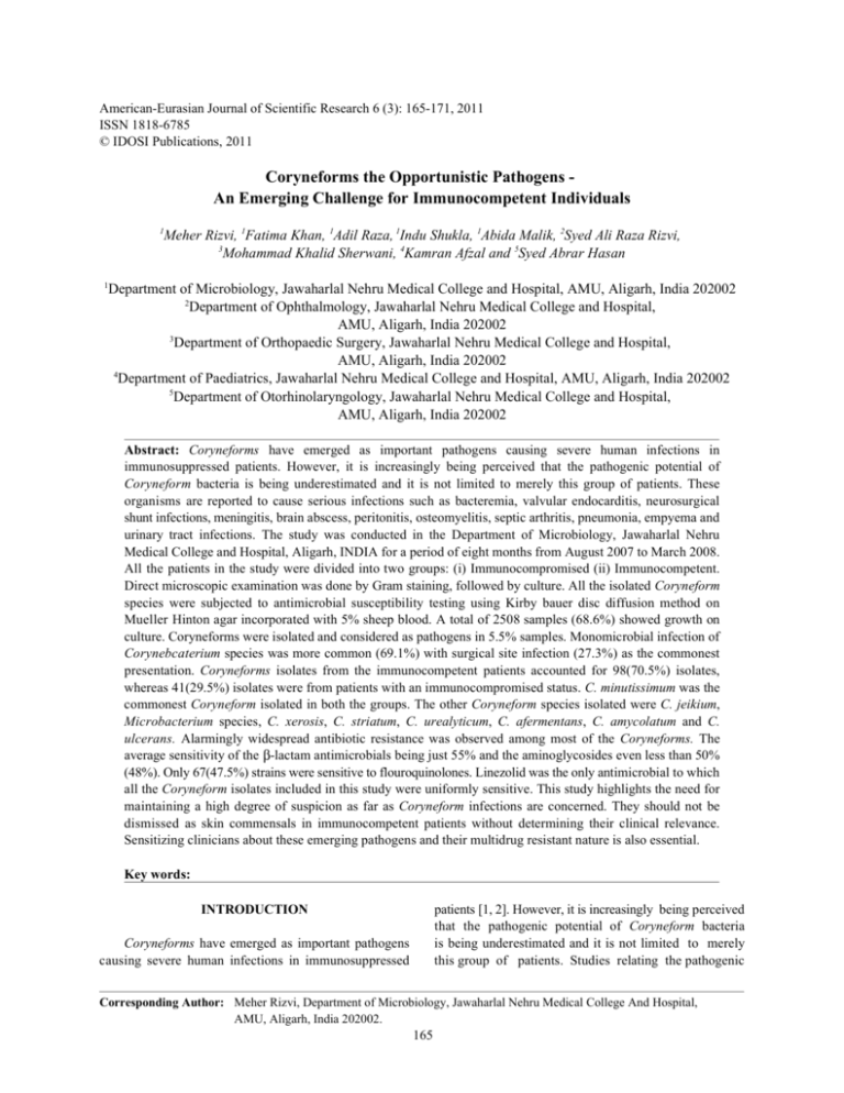
American-Eurasian Journal of Scientific Research 6 (3): 165-171, 2011 ISSN 1818-6785 © IDOSI Publications, 2011 Coryneforms the Opportunistic Pathogens An Emerging Challenge for Immunocompetent Individuals 1 1 Meher Rizvi, 1Fatima Khan, 1Adil Raza, 1Indu Shukla, 1Abida Malik, 2Syed Ali Raza Rizvi, 3 Mohammad Khalid Sherwani, 4Kamran Afzal and 5Syed Abrar Hasan Department of Microbiology, Jawaharlal Nehru Medical College and Hospital, AMU, Aligarh, India 202002 2 Department of Ophthalmology, Jawaharlal Nehru Medical College and Hospital, AMU, Aligarh, India 202002 3 Department of Orthopaedic Surgery, Jawaharlal Nehru Medical College and Hospital, AMU, Aligarh, India 202002 4 Department of Paediatrics, Jawaharlal Nehru Medical College and Hospital, AMU, Aligarh, India 202002 5 Department of Otorhinolaryngology, Jawaharlal Nehru Medical College and Hospital, AMU, Aligarh, India 202002 Abstract: Coryneforms have emerged as important pathogens causing severe human infections in immunosuppressed patients. However, it is increasingly being perceived that the pathogenic potential of Coryneform bacteria is being underestimated and it is not limited to merely this group of patients. These organisms are reported to cause serious infections such as bacteremia, valvular endocarditis, neurosurgical shunt infections, meningitis, brain abscess, peritonitis, osteomyelitis, septic arthritis, pneumonia, empyema and urinary tract infections. The study was conducted in the Department of Microbiology, Jawaharlal Nehru Medical College and Hospital, Aligarh, INDIA for a period of eight months from August 2007 to March 2008. All the patients in the study were divided into two groups: (i) Immunocompromised (ii) Immunocompetent. Direct microscopic examination was done by Gram staining, followed by culture. All the isolated Coryneform species were subjected to antimicrobial susceptibility testing using Kirby bauer disc diffusion method on Mueller Hinton agar incorporated with 5% sheep blood. A total of 2508 samples (68.6%) showed growth on culture. Coryneforms were isolated and considered as pathogens in 5.5% samples. Monomicrobial infection of Corynebcaterium species was more common (69.1%) with surgical site infection (27.3%) as the commonest presentation. Coryneforms isolates from the immunocompetent patients accounted for 98(70.5%) isolates, whereas 41(29.5%) isolates were from patients with an immunocompromised status. C. minutissimum was the commonest Coryneform isolated in both the groups. The other Coryneform species isolated were C. jeikium, Microbacterium species, C. xerosis, C. striatum, C. urealyticum, C. afermentans, C. amycolatum and C. ulcerans. Alarmingly widespread antibiotic resistance was observed among most of the Coryneforms. The average sensitivity of the -lactam antimicrobials being just 55% and the aminoglycosides even less than 50% (48%). Only 67(47.5%) strains were sensitive to flouroquinolones. Linezolid was the only antimicrobial to which all the Coryneform isolates included in this study were uniformly sensitive. This study highlights the need for maintaining a high degree of suspicion as far as Coryneform infections are concerned. They should not be dismissed as skin commensals in immunocompetent patients without determining their clinical relevance. Sensitizing clinicians about these emerging pathogens and their multidrug resistant nature is also essential. Key words: INTRODUCTION patients [1, 2]. However, it is increasingly being perceived that the pathogenic potential of Coryneform bacteria is being underestimated and it is not limited to merely this group of patients. Studies relating the pathogenic Coryneforms have emerged as important pathogens causing severe human infections in immunosuppressed Corresponding Author: Meher Rizvi, Department of Microbiology, Jawaharlal Nehru Medical College And Hospital, AMU, Aligarh, India 202002. 165 Am-Euras. J. Sci. Res., 6 (3): 165-171, 2011 potential of Coryneforms to the immunocompetent individuals have proved them as well established pathogens [3,4]. Several species such as Corynebacterium xerosis, Corynebacterium amycolatum, Corynebacterium striatum, Corynebacterium minutissimum, Corynebacterium pseudodiphtheriticum, Corynebacterium matruchotii, Corynebacterium aquaticum, Corynebacterium genitalium and Corynebacterium pseudogenitalium have been related to human infections. Corynebacterium jeikeium and Corynebacterium urealyticum are well established human pathogens exhibiting resistance to several antibiotics [5, 6]. These organisms are reported to cause serious infections such as bacteremia, valvular endocarditis, neurosurgical shunt infections, meningitis, brain abscess, peritonitis, osteomyelitis, septic arthritis, pneumonia, empyema and urinary tract infections [7]. However, reports of the epidemiology of Coryneforms from developing countries are few. Coryneforms appear to be significant pathogens of tropical countries rather than of temperate areas [8]. There is sparse data from India from where there are only a few case reports [9, 10]. Therefore, this broad based study was designed to study the role of Coryneforms as pathogens in immunocompetent along with the immunocompromised individuals and to evaluate the antimicrobial susceptibilities of different genera and species of Coryneforms in diverse clinical settings. considered to be immunocompromised when they had history of HIV, malignancy, diabetes mellitus or any other chronic disease. Patients without any such history, but age greater than 60 years or a differential cell count showing leucopenia were considered immunocompromised. A total of 987 patients were enrolled in this group. (ii) Immunocompetent: patients without an underlying chronic disease and didn’t give any of the above mentioned history were considered to be immunocompetent. A total of 2666 patients were included in this group. Direct microscopic examination was done by Gram staining, followed by culture on 5% sheep blood agar, MacConkey agar and enrichment was done in brain heart infusion broth with 1% serum. Incubation was done for 18-24 hours at 37°C. Isolated Corynebacteria species were considered pathogenic when at least one of the following criteria were met: (I) Clinical history of inflammation, tenderness, purulent discharge with or without fever, (ii) in cases of superficial samples, isolation at least 3 times in samples taken at 3 different times and the presence of polymorphonuclear neutrophils on Gram’s staining, (iii) isolation from a deep sample, (iv) in any kind of sample, the presence of Gram positive bacilli within polymorphonuclear neutrophils on gram’s staining [11]. Identification of Coryneform species was done using standard biochemical methods [12]. All the isolated Coryneform species were subjected to antimicrobial susceptibility testing using Kirby bauer disc diffusion method on Mueller Hinton agar incorporated with 5% sheep blood [13]. The antimicrobial agents used were ampicillin 10µg, oxacillin 1µg, cefazolin 30µg, erythromycin 15µg, gentamicin 10µg, amikacin 30µg, chloramphenicol 30µg, tetracycline 30µg, ofloxacin 5µg, vancomycin 30µg, teicoplanin 30µg and linezolid 30µg. In Staphylococcal isolates oxacillin (1µg) for the detection of MRSA. Among the Gram negative bacilli screening of possible ESBL production was done by using ceftriaxone (30µg) and cefoperazone (75µg). Those isolates with zone diameters less than 25mm for ceftriaxone and less than 22mm for cefoperazone were subsequently confirmed for ESBL production. Confirmation was done by noting the potentiation of the activity of cefoperazone in the presence of cefoperazone sulbactum [14]. Detection of AmpC betalactamase was done on isolates resistant to ceftriaxone (30µg), cefixime (15µg), cefoperazone (75µg) and cefoperazone sulbactum (75/75µg). Induction of AmpC synthesis was based on the disc approximation assay using imipenem as inducer [14]. MATERIALS AND METHODS The study was conducted in the Department of Microbiology, Jawaharlal Nehru Medical College and Hospital, Aligarh, INDIA for a period of eight months from August 2007 to March 2008. Detailed clinical history was elicited from the patients and depending on the type of infection; various specimens like cerebrospinal fluid (meningitis and encephalitis), pus, urine (urinary tract infection- UTI), sputum (lower respiratory tract infectionLRTI), throat swabs (upper respiratory tract infectionURTI), eye swabs (conjunctivitis), ear swabs (chronic suppurative otitis media- CSOM), cervical swabs (cervicitis), surgical wound swabs (surgical site infection), joint fluid (synovitis), ascetic fluid, pleural fluid, drains and catheter samples were included in the study making a total of 3653 samples. Empiric treatment was started whenever necessary. All the patients in the study were divided into two groups: (I) Immunocompromised: patients were 166 Am-Euras. J. Sci. Res., 6 (3): 165-171, 2011 Ethical clearance was obtained from the Institutional Ethical Committee of Jawaharlal Nehru Medical College. Informed and written permission was obtained from all patients. Statistical analysis was done using the Student’s t test, chi square test and unpaired Student’s t test. RESULTS A total of 2508 samples (68.6%) showed growth on culture. Staphylococcus aureus (27.9%) was the commonest pathogen isolated during the study, followed by Escherichia coli (21.1%), Pseudomonas aeruginosa (15.9%) and Klebsiella pneumoniae (11.6%). Coryneforms were isolated in (6.8%) of samples, however, (5.5%) of these strains were considered as pathogens and were included in the study (Figure 1). Monomicrobial infection of Corynebcaterium species was more common with 96 (69.1%) isolates seen in pure culture and 43 (30.9%) Coryneform isolates were associated with polymicrobial infection (Table 1). Coryneforms were isolated in 3.7% of the immunocompetent individuals and in 4.1% of the immunocompromised individuals. Coryneforms isolates from the immunocompetent patients accounted for 98(70.5%) isolates, whereas 41(29.5%) isolates were from patients with an immunocompromised status (Table 2). Amongst the immunocompromised patients, 19(46.3%) were ICU patients, 13(31.7%) had malignancies, 5(12.2%) were neonates admitted in nursery (one with symptoms of meningitis, one with meningoencephalitis and 3 were preterm babies) and 4(9.7%) had diabetes mellitus. Fig. 1: Distribution of Various Pathogens Isolated from the Clinical Samples CONS- Coagulase negative Staphylococcus species Surgical site infection (27.3%) was the commonest presentation in patients with Coryneform infection, followed by device related infections (11.5%), soft tissue infections (10.1 %) and chronic bone and joint infection (8.6%) and UTI (8.6 %). The lowest isolation rate of Coryneform species was from patients with meningitis and breast abscess (1.4%) (Table 3 Please revise Table 3). Amongst both the immunocompetent as well as immunocompromised individuals C. minutissimum was the commonest Coryneform isolated (22.3%). The other Coryneform species isolated were C. jeikium (17.9%), Microbacterium species (8.6%), C. xerosis (7.9%), C. striatum (7.2%), C. urealyticum (6.5%), C. afermentans (5.7%), C. amycolatum (2.9%) and C. ulcerans (1.4%) (Table 2). In most of the patients with surgical site infections, C. minutissimum was the commonest isolate Table 1: Distribution of Coryneform Species in Pure and Mixed Cultures Coryneform species isolated No. (%) In Pure culture Along with one organism Along with two organisms Total 96 (69.1) 23 (16.6) 20 (14.4) 139 (100) Table 2: Isolation of Corynefrom Species in Relation to Immune Status of the Patient Coryneform species Immunocompetent No. (%) C.minutissimum C. jeikeium Microbacterium sp C.xerosis C.striatum C.urealyticum C.afermentans C. amycolatum C.ulcerans Others Total 17(54.8) 12(48) 10(83.3) 8(72.7) 8(80) 9(100) 8(100) 3(75) 2(100) 21(77.8) 98 (70.5) Immunocompromised No. (%) 14(45.2) 13 (52) 2(16.7) 3(27.3) 2(20) 1(25) 6(22.2) 41(29.5) 167 Total No. (%) 31(22.3) 25(18.0) 12(8.6) 11(7.9) 10(7.2) 9(6.5) 8(5.7) 4(2.9) 2(1.4) 27(19.4) 139(100) Am-Euras. J. Sci. Res., 6 (3): 165-171, 2011 Table 3: Isolation of Coryneform Species in Relation to Clinical Presentation in Patients Coryneform species Osteomyelitis Chronic bone and Surgical Urinary joint infection site infections Tract Infections Breast Meningitis Device related abscess Peritonitis infection C. minutissimum 3 1 15 1 - 1 - C. jeikeium 5 4 5 1 - 1 5 2 Microbacterium sp - 1 7 - - - - 1 C. xerosis - 2 - 5 - - 1 1 C. striatum - 2 4 - - - - 3 C. urealyticum - - 2 3 - - 2 - C. afermentans - - - - - - - 4 C. amycolatum 1 - 1 - - - - 2 C. ulcerans - - - - - - - - Others 1 2 4 2 2 - - 2 Total 10(7.2) 12(8.6) 38(27.3) 12(8.6) 2(1.4) 2(1.4) 8(5.7) 16(11.5) Cervicitis Umblical tip Total No.(%) 1 to be continued Coryneform Soft tissue UpperRespiratory Lower Respiratory chronic suppurative species infection Tract Infection Tract Infection otitis media Conjunctivitis C. minutissimum 8 - - - - 1 - 31(22.3) C. jeikeium - - - - - - 2 25(18.0) Microbacterium sp - - - 1 1 - 1 12(8.6) C. xerosis 1 - - - - - 1 11(7.9) C. striatum 1 - - - - - - 10(7.2) C. urealyticum 1 - - - - - 1 9(6.5) C. afermentans - - - 4 - - - 8(5.7) C. amycolatum - - - - - - - 4(2.9) C. ulcerans - 1 1 - - - - 2(1.4) Others 3 4 2 2 2 - 1 27(19.4) Total 14(10.1) 5 (3.6) 3 (2.1) 7 (5.0) 3 (2.1) 1 (0.7) 6 (4.3) 139 (100) Table 4: Antibiotic Susceptibility Pattern of Clinically Important Coryneforms Coryneform species To t a l Ampicillin Oxacillin Cefazolin Erythromycin Gentamicin Amikacin Ofoxacin Vancomycin Teicoplanin Linezolid No.(%) C. minutissimum 8 10 9 15 4 8 6 24 24 31 31(22.3) C. jeikeium 8 8 0 9 8 12 19 22 22 25 25(18.0) Microbacterium sp 0 5 4 5 4 6 3 11 11 12 12(8.6) C. xerosis 11 11 9 11 9 11 11 11 11 11 11(7.9) C. striatum 4 5 2 7 4 3 3 10 10 10 10(7.2) C. urealyticum 5 9 9 1 2 3 2 9 9 9 9(6.5) C. afermentans 8 8 6 8 8 8 8 8 8 8 8(5.7) C. amycolatum 3 4 3 3 2 3 2 4 4 4 4(2.9) C. ulcerans 2 2 2 2 2 2 2 2 2 2 2(1.4) Others 25 27 27 22 16 21 11 27 27 27 27(19.4) Total 74(52.5)53.2 89(63.1) 71(50.4) 83(58.9) 59(41.8) 77(54.6) 67(47.5) 128(92.1) (39.5%), while in patients with osteomyelitis, C. jeikium was the most common (50% revise??). UTI was predominantly caused by C. xerosis (41.7%) and C. urealyticum (25%) (Table 3). Results of the antimicrobial susceptibility test showed that among the S. aureus isolates, 410 (58.6%) were MSSA and 290 (41.4%) were MRSA. In case of the Gram-negative bacilli, 750(50.5%) were ESBL-producers and 260(17.5%) were AmpC producers. 128(92.1) 139(100) 139(100) Alarmingly widespread antibiotic resistance was observed among most of the Coryneforms (Table 4). Amongst the -lactam group of antimicrobials, oxacillin was found to have the best susceptibility profile with (63.1%) sensitivity, followed by ampicillin (52.5%) and cefazolin (50.4%). Amikacin was better among the aminoglycosides with around 13% higher sensitivity than gentamicin [Amikacin- (54.6%), gentamicin- (41.8%)]. Flouroquinolones had even poorer susceptibility than the 168 Am-Euras. J. Sci. Res., 6 (3): 165-171, 2011 other group of drugs with only 67(47.5%) strains being sensitive. The susceptibility to glycopeptides (teicoplanin and vancomycin) though not 100% but was significantly higher 128(92.1%) than the other antimicrobials. Maximum overall resistance was observed in C. minutissimum and C. jeikeium. Glycopeptide resistance was observed in 7(22.6%) strains of C. minutissimum, 3 (12%) strains of C.jeikium and 1 (8.3%) isolate of Microbacterium sp. Linezolid was the only drug to which all the Coryneform strains were uniformly sensitive (Table 4). Patients with Coryneform infection recovered after empiric treatment was replaced by specific antimicrobials on the basis of susceptibility profile. DISCUSSION Corynebacterium species are widely distributed in the environment and are members of skin and mucous membranes [15, 16]. However, in the recent years, recognition of infections with nondiphtheritic Corynebacterium species has increased and much new information has become available on many of them. They are usually considered opportunistic pathogens in patients with poor general condition [1, 3]. Few studies have dealt with Coryneform infection in healthy immunocompetent people [3, 4]. In this study, we assessed the role of Coryneform infection among both immunocompetent as well as immunocompromised individuals. Continue with the following paragraph. Coryneforms were isolated in (6.8%) of the total samples processed. However, only (5.5%) of these were considered pathogenic and were included in the study. Significantly large number of Coryneforms [nearly two-third 98(70.5%)] were isolated from immunocompetent patients. Immunocompromised patients accounted for 41(29.5%) of the Coryneform isolates. This finding highlights the role of Coryneforms as emerging pathogens in otherwise healthy individuals. C. minutissimum was the most common isolate in our study and it predominantly caused infection in immunocompetent people (22.3%). The other commonly isolated Coryneforms were C. jeikeium, Microbacterium sp., C. xerosis and C. striatum. C.jeikeium was isolated as the most common corynefrom from orthopaedics and other surgical site infections in another study from our hospital from 2007-2009 [17]. Amongst these, C. jeikeium infection was associated more commonly with immunosuppressed patients, while the others infected mostly immunocompetent patients. 169 However other authors have quoted C.striatum as asn emerging nosocomial pathogen particularly in the immunocompromised individuals[18, 19]. In the present study, the majority of the patients with Coryneform infection had surgical site infection (27.3%) and orthopedic related complaints (15.8%). Two unusual cases of meningitis in immunocompetent patients were noticed in our study. In one the incriminatory pathogen was Rhodococcus equii and Corynebacterium aquaticum in the other. Both were vancomycin sensitive. One study has reported vancomycin resistant C. aquaticum [8]. This study highlights the alarming problem of drug resistance among Coryneforms. The average sensitivity of the -lactam antimicrobials being just 55% and the aminoglycosides even less than 50% (48%). The fluoroquinolones were found to have extremely poor activity against the Coryneforms which another study had also reported [20]. Along with resistance to other antimicrobial agents, vancomycin and teicoplanin resistance was observed in 7 strains of C. minutissimum (22.6%), 3(12%) isolates of of C. jeikeium and one (8.3%) strain Microbacterium sp. Multidrug resistant character of these bacteria have also been observed in another study [8]. Whereas some studies have quoted uniform sensitivity to the glycopeptides antibiotics [18, 21]. C. minutissimum and C. jeikeium were the least sensitive to the various group of drugs followed by Microbacterium sp, C. urealyticum, C. amycolatum and C. striatum. Their multidrug resistant character yet again stresses the need to report these Coryneforms. It was observed that those bacteria which were vancomycin resistant were also predominantly resistant to other antimicrobials. Linezolid was the only antimicrobial to which all the Coryneform isolates included in this study were uniformly sensitive. However until definitive results of susceptibility testing are available vancomycin will continue to be the most recommended treatment for severe Corynebacterium infection [22]. This study highlights the need for maintaining a high degree of suspicion as far as Coryneform infections are concerned. They should not be dismissed as skin commensals in immunocompetent patients without determining their clinical relevance. We believe that for routine laboratory testing the simple criteria outlined by Funke [5] for clinical relevance should be followed. Sensitizing clinicians about these emerging pathogens and their multidrug resistant nature is also essential. Another cause of concern is the possibility of transfer of drug resistance to Staphylococcus as they often share some ecological niche. Am-Euras. J. Sci. Res., 6 (3): 165-171, 2011 Our assessment of Corynebacterium isolates showed that several Corynebacterium species exhibiting unpredictable antimicrobial resistance might be encountered in patients with varied clinical manifestations. Much more work needs to be done on this enlarging group of this emerging bacteria in order to understand their epidemiology in immunocompetent people, their resistance profile and their virulence potential. 9. 10. 11. REFERENCES 1. 2. 3. 4. 5. 6. 7. 8. Mikucka, A., E. Gospodarek and M. Bialek, 1997. Opportunistic infections with Coryneform. Medical Science Monitor, 3: 154-157. Make references like this style. Young, V.M., W.F. Meyers, M.R. Moody and S.C. Schimpff, 1981. The emergence of Coryneform bacteria as a cause of nosocomial infections in compromised hosts. American Journal of Medicine, 70: 646-650. Watkins, D.A., A. Chahine and R.J. Creger, 1993. Corynebacterium striatum: a diphtheroid with pathogenic potential. Clinical Infectious Diseases, 17: 21-25. Roux, V., M. Drancourt, A. Stein, P. Riegel, D. Raoult and B. La Scola, 2004. Corynebacterium Species Isolated from Bone and Joint Infections identified by 16Sr RNA Gene sequence analysis. Journal of Clinical Microbiology, pp: 2231-2233. Funke, G. and K.A. Bernard, 1999. Coryneform Gram Positive Rods. In P.R. Murray, E.J. baron, M.A. Pfaller, C. Tenover, R.H. Yolken (eds.). Manual of Clinical Microbiology, 7th ed. ASM Press. Washington, DC., pp: 319-345. Claeys, G., G. Vershchraegen, L. DeSmet, R. Verdonk and H. Claessens, 1986. Corynebacterium JK (Johnson-Kay strain) infection of a Kuntsner-nailed tibial fracture. Clinical Orthopaedics and Related Research, 202: 227-229. Brown, A.E., 2000. Other Corynebacteria and Rhodococcus. In G.L. Mandell, J.E. Bennet and R. Dolin, eds. Principles and practice of infectious diseases. 5th edn, Philedelphia: Churchill Livingstone, pp: 2198-2208. Camello, T.C.F., G.A.L. Mattos, L.C.D. Formiga and E.A. Marquez, 2003. Nondiphtherial Corynebacterium species isolated from clinical specimens of patients in a university hospital, Reo De Janeiro, Brazil. Brazilian Journal of Micorbiology, 34: 39-44. 12. 13. 14. 15. 16. 17. 18. 19. 170 Ciraj, A.M., K. Rajani, G. Sreejith, K.L. Shobha and P.S. Rao, 2006. Urinary Tract Infection due to Arcanobacterium haemolyticum. Indian Journal of Medical Microbiology, 24: 300. Parija, S.C., V. Kaliaperumal, S.V. Kumar, S. Sujatha and V. Babu, 2005. Arcanobacterium haemolyticum associated with pyothorax: case report. BMC Infectious Diseases, pp: 5-68. Horan, T.C., M. Andrus and M. Dudeck, 2008. CDC/NHSN surveillance definition of healthcare-associated infection and criteria for specific types of infections in acute care settings. American Journal of Infection Control, 36: 309-332. Tallman, P., E. Muscare, P. Carson, W.H. Eaglstein and V. Falanga, 1997. Initial rate of healing predicts complete healing of venous ulcers. Archives of Dermatology, 133: 1231-1234. Van Rijswijk, L., 1993. Full thickness leg ulcers: patient demographics and predictors of healing. Multi Center Leg Ulcer Study Group. The Journal of Family Practice, 36: 625-632. Rizvi, M., N. Fatima, M. Rashid, I. Shukla, A. Malik, A. Usman and S. Siddiqui, 2009. Extended spectrum AmpC and metallo-beta-lactamases in Serratia and Citrobacter spp. in a disc approximation assay. Journal of Infection in Developing Countries, 3(4): 285-94. Coyle, M.B. and B.A. Lipsky, 1990. Coryneform bacteria in infectious diseases: Clinical and laboratory aspects. Clinical Microbiology Reviews, 3: 227-246. Clarridge, J.E. and C.A. Spigel, 1995. Corynebacterium and miscellaneous irregular gram positive rods, Erysipelothrix and Gardnerella. In: P.R. Murray, E.J. Baron, M.A. Pfaller, F.C. Tenover and R.H. Yolken (eds), Manual of Clinical Microbiology. Washington DC. Am Soc. Microbiol., pp: 357-378. Rizvi, M., F. Khan, R. Adil, I. Shukla and A.B. Sabir, 2011. Emergence of coryneforms in osteomyelitis and orthopaedic surgical site infections. AMJ., 4(7): 412-418. Otsukaab, Y., K. Ohkusuua, Y. Kawamuraa, S. Babaa, T. Ezakia and S. Kimurao, 2006. Emergence of multidrug resistant Corynebacterium striatum as a nosocomial pathogen in long term hospitaised patients with underlying diseases. Diagnostic Microbiol. Infect Dis., 54(2): 109-114. Marull, J. and P.A. Casares, 2008. nosocomial valve endocarditis due to Corynebacterium striatum: a case report. Cases Journal, 1: 388. Am-Euras. J. Sci. Res., 6 (3): 165-171, 2011 20. Soriano, F.R., R. Hernandez-Roblas, J. Zapardiel, J.L. Rodiriguez-Tudda, P. Aviles and M. Romero, 1989. Increasing incidence of Corynebacterium. EM Journal of Clinical Microbiological Infectious Diseases, 8: 117-118. 21. Funke, G., V. Punter and A. Von-Graevemitz, 1996. Antimicrobial susceptibility patterns of some recently established coryneform bacteria. Antimicrob Agents chemoth, 40(12): 2874-2878. 22. Williams, D.Y., S.T. Selepak and V.J. Gill, 1993. Identification of clinical isolates of non diphtherial Corynebacterium species and their antibiotic susceptibility patterns. Diagnostic Microbiology and Infectious Diseases, 17: 23-28. 171
