Circulatory System Review - Le site web de M. St Denis
advertisement
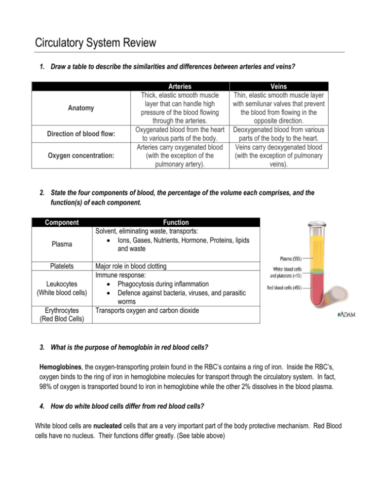
Circulatory System Review 1. Draw a table to describe the similarities and differences between arteries and veins? Anatomy Direction of blood flow: Oxygen concentration: Arteries Thick, elastic smooth muscle layer that can handle high pressure of the blood flowing through the arteries. Oxygenated blood from the heart to various parts of the body. Arteries carry oxygenated blood (with the exception of the pulmonary artery). Veins Thin, elastic smooth muscle layer with semilunar valves that prevent the blood from flowing in the opposite direction. Deoxygenated blood from various parts of the body to the heart. Veins carry deoxygenated blood (with the exception of pulmonary veins). 2. State the four components of blood, the percentage of the volume each comprises, and the function(s) of each component. Component Plasma Platelets Leukocytes (White blood cells) Erythrocytes (Red Blod Cells) Function Solvent, eliminating waste, transports: Ions, Gases, Nutrients, Hormone, Proteins, lipids and waste Major role in blood clotting Immune response: Phagocytosis during inflammation Defence against bacteria, viruses, and parasitic worms Transports oxygen and carbon dioxide 3. What is the purpose of hemoglobin in red blood cells? Hemoglobines, the oxygen-transporting protein found in the RBC’s contains a ring of iron. Inside the RBC’s, oxygen binds to the ring of iron in hemoglobine molecules for transport through the circulatory system. In fact, 98% of oxygen is transported bound to iron in hemoglobine while the other 2% dissolves in the blood plasma. 4. How do white blood cells differ from red blood cells? White blood cells are nucleated cells that are a very important part of the body protective mechanism. Red Blood cells have no nucleus. Their functions differ greatly. (See table above) 5. List three major proteins found in blood and list their function. Protein Function Albumins 1. Maintain osmotic pressure by drawing water back into capillaries to help maintain body fluid levels. 2. Transport smaller molecules such as hormones and ions Globulins 1. Help protect again invading microbes by binding to foreign substances 2. Transport hormones and fat soluble vitamins Fibrinogens 1. Converted into fibrin networks that form blood clots. 6. What is anemia. What is the cause of this condition? Anemia is a condition resulting in a decrease in the ability to transport oxygen in the blood. A deficiency in either hemoglobine or in red blood cells decreases the oxygen transported to the tissue. This condition is characterised by weakness and fatigues. People suffering from anemia often: i. Are pale and end to faint easily ii. Become short of breath easily iii. Have an increased heart rate i. This is to offset the reduction in the oxygen in the oxygen carrying capacity of the blood. 7. “Oxygenated blood is found in all arteries of the body”. Is this statement true or false? Provide a reason to explain your answer. This statement is false. In the pulmonary circuit, the blood found in the arteries is low in oxygen and high in carbon dioxide. This is the only location where arteries contain deoxygenated blood. 8. Why do you think the left ventricle has more muscle than the right ventricle does? The left ventricle is thicker and more muscular than the right ventricle because it pumps blood at a higher pressure. It must send the blood to more locations around the body, whereas the right ventricle pumps blood to the lungs. 9. Draw a schematic diagram of the heart. Label all of the heart’s parts 10. Which chamber receives blood from the tissue of the body? The right atrium receives deoxygenated blood from the tissue of the body 11. Explain how the structure of a vein contributes to its function in the circulatory system. Valves are found in the veins that steer the blood towards the heart as they only open in one direction. This is needed since the smooth muscle found in the veins does not rival the ones found in the arteries. Skeletal muscles also aid the blood flow. The pressure in the veins increase as the sequential skeletal muscles contractions push against the vein and reduce its diameter allowing the blood to return towards the heart. 12. How does exercise change your heart rate? Provide and explanation for any changes. Your heart rate changes during exercise, generally increasing in relation to how hard you're exercising. When you start to move during exercise, your muscles require more oxygen. They get that oxygen from your blood, which your heart starts to pump at a higher rate to fuel your muscles. 13. Differentiate between systolic and diastolic blood pressure Systolic Pressure: The peak pressure at the moment the ventricles contract. This is the higher number of the two in a blood pressure reading. Normal systolic pressure for a young adult is said to be approx 120 mm Hg. Diastolic Pressure: The pressure at the moment the heart relaxes to let the heart ventricles fill again. This is the lower number of the tow in a blood pressure reading. Normal diastolic pressure for a young adult is said to be between 70 and 80 mm Hg. 14. Explain why we say that the circulatory system contains two circuits. Two types of circulation in the body The first goes to the lungs and is called the pulmonary circuit or the pulmonary circulation. Deoxygenated blood leaves the right ventricle of the heart and travels through the pulmonary artery to the lungs where blood is oxygenated. Blood then returns to the left atrium of the heart by pulmonary veins. The other main circulation in the body is called the systemic circuit or the systemic circulation. In this circulation, blood travels from the left ventricle of the heart and goes to tissues all over the body. 15. What are the functions of the valves in the heart? The human heart contains four valves that control the direction of blood flow, ensuring a steady flow from the atria to the ventricles and from the ventricles the blood vessels. These valves are one way valves that open when blood pressure builds on one side and close when it increases on the other. Atrioventricular Valves The right atrioventricular valve between the right atrium and the right ventricle is called the tricuspid valve because it contains three flaps. The left atrioventricular valve between the left atrium and the left ventricle is called the bicuspid valve because it contains three flaps. Semilunar Valves Between the ventricles and the arteries are the semilunar valves. These valves consist of three semicircular flaps of tissue. When the ventricles contract, this forces the semilunar valves open. Blood flow from the ventricles to the large arteries, and the backflow of blood causes the valve to close preventing any blood to return to the ventricles. 16. What are the function of capillaries Capillaries are the smallest blood vessels in the body, and are the blood vessels responsible for actually delivering oxygen and other nutrients to the tissues. Structure: Capillaries are very small. They are so small that you cannot see them without a microscope. In most cases, the walls of capillaries are only one or two cells thick, and they are so narrow that blood cells have to line up in single file to pass through them. Function: Capillaries are the site where oxygen and other nutrients in the blood are actually delivered to the tissues of the body. Capillaries are so small that these substances actually pass right through them via a process known as diffusion. 17. Explain how the circulatory system depends on the respiratory system. In simple terms, the respiratory system and the circulatory system rely on each other. The lungs bring the oxygen to the blood supply and the blood supply then sends the oxygen throughout the body to all the vital organs and tissues. The blood supply also picks up the leftover blood that has used up all its oxygen and brings it back to the lungs to start the process over again. 18. What is stroke volume? How do you measure it? Does it change/vary? Explain. Stroke Volume: the volume of blood ejected from the left ventricle in a single beat. Stroke Volume: Typical SV rest values: ~ 70 ml/beat During exercise SV ’s: ~ 150 – 300 ml/beat (depends on intensity) SV (ml) = LVEDV(ml) – LVESV(ml) LVEDV: Left ventricular end-diastolic volume The amount of blood remaining in the left ventricle when it is relaxed i.e. the volume when the ventricle is “full” LVESV: Left ventricular end-systolic volume The amount of blood remaining in the left ventricle after it contracts i.e. the volume when the ventricle is “empty” SV is Regulated by 3 factors Strength of ventricular contractions (larger contraction = SV by LVESV) (heart pumps harder = less blood in ventricle) Aortic blood pressure (larger BP = SV) LVEDV (Larger LVEDV = SV) 19. Why is it important to be aware of your MHP and your THR? The maximum heart rate (MHR) is the highest heart rate an individual can achieve without severe problems through exercise stress, and depends on age. Your Target Heart Rate is a set heart rate that you want to achieve while exercising in order to facilitate cardio-vascular improvement, and maximize benefits. By knowing these two important rates, and individual can track his/her own rate in order to achieve significant health benefits without being at risk of severe heart risks due to exercise. 20. How does the cardiovascular system respond to physical activity? HR ’s Heart contractility ’s Heart pumps harder = _’ed blood pumped per beat Blood vessel diameter changes Vasoconstriction in non-working muscle Vasodilation in working muscle Blood pressure ’s getting blood where it needs to go quickly Distribution of blood changes 21. Name the four factors that contribute to an increase of venous return. 1. Vasoconstriction: Pushes blood back to heart Directs blood away from areas not in use 2. Skeletal muscle pump: Muscles contract and squeeze blood up towards heart 3. Thoracic pump Pressure increases in thoracic cavity pushing blood back to heart 4. Sympathetic Drive Nervous stimulation of the heart causes an _ in HR and force of contraction
