The Vision System and the Brain I
advertisement
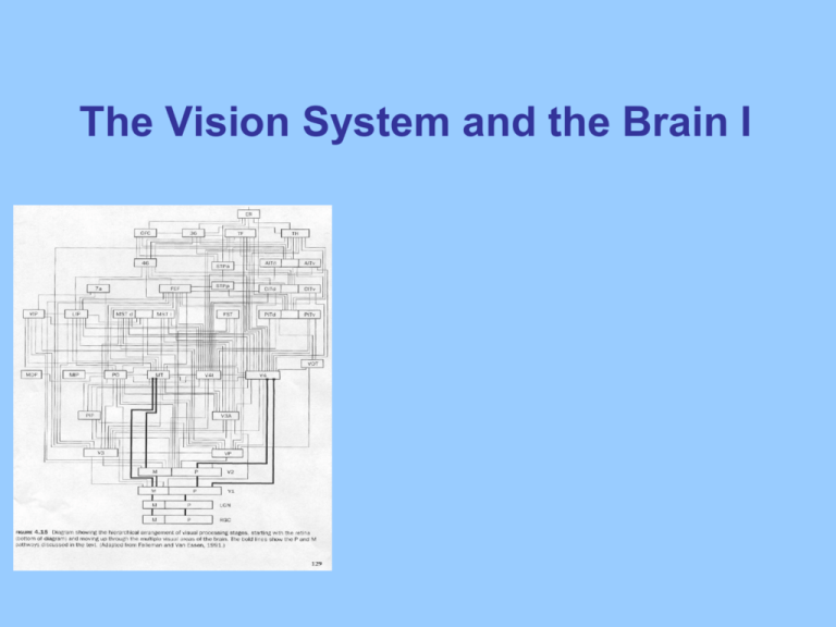
The Vision System and the Brain I Why is vision special? • For humans, who are diurnal, vision is vital sense • Can perceive information at a distance • Localise objects accurately • Vision tends to dominate over other senses • Approx 1/3 of cerebral cortex dedicated to visual analysis and perception • Most widely researched sensory system Visual system: complex processor • The visual system has to: – translate discrete points of light falling onto our photoreceptors in the retina into meaningful objects that we recognise – discriminate objects from other aspects of the visual scene i.e. background environment – recognise these objects in different orientations, even if it has only ever seen a lion from the front • Today: how the brain begins to represent sensory information & processes it to form integrated percepts Parallel processing in the visual system • What do neural signals in the visual pathway represent? • The central hypothesis is that visual information is distributed across subsystems • Consequently, perception is analytic – some processes represent shape, others colour, movement etc. We perceive objects as unified wholes – but how? We do not have the impression that our perception of the red car driving past was created in a piecemeal fashion (I.e. the colour, motion and shape) are processed separately. However, evidence from cognitive neuroscience provides compelling support for the idea that perception DOES operate in an analytic manner – this is called the feature-extraction hypothesis. Visual information is contained in the light reflected from objects As light passes through the lens, the image is inverted and an optical image is focused onto the back surface of the eye, the “retina”. The eye as a camera • To produce optimal images, eye has many features of a camera – e.g. cornea and lens to focus light • Light travels in a straight line until it hits a change in the density of the medium which causes light rays to bend (refraction) • The cornea has a fixed curvature and bends light rays – responsible for forming the image on the retina • Focus is adjusted by changes in the shape of the lens controlled by ciliary muscles inside the eye • Myopia (short-sightedness) results from steeper cornea/longer than normal eyeball, so image is focused in front of the retina Visual processing begins in the retina Receptor cells are highly specialised, and sense selective changes in the environment, e.g., light (photons) and sound waves. Stimulation leads to changes in receptor molecules, which open/close ion channels. Vision: photoreceptors on the retinal surface contain pigments that are sensitive to light Photoreceptors: rods and cones Photoreceptors contain specialised membranes with pigment molecules for phototransduction. There are two types of photoreceptors: (i) Cones (100M) contain 3 pigments and contribute to high acuity colour and daytime vision. Photopic system (Gk photos light) Fovea 1 (ii) Rods (10M) contain rhodopsin, which is very sensitive, and contribute to night vision. Scotopic system (Gk skotos darkness) Spectral sensitivity functions for rods + cones Retinal cells sense light information from a specific point of the world (receptive field). Rods and cones respond to point of light in a particular range of wavelengths. Anatomy of the retina Changes in Glutamate release in photoreceptors cause graded currents in bipolar cells. Stimulation of ganglion cells by bipolar cells results in action potentials, which travel through the optic nerve into the brain. Photoreceptors: pigments and receptive fields rods and cones ganglion cells Retinal cells sense light information from a specific point of the world (receptive field). Rods and cones respond to points of light in a particular range of wavelengths. Inputs from photoreceptors are integrated into opponent center-surround RFs in ganglion cells. Optic nerve The axons of the ganglion cells form the optic nerve which carried visual signals from the retina into the brain. Some axons cross at the optic chiasm. The result is that each visual field is represented contralaterally (opposite side) in the brain. Visual receptive fields (RF’s) • The whole area of the world that you can see at any one time is called your visual field • The part that you see to your left is your left visual field, and the space on the right is the right visual field • The part of the visual field to which any one neuron responds is that neuron’s receptive field. Concentric receptive fields Ganglion cells (like those in the LGN further along the visual pathway) have concentric receptive fields with either a centre-off or centre-on surround. Cell fires rapidly when light hits centre, is inhibited when light is over the surround, and halfway across –no activity. As such, these cells are ideal for signalling changes in illumination (contrast) such as those that arise from stimulus edges. Overview of visual transduction • Output from photoreceptors is first processed in bipolar cells and from there to the ganglion cells. • 260 million photoreceptors and only 1 million ganglion cells – so already substantial compression of information • Axons of the ganglion cells form a bundle call the optic nerve Lateral geniculate nucleus Visual signals from the retina into the LGN are segregated according to (1) the type of ganglion cell and (2) the eye of origin. The LGN contains 6 layers of cells. The inner 2 contain large cells (magnocellular) and the outer 4 contain small cells (parvocellular). In between layers (interlaminar) are other small cells (koniocellular). These layers receive input from different types of ganglion cells. Input from each eye alternates within these types of layers. Receptive fields in the LGN are similar to those in the retina. Lateral geniculate nucleus (LGN) 90% fibres from optic nerve terminate in LGN. Projections are not random but rather terminate selectively in one of the six layers that comprise LGN. Ipsilateral eye projects to layers 2, 3, 5. Contralateral eye projects to 1, 4, 6. Layers 1&2 : magnocellular system. Anatomy of the visual system In the main pathway, retinal signals travel to the lateral geniculate nucleus (LGN) of the thalamus and then to primary visual cortex (V1). There are also alternative pathways, e.g., through the superior colliculus (SC). Before entering the brain, each optic nerve splits into 2 parts Three kinds of cells in LGN Parvocellular neurons Form “P” Pathway Magnocellular neurons Form “M” pathway Koniocellular neurons Cell bodies Smaller Larger Small Receptive fields Smaller Larger Mostly small, variable Retinal location In and near fovea Throughout the retina Throughout the retina Colour sensitive Yes No Some are Respond to Detailed analysis of stationary objects Movement and broad outlines of shape Varied and not yet fully described Primary visual cortex (V1) The first cortical synapses for neurons carrying visual information are in the medial portion of the occipital lobe – Area 17 in Brodmann’s map. This area is located medially (midline) and buried below the superficial layer of the cortex along the calcarine sulcus. The cytoarchitecture is quite regular and stippled – thus giving rise to the name “striate” cortex. Also referred to as primary visual cortex. This name reflects the hypothesis that this is the first visual processing area in the cortex. Anatomy of V1 The segregation of the M and P pathways is maintained in the cortex. Axons from both regions terminate in layer 4 of the striate cortex but (a) the terminal zones of axons in this layer are offset from one another and (b) p pathway involves a 2nd synapse carrying information into the more superficial layers 2 and 3. V1: blobs and interblobs Different types of inputs to V1 form a pattern of “blobs” revealed by staining for cytochrome oxidase (mitochondrial enzyme). Blobs extend through layers 2, 3 and (less so) 5, 6. Parvocellular and koniocellular inputs linked to colour processing form blobs. Parvocellular inputs concerned with shape analysis and magnocellular inputs form interblob regions. Anatomy of the visual system In the main pathway, retinal signals travel to the lateral geniculate nucleus (LGN) of the thalamus and then to primary visual cortex (V1). There are also alternative pathways, e.g., through the superior colliculus (SC). Before entering the brain, each optic nerve splits into 2 parts V1 - Columnar organisation Neurons with similar selectivities cluster in columns, according to dominant eye and preferred orientation. Organisation revealed by 2DG and optical imaging. Orientation columns regularly interrupted by blobs. Hypercolumns (cortical modules) contain full set of orientation, for both eyes. Occular Dominance Columns Orientation Selectivity V1 - Blindsight DB—patient at UL—had migranes for years.. and was found to have a tumour on left side of V1 which was removed which reduced migranes but also left him apparently BLIND in right visual pathway. Wieskrantz—tested DB..and he could see on his right VF! Lesions to V1 result in Blindsight. The person is phenomenologically blind. However, many residual unconscious visual functions remain. The individual can point to stimuli s/he cannot see, and guess about many aspects of the stimulus with performance that is much better than chance. Blindsight illustrates the existence of parallel circuitry for the processing of visual information that can guide action. It also raises interesting questions regarding the nature of visual awareness. Extrastriate visual cortex Visual information in extrastriate cortex forms 2 largely independent parallel streams. Ventral stream (to inferior temporal) computes stimulus appearance (“what”) and contains input from parvocellular, koniocellular and magnocellular cells. Dorsal stream (to posterior parietal) computes stimulus location and movement (“where”) from magnocellular inputs mainly. Ventral visual areas IT face-selective cells achromatopsia V4 is selective for colour and form. RFs large and more complex than blobs (V1) and thin stripes (V2). Neurons respond to many wavelengths and compensate for illumination (colour constancy). Lesions can contribute to achromatopsia. IT is downstream from V4. RFs are huge, selective for objects or complex shapes (*faces), and invariant to location or backgrounds. Lesions ~ agnosia or prosopagnosia (object- or face-recognition deficits). Neuronal activity modulated by attention and memory. What does an agnosic experience? Dorsal visual areas akinetopsia hemispatial neglect MT is selective for motion. RFs large and more complex than interblobs (V1). Lesions contribute to akinetopsia (motion blindness) . PPC is downstream from MT. RFs are large and map spatial location along different reference frames for different types of movements. Lesions result in Balint’s syndrome (bilateral occipital-parietal lesions) or hemispatial neglect (inferior parietal lobe). Neuronal activity modulated by attention and memory. W. W. Norton In summary … Vision is one example of transducing energy into perception Vision exemplifies general scheme for perception periphery>thalamus>primary sensory area >associative unimodal areas… (feedback) Visual system exemplifies functional specialisation starts from sensory receptor cells occurs within brain regions (retina, LGN, V1) occurs between brain regions (V4, IT, MT, PPC) occurs between brain circuits (ventral, dorsal) Visual system also exemplifies concurrent processing massive anatomical interconnectivity residual abilities survive perceptual deficits
