Forensic Anthropology Report: Skeletal Remains Analysis
advertisement
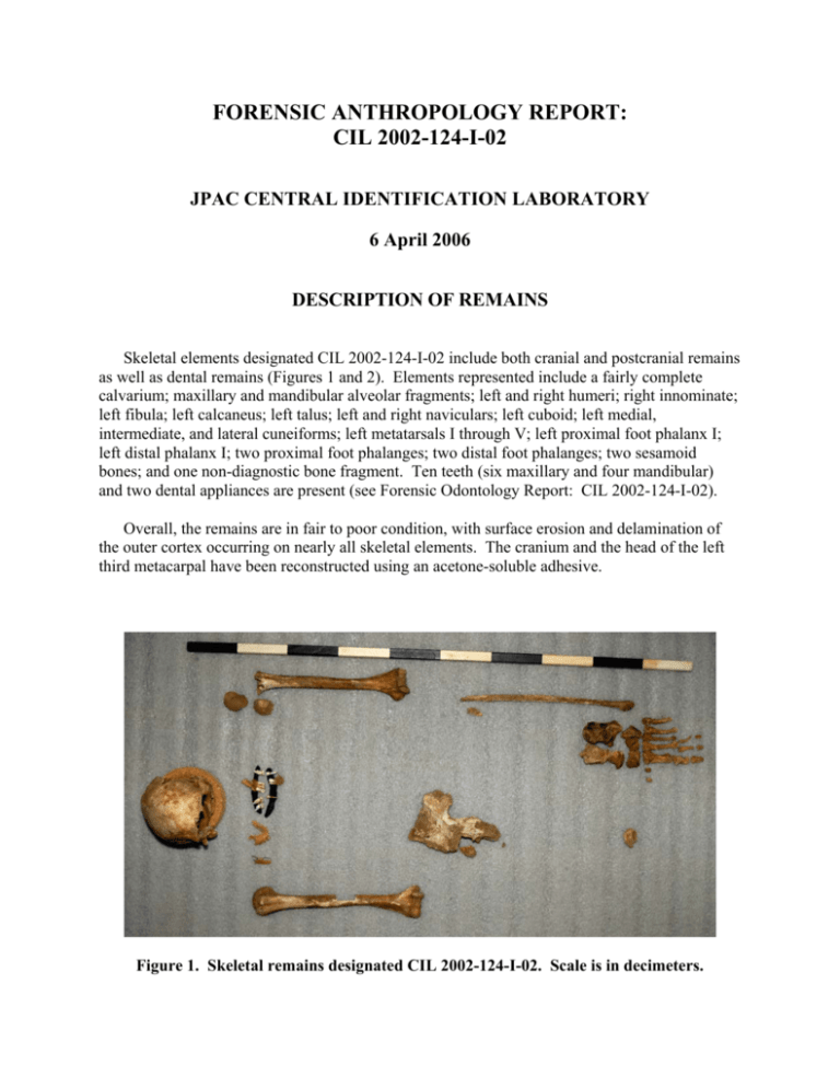
FORENSIC ANTHROPOLOGY REPORT: CIL 2002-124-I-02 JPAC CENTRAL IDENTIFICATION LABORATORY 6 April 2006 DESCRIPTION OF REMAINS Skeletal elements designated CIL 2002-124-I-02 include both cranial and postcranial remains as well as dental remains (Figures 1 and 2). Elements represented include a fairly complete calvarium; maxillary and mandibular alveolar fragments; left and right humeri; right innominate; left fibula; left calcaneus; left talus; left and right naviculars; left cuboid; left medial, intermediate, and lateral cuneiforms; left metatarsals I through V; left proximal foot phalanx I; left distal phalanx I; two proximal foot phalanges; two distal foot phalanges; two sesamoid bones; and one non-diagnostic bone fragment. Ten teeth (six maxillary and four mandibular) and two dental appliances are present (see Forensic Odontology Report: CIL 2002-124-I-02). Overall, the remains are in fair to poor condition, with surface erosion and delamination of the outer cortex occurring on nearly all skeletal elements. The cranium and the head of the left third metacarpal have been reconstructed using an acetone-soluble adhesive. Figure 1. Skeletal remains designated CIL 2002-124-I-02. Scale is in decimeters. Forensic Anthropology Report: CIL 2002-124-I-02 Figure 2. Skeletal inventory for CIL 2002-124-I-02; gray denotes skeletal elements that are present, and blue denotes the presence of non-seriated elements. Page 2 of 8 Forensic Anthropology Report: CIL 2002-124-I-02 MINIMUM NUMBER OF INDIVIDUALS One. These remains were segregated from a larger assemblage of skeletal remains with a minimum number of four individuals. The remains present for analysis in this report are those that could be associated to this individual via mitochondrial DNA (mtDNA) sequence matches, odontological association, osteometric comparisons (Byrd and Adams 2003), visual pairmatching, and taphonomic indicators, as well as through field context (as determined by consultation with the Recovery Leader/Anthropologist). There is no duplication of elements or any other evidence of multiple individuals in this set of remains. SEX Male. Gross morphological and metric analyses were used to determine sex. Non-metric cranial characteristics include a small supraorbital ridge, a blunt supraorbital margin, and a large mastoid process. These traits, with the exception of the first, are more consistent with male morphology (Bass 1995; Buikstra and Ubelaker 1994; Rogers 2005). Postcranially, the pelvic region provides the most diagnostic traits for sex determination. Although most of the key areas for observation in this region are eroded or absent due to postmortem damage, a few diagnostic traits are present that suggest probable male morphology (Bass 1995; Buikstra and Ubelaker 1994). The acetabulum is directed laterally, the lunate surface is broad and deep, and the auricular surface is flat. The greater sciatic notch is deep and forms an acute angle. Due to the paucity of conventional postcranial traits used for sex determination, non-conventional traits were also assessed. The distal humerus morphology demonstrates two features associated with male morphology (asymmetrical trochlea and triangular olecranon fossa shape) and two features that were indeterminate (slight trochlear constriction and slightly elevated medial epicondyle) (Ceri et al. 2005; Rogers 1999). Six postcranial measurements were taken and evaluated against a discriminant model in FORDISC 2.0 (Ousley and Jantz 1996). The discriminant model has a correct classification rate of 95.0% when applied to the reference sample of 339 individuals. The remains were classified by the model as male with a posterior probability of 0.997 and a typicality of 0.580. Overall, the evidence indicates that this individual’s size and morphology are consistent with males. AGE Adult or older juvenile, ≥16 years of age. The estimation of age is based on dental development and overall appearance of skeletal elements. All secondary centers of epiphyseal fusion that were available appear to be completely fused. Gross examination of the distal root of tooth #1 exhibits complete apical closure. This stage of tooth formation occurs on average at 20.2 years, or between 16 and 24.4 years (within two standard deviations) for males (Mincer et al. 1993). Page 3 of 8 Forensic Anthropology Report: CIL 2002-124-I-02 RACE Indeterminate. Gross morphological observations and metric analyses were used to assess race. Only part of the skull, the cranial vault, was available for analysis. The presence of an inion hook, absence of wormian bones, and rugged muscle markings are common in caucasoids (Rhine 1990). Small brow ridges, elliptic external auditory meatus, and complex cranial sutures are common in mongoloids (Rhine 1990). The lack of more diagnostic cranial elements for the determination of ancestry and the distribution of observed features between caucasoids and mongoloids results in an indeterminate assessment. Six postcranial measurements were taken and evaluated against a discriminant model including White and Black male populations in FORDISC 2.0 (Ousley and Jantz 1996). The discriminant model produced a correct classification rate of 77.6% when applied to the reference sample of 210 individuals. The remains were classified by the model as a White male with a posterior probability of 0.774 and a typicality of 0.718. Determination of ancestry using postcranial remains is less reliable than that based on the cranium, and the analysis is further weakened given the paucity of postcranial remains. Only measurements from the humerus and calcaneus were available for metric analysis. Thus, an analysis of ancestry for this individual is indeterminate. STATURE 65.5 to 71.7 inches. The total length of the right humerus (338 mm) was used to estimate stature using the White male model of Trotter and Gleser (1952), which was calculated using FORDISC 2.0 (Ousley and Jantz 1996). The White male equation was selected because it provides a more robust model due to the larger sample size. This model yields a point estimate of 68.6 inches with a 95% prediction interval of 65.5 to 71.7 inches. TRAUMA Perimortem trauma was identified though fracture angle, shape, edge color, and edge morphology (Galloway 1999; Ubelaker and Adams 1995). The left distal humerus exhibits a complete fracture that originates at the trochlea and terminates superior to the medial supracondylar crest (Figure 3). On the posterior aspect of the bone, the fracture follows the morphology of the olecranon fossa (Figure 4). On the anterior aspect, the fracture follows the morphology of the coronoid fossa. The fracture line aligns with the bone grain, which can be another indicator of perimortem trauma. The fracture edges are sharp and exhibit the same color patterns as the surrounding cortical bone. Overall morphology suggests that the fracture is a Type II or high medial condylar fracture (Galloway 1999). This fracture is most common in falls, but can be the result of a direct blow to the joint surface. Page 4 of 8 Forensic Anthropology Report: CIL 2002-124-I-02 Figure 3. Fracture of the left distal humerus. View is anterior. Figure 4. Fracture of the left distal humerus. View is posterior. Page 5 of 8 Forensic Anthropology Report: CIL 2002-124-1-02 OBSERVATIONS The overall condition of the remains indicates both surface and subsurface exposure during the postmortem interval. The remains display a range of color from white to dark brown as a result of weathering and sediment staining. There is cortical delamination and postmortem breakage present on nearly all of the skeletal elements. The amount of postmortem breakage is extensive, especially on the cranium. The anterior aspect of the right innominate, superior to the arcuate and iliopectinal lines, exhibits a mosaic splitting pattern associated with prolonged surface exposure (Behrensmeyer 1978). Inferior to the arcuate and iliopectinal lines and on the posterior aspect of the innominate, the bone color demonstrates evidence of prolonged contact with soil. Numerous parallel striations indicative of rodent gnawing activity are present on the broken surface of the ilium (Haglund 1997) (Figure 5). Two skeletal anomalies were noted during examination. An acetabular fold is present on the right innominate. The fold is located on the supramedial surface of the acetabular roof. The etiology of this trait is unknown, but it is believed to be an epigenetic or biomechanical trait (Mafart 2005). The second anomaly observed involves a rare coalition of the navicular and cuboid bones (Talkhani and Laing 1999). While a complete bony bridge is not present, both tarsals have a bony extension, producing a small gap between the articular surfaces. The articular surfaces of these extensions are rugose and porotic, suggesting that a fibrous (syndesmosis) or cartilaginous (synchondrosis) union existed. This type of tarsal coalition is usually asymptomatic; however, in some cases the individual may feel pain or stiffness in the foot (Talkhani and Laing 1999). Specimens sampled for mtDNA include teeth (#I, #2, #9, #11, #15, #20, and #22), the right humeral diaphysis, and the petrous portion of the right temporal bone. For analytical purposes, each element or fragment in the assemblage has been labeled in pencil with a code that corresponds to its original field provenience. CONCLUSIONS The human skeletal remains designated CIL 2002-124-1-02 consist of one male individual, greater than or equal to 16 years of age, with a stature between 65.5 and 71.7 inches. There is a perimortem fracture present on the distal left humerus. CHRISTIAN M. CROWDER, PhD Anthropologist Page 6 of 8 Forensic Anthropology Report: CIL 2002-124-I-02 Figure 5. Shallow grooves on the ilium indicative of rodent gnaw marks. REFERENCES Bass, W. M. 1995 Human Osteology: A Laboratory and Field Manual. 4th ed. Special Publication No. 2 of the Missouri Archaeological Society, Columbia, MO. Behrensmeyer, A. K. 1978 Taphonomic and ecologic information from bone weathering. Palaeobiology 4:150162. Buikstra, J. E. and D. H. Ubelaker (editors) 1994 Standards for Data Collection from Human Skeletal Remains. Arkansas Archeological Survey Research Series No. 44, Fayetteville, AR. Byrd, J. E. and B. J. Adams 2003 Osteometric sorting of commingled human remains. Journal of Forensic Sciences 48:717-724. Ceri, G. F, H. Schutkowski, and D. A. Weston 2005 The distal humerus—A blind test of the Rogers’ sexing technique using a documented skeletal collection. Journal of Forensic Sciences 50:1289-1293. Page 7 of 8 Forensic Anthropology Report: CIL 2002-124-I-02 Galloway, A. (editor) 1999 Broken Bones: Anthropological Analysis of Blunt Force Trauma. Charles C Thomas, Springfield, IL. Haglund W. D. 1997 Rodents and human remains. In Forensic Taphonomy: The Postmortem Fate of Human Remains, edited by W. D. Haglund and M. H. Sorg, pp. 405-414. CRC Press, Salem, MA. Mafart, B. 2005 Description, significance and frequency of the acetabular crease of the hip bone. International Journal of Osteoarchaeology 15:208-215. Mincer, H. H., E. F. Harris, and H. E. Berryman 1993 A.B.F.O. study of third molar development and its use as an estimator of chronological age. Journal of Forensic Sciences 38:379-390. Ousley, S. and R. Jantz 1996 FORDISC 2.0. University of Tennessee, Knoxville, TN. Rhine, S. 1990 Non-metric skull racing. In Skeletal Attribution of Race: Methods for Forensic Anthropology, edited by G. W. Gill and S. Rhine, pp. 9-20. Maxwell Museum Anthropological Papers No. 4, Albuquerque, NM. Rogers, T. L. 2005 Determining the sex of human remains through cranial morphology. Journal of Forensic Sciences 50:1-8. Rogers, T. L. 1999 A visual method for determining sex of skeletal remains using the distal humerus. Journal of Forensic Sciences 44:57-60. Talkhani, I. S. and P. Laing 1999 Cuboid-navicular coalition in an adult: A case report. Foot and Ankle Surgery 5:151154. Trotter, M. and G. C. Gleser 1952 Estimation of stature from long bones of American Whites and Negroes. American Journal of Physical Anthropology 10:463-514. Ubelaker, D. H. and B. J. Adams 1995 Differentiation of perimortem and postmortem trauma using taphonomic indicators. Journal of Forensic Sciences 40:509-512. Page 8 of 8
