The zymogen-enteropeptidase system: A practical approach to study
advertisement
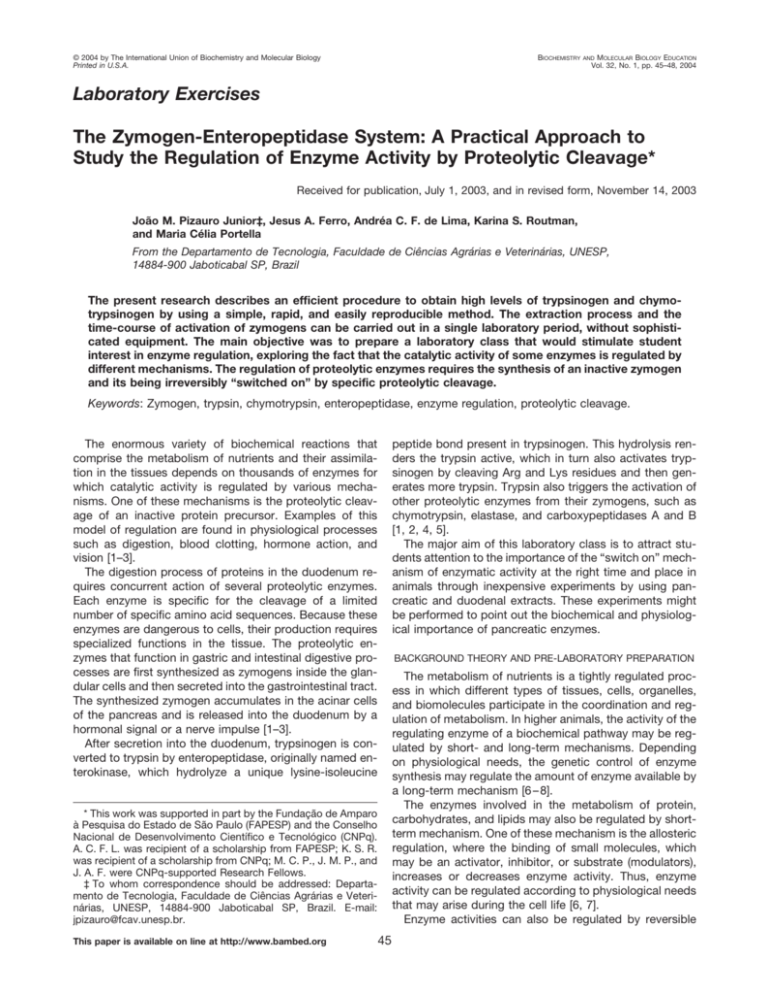
© 2004 by The International Union of Biochemistry and Molecular Biology Printed in U.S.A. BIOCHEMISTRY MOLECULAR BIOLOGY EDUCATION Vol. 32, No. 1, pp. 45–48, 2004 AND Laboratory Exercises The Zymogen-Enteropeptidase System: A Practical Approach to Study the Regulation of Enzyme Activity by Proteolytic Cleavage* Received for publication, July 1, 2003, and in revised form, November 14, 2003 João M. Pizauro Junior‡, Jesus A. Ferro, Andréa C. F. de Lima, Karina S. Routman, and Maria Célia Portella From the Departamento de Tecnologia, Faculdade de Ciências Agrárias e Veterinárias, UNESP, 14884-900 Jaboticabal SP, Brazil The present research describes an efficient procedure to obtain high levels of trypsinogen and chymotrypsinogen by using a simple, rapid, and easily reproducible method. The extraction process and the time-course of activation of zymogens can be carried out in a single laboratory period, without sophisticated equipment. The main objective was to prepare a laboratory class that would stimulate student interest in enzyme regulation, exploring the fact that the catalytic activity of some enzymes is regulated by different mechanisms. The regulation of proteolytic enzymes requires the synthesis of an inactive zymogen and its being irreversibly “switched on” by specific proteolytic cleavage. Keywords: Zymogen, trypsin, chymotrypsin, enteropeptidase, enzyme regulation, proteolytic cleavage. peptide bond present in trypsinogen. This hydrolysis renders the trypsin active, which in turn also activates trypsinogen by cleaving Arg and Lys residues and then generates more trypsin. Trypsin also triggers the activation of other proteolytic enzymes from their zymogens, such as chymotrypsin, elastase, and carboxypeptidases A and B [1, 2, 4, 5]. The major aim of this laboratory class is to attract students attention to the importance of the “switch on” mechanism of enzymatic activity at the right time and place in animals through inexpensive experiments by using pancreatic and duodenal extracts. These experiments might be performed to point out the biochemical and physiological importance of pancreatic enzymes. The enormous variety of biochemical reactions that comprise the metabolism of nutrients and their assimilation in the tissues depends on thousands of enzymes for which catalytic activity is regulated by various mechanisms. One of these mechanisms is the proteolytic cleavage of an inactive protein precursor. Examples of this model of regulation are found in physiological processes such as digestion, blood clotting, hormone action, and vision [1–3]. The digestion process of proteins in the duodenum requires concurrent action of several proteolytic enzymes. Each enzyme is specific for the cleavage of a limited number of specific amino acid sequences. Because these enzymes are dangerous to cells, their production requires specialized functions in the tissue. The proteolytic enzymes that function in gastric and intestinal digestive processes are first synthesized as zymogens inside the glandular cells and then secreted into the gastrointestinal tract. The synthesized zymogen accumulates in the acinar cells of the pancreas and is released into the duodenum by a hormonal signal or a nerve impulse [1–3]. After secretion into the duodenum, trypsinogen is converted to trypsin by enteropeptidase, originally named enterokinase, which hydrolyze a unique lysine-isoleucine BACKGROUND THEORY AND PRE-LABORATORY PREPARATION The metabolism of nutrients is a tightly regulated process in which different types of tissues, cells, organelles, and biomolecules participate in the coordination and regulation of metabolism. In higher animals, the activity of the regulating enzyme of a biochemical pathway may be regulated by short- and long-term mechanisms. Depending on physiological needs, the genetic control of enzyme synthesis may regulate the amount of enzyme available by a long-term mechanism [6 – 8]. The enzymes involved in the metabolism of protein, carbohydrates, and lipids may also be regulated by shortterm mechanism. One of these mechanism is the allosteric regulation, where the binding of small molecules, which may be an activator, inhibitor, or substrate (modulators), increases or decreases enzyme activity. Thus, enzyme activity can be regulated according to physiological needs that may arise during the cell life [6, 7]. Enzyme activities can also be regulated by reversible * This work was supported in part by the Fundação de Amparo à Pesquisa do Estado de São Paulo (FAPESP) and the Conselho Nacional de Desenvolvimento Cientı́fico e Tecnológico (CNPq). A. C. F. L. was recipient of a scholarship from FAPESP; K. S. R. was recipient of a scholarship from CNPq; M. C. P., J. M. P., and J. A. F. were CNPq-supported Research Fellows. ‡ To whom correspondence should be addressed: Departamento de Tecnologia, Faculdade de Ciências Agrárias e Veterinárias, UNESP, 14884-900 Jaboticabal SP, Brazil. E-mail: jpizauro@fcav.unesp.br. This paper is available on line at http://www.bambed.org 45 46 BAMBED, Vol. 32, No. 1, pp. 45–48, 2004 FIG. 1. Schematic representation for the activation process of pancreatic proteolytic enzymes triggered by enteropeptidase as described by Orten et al. [5]. covalent modifications, such as the addition of phosphate groups to serine, threonine, or tyrosine residues. Covalent modification is a highly efficient pathway to control not only enzyme in metabolic reactions, such as glycogen synthesis and break down, but also many other cellular functions, including cell growth and differentiation [6 – 8]. Another important mechanism to regulate protein function is the proteolytic activation that can act like switches, turning proteins “on” and “off.” The proteolytic activation is a special form of covalent modification in which the inactive precursor of the enzyme is activated by an irreversible cleavage. Enzymes active in digestive and blood clotting processes are switched on by proteolytic cleavage. In general, these proenzymes are synthesized as slightly longer polypeptide chains than true active enzymes, and usually they are part of a cascade of mechanisms. As observed in Fig. 1, after secretion in the duodenum, trypsinogen is converted to trypsin by enteropeptidase, which hydrolyzes a unique lysine-isoleucine peptide bond in trypsinogen. The hydrolysis renders trypsin active, which also has the ability to activate trypsinogen by cleaving Arg and Lys residues and then generating more trypsin [5]. In addition, trypsin also triggers a cascade activation of other proteolytic enzymes, and the resulting mixture of enzymes includes endopeptidases (trypsin, chymotrypsin, and elastase) and exopeptidases (carboxypeptidases A and B). Thus, the formation of trypsin by enteropeptidase is the essential activation step [2]. General Note—In this experiment, we describe a methodology adapted to the use of trypsinogen, chymotrypsinogen, and enteropeptidase from broilers, pigs, fishes, and ruminants due to the facility of obtaining these animals. Alternatively, other animals can be used such as guinea pigs, mice, or rabbits. Generally, this experiment uses some hazardous reagents, like acetic acid, p-nitroanilide, N,N-dimethylformamide, and dimethylsulfoxide. These reagents should be manipulated according to their handling instructions and disposed in accordance with local safety instructions. EXPERIMENTAL PROCEDURES Buffers and Substrate Solutions 0.05 M Tris Buffer, pH 8.2, Containing 50 mM of CaCl2— Dissolve 6.057 g of tris(hydroxymetil)-aminometane and 7.351 g CaCl2䡠2H2O in ⬃900 ml of CO2 free water and adjust to pH 8.2 with diluted HCl. Finally, using a volumetric flask, make up to 1 liter with distilled water. Bring to 37 °C immediately before using. BAPNA Solution—Dissolve 40 mg of benzoyl-DL-arginine-pnitroanilde hydrocloride (BAPNA) in 1 ml dimethylsufoxide. This solution is stable at 4 °C for 2 wk. Prepare fresh daily by dilution of 1 ml of this solution to 100 ml with 0.05 M Tris buffer, pH 8.2, containing 50 mM CaCl2. 0.2 M Tris Buffer, pH 7.6, Containing 50 mM of CaCl2—Dissolve 24.228 g of tris(hydroxymetil)-aminometane and 7.351g CaCl2䡠 2H2O in 900 ml of CO2 free water, and adjust to pH 7.6 with diluted HCl. Finally, using a volumetric flask, make up to 1 liter with distilled water. Bring to 37 °C immediately before using. GAPNA Solution—Dissolve 20 mg N-glutarayl-L-phenylalanine-p-nitroanilde (GAPNA) in 1 ml dimethylformamide. This solution is stable at 4 °C for 2 wk. Prepare fresh daily by dilution of 1 ml of this solution to 25 ml with 0.2 M Tris buffer, pH 7.6, containing 50 mM CaCl2 [10]. Preparation of the 1 mM p-Nitroaniline Solution—Dissolve 13.814 mg p-nitroanilde (pNA) in 1 ml dimethylsufoxide. Finally, using a volumetric flask, make up to 1 liter with Tris buffer. Prepare fresh daily. Standard Curve of p-Nitroaniline—Prepare a set of 3-fold numbered tubes, place 0.0, 0.2, 0.4, 0.6, 0.8, 1.0, 1.4, and 1.6 ml of 0.1 mM p-nitroaniline and 0.2 ml of 30% acetic acid in each tube. In each tube, adjust the total volume to 2.0 ml with the appropriate amount of Tris buffer, pH 8.2, containing 50 mM of CaCl2 to give seven calibration standards and one blank tube with reagent grade water only. Measure the absorbance of each tube at 410 nm using control (water blank). Then plot absorbance (y-axis) against the number of mol p-nitroaniline (x-axis), labeling the graph appropriately. Do a curve fitting by linear regression to obtain the ⑀ value (x M⫺1 cm⫺1), which corresponds to the angular coefficient of the curve. Sources of Proteolytic Enzymes The broilers were obtained from a commercial hatchery. In order to obtain pancreatic glands with high levels of proenzymes and low levels of trypsin in the duodenal extract, animals were starved overnight. Adult Hubbard broilers of both sexes were slaughtered. The pancreas were removed, rinsed in ice-cold 0.9% (w/v) NaCl solution, and homogenized with 0.5 M Tris-HCl buffer, pH 8.0, containing 0.05 M CaCl2 (20 ml/g), in an OMNI-GLH-2511 tissue homogenizer (OMNI International, Inc., Warrenton, VA) at 4 °C. The homogenate was centrifuged at 14,000 ⫻ g for 30 min at 4 °C, and the supernatant was diluted 10-fold prior to the activation assay. The same procedure was used for a 100-day-old goat of about 20 kg live weight and a 21-day-old piglet of about 7 kg live weight. 47 FIG. 2. Time-course of activation of chicken trypsinogen and chymotrypsinogen by enteropeptidase at 37 °C. Results represent the means of three determinations as described under “Experimental Procedures.” The hepatopancreas and digestive tract of a 2-year-old common carp (Cyprinus carpio) of 1.6 kg live weight were used due to the existence of a diffuse pancreas. Preparation of Crude Duodenal Enteropeptidase For preparation of crude duodenal enteropeptidase, the duodenum was removed and opened right after slaughtering. The mucous membrane was gently scraped off with a knife and homogenized with 0.05 M Tris-HCl buffer, pH 8.0, containing 0.05 M CaCl2 (10 ml of buffer/g of tissue) in a high-speed shearing homogenizer at 4 °C [9]. The homogenate was then centrifuged at 14,000 ⫻ g for 30 min at 4 °C, and the supernatant was used in the activation process of trypsinogen [9]. For activation of chymotrypsinogen, the crude duodenal enteropeptidase was homogenized with 0.2 M Tris-HCl buffer, pH 7.6, containing 0.05 M CaCl2. Alternatively, duodenal extract can be substituted by 0.08 units of enterokinase/mg protein pancreatic extract [10]. Activation of Trypsinogen from Pancreas Samples The supernatant (5.0 ml) of pancreatic tissue, diluted 10-fold with 0.05 M Tris-HCl buffer, pH 8.0, containing 0.05 M CaCl2, was mixed with an equal volume of crude duodenal enteropeptidase. The activation of trypsinogen was carried out at 37 °C. At intervals of 0, 5, 10, 15, 20, 25, 30, 35, and 40 min, aliquots of 0.8 ml were removed and immediately transferred to labeled tubes (13 ⫻ 130 mm) with 1.0 ml of BAPNA solution [11], previously incubated at 37 °C for 4 –5 min in order to achieve temperature equilibration, mixed well, and then incubated at 37 °C for 5 min. After incubation the reaction was stopped by the addition of 0.2 ml of 30% acetic acid. The absorbance in each assay tube was determined at 410 nm against a control tube. The amount of p-nitroaniline liberated was calculated. Controls without the added supernatant of pancreatic tissue were included in each experiment to determine a nonenzymatic hydrolysis of the substrate plus the amount of trypsin that may be present in the crude duodenal extract. One enzyme unit (U) was defined as the amount of enzyme necessary to release 1.0 nmol of product per minute at 37 °C. Any turbidity detected was removed by centrifugation prior to absorbance determination. TABLE I Trypsin and chymotrypsin activities in activated extracts of pancreatic tissue from different species, assayed at 37 °C Results represent the means of three determinations ⫾ SD as described in “Experimental Procedures.” Animal Trypsin (U/mg) Chymotrypsin (U/mg) Chicken Pig Carp Goat 43.8 ⫾ 1.71 24.9 ⫾ 0.94 419.6 ⫾ 4.90 65.5 ⫾ 1.23 4.5 ⫾ 0.43 18.2 ⫾ 0.74 6.6 ⫾ 0.39 5.2 ⫾ 0.74 with equal volume of crude duodenal enteropeptidase (homogenized with 0.2 M Tris-HCl buffer, pH 7.6, containing 0.05 M CaCl2), and the activation was carried out at 37 °C. Procedures for the activation of chymotrypsinogen from pancreas sample were identical to those used for the activation of trypsinogen. Briefly, at intervals of 0, 5, 10, 15, 20, 25, 30, 35, and 40 min, aliquots of 1.0 ml were removed and immediately transferred to tubes with 0.3 ml of GAPNA solution [12]. The reaction was carried out for 5 min and stopped with 0.2 ml of 30% acetic acid. Absorbance determination and control (blank) assay were identical to those described for trypsin analysis. One enzyme unit (U) was defined as the amount of enzyme releasing 1.0 nmol of product per minute at 37 °C. Determination of Protein Concentration Protein concentration was determined as described by Hartree [13] using bovine serum albumin as a standard. Student Pitfalls This is a very easy class experiment, but some problems such as pancreas and hepatopancreas identification may occur. Some doubts can arise in calculating the conversion factor for enzyme unit. This factor is calculated by linear regression of the standard curve and corresponds to its angular coefficient. An absorbance value multiplied by this factor gives the p-nitroaniline concentration present in the tube, which should be used to calculate the amount of product liberated in the reaction medium and enzyme unit (U). RESULTS AND DISCUSSION Activation of Chymotrypsinogen from Pancreas Samples Five microliters of the pancreas samples, diluted 10-fold with 0.2 M Tris-HCl buffer, pH 7.6, containing 0.05 M CaCl2, was mixed The laboratory class described above involved a demonstration in which zymogens can be obtained from pancreatic tissue prior to activation, enabling the activation 48 BAMBED, Vol. 32, No. 1, pp. 45–48, 2004 kinetics and the assay of trypsin and chymotrypsin to be carried out in vitro. This experimental approach also provided an efficient procedure to obtain high levels of trypsinogen and chymotrypsinogen by using a method that is simple, rapid, and easy to reproduce. The activation kinetics of trypsinogen and chymotrypsinogen from chicken pancreatic homogenates by enteropeptidase was maximal at 15 min and remained constant for at least 40 min (Fig. 2). This feature suggests that the 20-min incubation used was sufficient for maximum activation without loss in activity. It should be emphasized that the addition of 50 mM calcium ions in the reaction medium is necessary for maximum activity. Calcium also protects both trypsin and chymotrypsin from inactivation [14]. Although the time course of activation of chicken trypsinogen and chymotrypsinogen by enteropeptidase is representative of all species used here, the data presented in Table I show that there are great differences in the proteolytic activities present in the pancreatic tissue among the species used. Taking into account that animals were starved overnight, the relative concentrations of proteolytic enzymes in the pancreatic tissue can be attributed to the fact that the content, synthesis, and differential enzyme production changes proportionally in response to change in dietary composition [15]. Carp, an omnivorous species of fish, showed the highest value of trypsin activity in the digestive tract. Hidalgo et al. [16] also found high proteolytic activity in the carp digestive tract, with values very similar to those found in trout. According to these authors, the classification of these fish as omnivorous or carnivorous cannot be justified by their proteolytic activity. They also stated that the production of proteolytic enzymes by pancreatic tissue is dependent on the type and quality of dietary protein ingested by the fish. According to Portella et al. [17], high levels of the proteolytic activities show that these enzymes are important to turn amino acids available for these species. To further clarify the physiological significance of our findings, additional studies must be performed to observe the possible regulation mechanisms of proteolytic enzyme synthesis in carp and in other animal species. Laboratory Time Our experience with different student classes has shown that two periods of 4 h are adequate to carry out the experiment, one period to obtain the zymogens and other period for activation and assay. Study Questions 1. Explain the different strategies or mechanisms used by cells to regulate enzyme catalytic activities (see books 1–3, 6 – 8). 2. Explain the importance of enzymology techniques in metabolic studies and in studies of enzyme regulation activities. 3. 4. 5. 6. 7. 8. What is the difference between trypsinogen and trypsin? Explain the importance of “switch on” mechanism in the uptake of essential amino acids. Explain the role of trypsin in triggering the activation of other proteolytic enzymes. Explain the biochemical and physiological importance of enteropeptidase in the uptake of essential amino acids. Explain the importance of calcium ions to maintain the activity and structure of trypsin. If trypsin is not synthesized by pancreas, explain what could occur with the growth and development of animals. Acknowledgments—We thank David George Francis and Eduardo Rossini Guimaraes for their helpful suggestions and critical review of the manuscript. REFERENCES [1] D. Voet, J. G. Voet (1995) in Biochemistry (D. Voet, J. G. Voet, eds) pp. 371– 410, John Wiley & Sons, Inc., New York. [2] L. Stryer (1995) in Biochemistry (L. Stryer, ed) pp. 237–262, W. H. Freeman and Company, New York. [3] D. L. Nelson, M. M. Cox (2000) in Lehninger Principles of Biochemistry (D. L. Nelson, M. M. Cox, eds) pp. 626 – 627, Worth Publishers, Inc., New York. [4] A. Krogdahl, H. Holm (1982) Activation and pattern of proteolytic enzymes in pancreatic tissue from rat, pig, cow, chicken, mink and fox, Comp. Biochem. Physiol. 72A, 575–578. [5] J. M. Orten, O. T. Neuhaus (1982) in Human Biochemistry (J. M. Orten, O. T. Neuhaus, eds) pp. 320 –374, The C. V. Mosby Company, St. Louis, MO. [6] The Medical Biochemistry Page (revised 2002) www-isu.indstate. edu/thcme/mwking/. [7] G. M. Cooper (2000) The Cell—A Molecular Approach, 2nd Ed, Sinauer Associates, Sunderland, MA. [8] H. Lodish, A. Berk, P. Matsudaira, C. A. Kaiser, M. Krieger, M. P. Scott, L. Zipursky, J. Darnell (2000) Molecular Cell Biology, 4th Ed, W. H. Freeman & Co., New York. [9] A. C. F. de Lima, J. M., Pizauro, M. Macari., E. B. Malheiros (2003) Efeito do uso de probiótico sobre o desempenho e atividade de enzimas digestivas de frangos de corte. R. Bras. Zootec. 32, 200 –207. [10] K. S. Routman, L. Yoshida, A. C. F. de Lima, M. Macari, J. M. Pizauro Júnior (2003) Intestinal and pancreatic enzyme activity of broilers exposed to thermal stress, Rev. Bras. Cienc. Avic. 5, 23–28. [11] M. L. Kakade, J. J. Rackis, J. G. McGhee (1974) Determination of trypsin inhibitor activity of soy products: A collaborative analysis of an improved procedure, Cereal Chem. 51, 376 –382. [12] B. F. Erlanger, F. Edel, A. G. Cooper (1966) The action of chymotrypsin on two new chromogenic substrates, Arch. Biochem. Biophys. 115, 206 –210. [13] E. F. Hartree (1972) Determination of protein. A modification of the Lowry method that gives a linear photometric response, Anal. Biochem. 48, 422– 427. [14] A. D. L. Gorril, J. W. Thomas (1967) Trypsin, chymotrypsin, and total proteolytic activity of pancreas, pancreatic juice, and intestinal contents from the bovine, Anal. Biochem. 19, 211–225. [15] P. M. Brannon (1990) Adaptation of the exocrine pancreas to diet, Annu. Rev. Nutr. 10, 88 –105. [16] M. C. Hidalgo, E. Urea, A. Sanz (1999) Comparative study of digestive enzymes in fish with different nutritional habits. Proteolytic and amilase activities, Aquaculture 170, 267–283. [17] M. C. Portella, J. M. Pizauro, M. B. Tesser, D. J. Carneiro (2002) in Book of Abstracts of the Annual Meeting of the Word Aquaculture Society & the China Society of Fisheries (Word Aquaculture Society, ed) p. 615, Shandong Huaxin Habios, Co., Ltd., Beljing, China.
