Frog Dissection Lab: Anatomy & Systems Assessment
advertisement
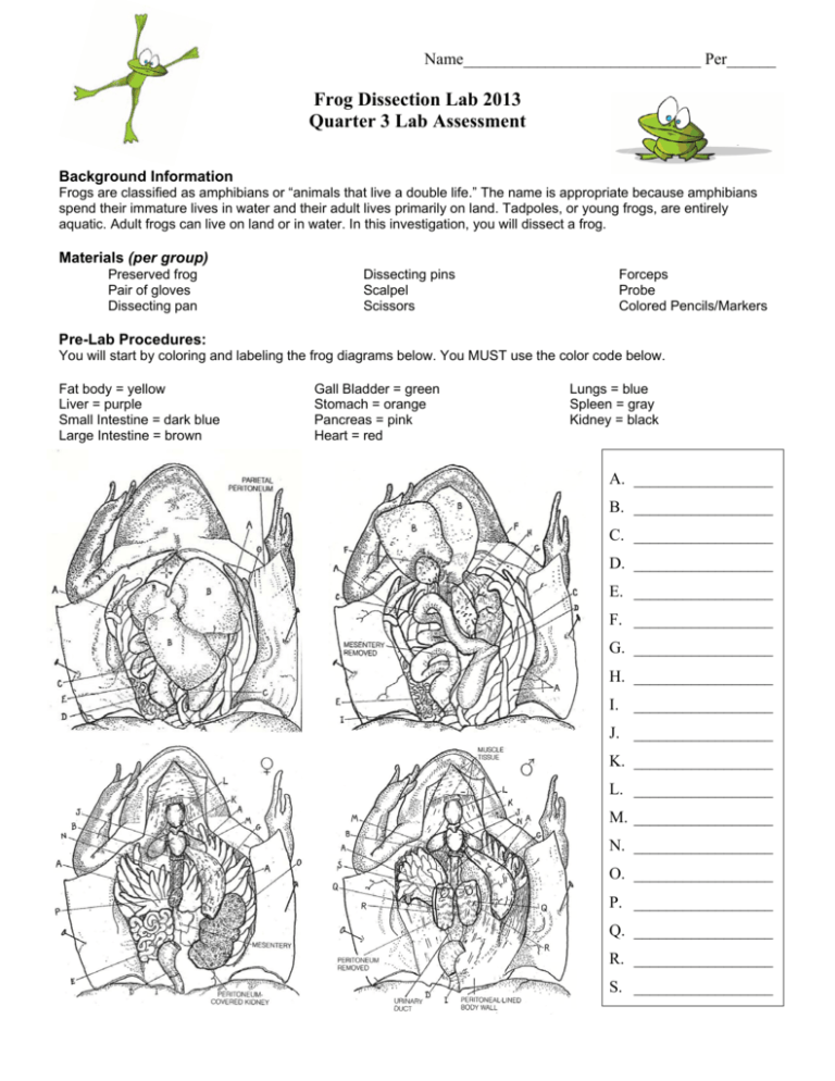
Name_____________________________ Per______ Frog Dissection Lab 2013 Quarter 3 Lab Assessment Background Information Frogs are classified as amphibians or “animals that live a double life.” The name is appropriate because amphibians spend their immature lives in water and their adult lives primarily on land. Tadpoles, or young frogs, are entirely aquatic. Adult frogs can live on land or in water. In this investigation, you will dissect a frog. Materials (per group) Preserved frog Pair of gloves Dissecting pan Dissecting pins Scalpel Scissors Forceps Probe Colored Pencils/Markers Pre-Lab Procedures: You will start by coloring and labeling the frog diagrams below. You MUST use the color code below. Fat body = yellow Liver = purple Small Intestine = dark blue Large Intestine = brown Gall Bladder = green Stomach = orange Pancreas = pink Heart = red Lungs = blue Spleen = gray Kidney = black A. _________________ B. _________________ C. _________________ D. _________________ E. _________________ F. _________________ G. _________________ H. _________________ I. _________________ J. _________________ K. _________________ L. _________________ M. _________________ N. _________________ O. _________________ P. _________________ Q. _________________ R. _________________ S. _________________ 2 Dissection Lab Procedures: External Features 1. Place the frog on its belly (ventral side) in the dissecting pan. 2. Examine the hind and front legs of the frog. The hind legs are strong and muscular and used for jumping and swimming. The forelegs provide balance and cushion the frog when it lands after jumping. Note the difference between the toes of the hind legs and those of the front legs. 3. Locate the large, bulging eyes. The frog has three eyelids. The two outer ones are the color of the frog’s body. The upper and lower lids do not move. The third eyelid is a transparent membrane that protects the eye while permitting the frog to see under water. It also keeps the eye moist when the frog is on land. 4. Behind each eye, find the circular eardrum, tympanum. Then locate the two openings into the nasal cavity. These nasal openings, or external nares, found towards the tip of the snout, will close when the frog is under water. 5. Feel the frog’s skin. It is smooth, moist, and thin. Because the skin is thin and moist, the frog can breathe directly through its skin as well as with his lungs. Flip the frog over to examine his belly. Notice the difference in coloring between the belly and the rest of the frog’s body. 6. Look at the thumb pads and try to determine if your frog is a male or a female. Internal Structures 1. Place the frog on its back (dorsal side) in the dissecting pan and pry open its mouth. If you cut the corners of the mouth (about 1-2 cm) with scissors, the mouth will open more easily. 2. Locate the tongue. Is it attached to the front or back of the mouth? In a live frog, the tongue is sticky and used to catch insects. 3. Gently run your finger along the inside of the upper jaw. The ridges that you feel are maxillary teeth. Two vomerine teeth can also be found in the upper jaw. They are located towards the front of the upper jaw, between and slightly behind the internal openings on of the nostrils. 4. Find the gullet (throat), the wide opening that leads to the esophagus. On both sides if the gullet, near the jaw hinges, are other openings. These are the openings to the Eustachian tube opening. Using your probe, find out where the Eustachian tubes lead. 3 Dissecting the Frog 1. Now you are ready to open the abdominal cavity. Place the frog on its back (dorsal side) in the dissecting pan. Secure it by placing the pins through the tip of the snout and each of its legs. 2. First your incision will be made along the middle of the belly-from the pelvis to the throat. Begin by lifting the belly skin with forceps and inserting the point at the midline near the pelvis. Carefully cut along the midline toward the throat. Note: Cut through the skin only. 3. At the top and bottom of this incision, make transverse cuts towards the forelegs and hind legs. 4. You are now ready to cut through the muscle layer. Repeat the incisions you made in steps 7 and 8, this time cutting through the muscle layer. Work carefully. CAUTION: Do not cut too deeply or you will damage the underlying organs. 5. The sternum, or breastbone, is located between the forelegs. Cut through this tough structure. Fold back the muscle layer and secure it with pins. 6. If your frog is a female, the body cavity may be full of black eggs and ovary tissue. If they are present, carefully remove the eggs and the tissue. Rinse out the body cavity with water. Digestive System 1. The largest organ in the abdominal cavity is the reddish brown liver. Find it and count the number of lobes (sections). 2. Locate the greenish sac attached to the liver. This is the gall bladder. It stores bile, which breaks down fats during digestion. 3. Beneath the liver, find the large, white stomach. It will be on the right side as you look at the frog. The stomach connects to the small intestine. The straight part of the small intestine (near the stomach) is called the duodenum; the remaining, coiled section of the small intestine is the ileum. Separate some of the coils of the ileum and you will see that they are connected by thin, transparent membranes. Such membranes are called mesenteries. 4. The small intestine eventually widens to form the large intestine. The large intestine is a straight tube leading to the anus. The lower portion of the large intestine is called the cloaca. 5. Two smaller organs are somewhat more difficult to find. In the mesentery along the inner curve of the stomach, locate the pinkish pancreas. In the mesentery of the coiled part of the small intestine, see if you can find a small, reddish, spherical structure. This is the spleen. 6. Using scissors, carefully remove the liver. Cut through the upper end of the stomach and the lower end of the large intestine. Then remove the stomach and intestines. Don’t throw away. 7. How long do you think the small intestine is? Record your guess. Then stretch out the small intestine and measure it. 8. Cut open the stomach and a section of intestine to examine the lining and internal features. 4 For extra diagrams of the following organ system – please refer to the diagrams on page 1. Respiratory System: 1. Locate the lungs, two reddish brown saclike structures. 2. OPTIONAL: Insert a clean medicine dropper (with the bulb removed) down the frog’s glottis (windpipe). Gently blow into the medicine dropper and watch the lungs inflate. (put tissue where you will place your mouth on the medicine dropper) Circulatory System: 1. Locate the heart between the lungs. Cut through the thin membrane that surrounds the heart. This will expose the heart for closer examination. 2. The frog’s heart has three chambers. Find the two upper chambers-the right and left atria (singular: atrium)-and the lower ventricle. Compare the thickness of the walls of the atria and the ventricle. 3. LABEL THE HEART DIAGRAM BELOW. Excretory System: 1. Find the two dark red kidneys attached to the back wall of the abdominal cavity. 2. Find the urinary bladder, which empties into the cloaca. The tubes leading from each kidney to the bladder are called ureters.
