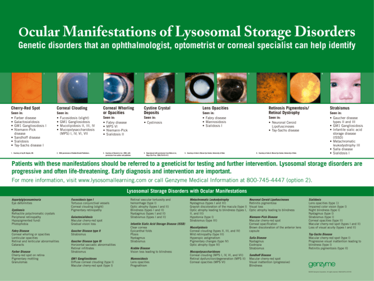
Ocular Manifestations of Lysosomal Storage Disorders
Genetic disorders that an ophthalmologist, optometrist or corneal specialist can help identify
1
2
Cherry-Red Spot
Corneal Clouding
Seen in:
• Farber disease
• Galactosialidosis
• GM1 Gangliosidosis I
• Niemann-Pick
disease
• Sandhoff disease
• Sialidosis
• Tay-Sachs disease I
Seen in:
• Fucosidosis (slight)
• GM1 Gangliosidosis
• Mucolipidosis II, III, IV
• Mucopolysaccharidosis
(MPS) I, IV, VI, VII
1. Courtesy of Jay M. Haynie, OD.
2. With permission of Hodder/Arnold Publishers.
3
4
Corneal Whorling
or Opacities
Cystine Crystal
Deposits
Seen in:
• Fabry disease
• MPS VI
• Niemann-Pick
• Sialidosis II
Seen in:
• Cystinosis
3. Courtesy of Desnick et al., 2003, with
permission from author and publisher.
4. Reproduced with permission from Kleta et al.,
Mayo Clin Proc. 2004;79:410-412.
5
6
Lens Opacities
Retinosis Pigmentosis/
Retinal Dystrophy
Seen in:
• Fabry disease
• Mannosidosis
• Sialidosis I
5. Courtesy of John A. Moran Eye Center, University of Utah.
Seen in:
• Neuronal Ceroid
Lipofuscinoses
• Tay-Sachs disease
6. Courtesy of John A. Moran Eye Center, University of Utah.
Strabismus
Seen in:
• Gaucher disease
types II and III
• GM1 Gangliosidosis
• Infantile sialic acid
storage disease
(ISSD)
• Metachromatic
leukodystrophy III
• Salla disease
• Sialidosis I
Patients with these manifestations should be referred to a geneticist for testing and further intervention. Lysosomal storage disorders are
progressive and often life-threatening. Early diagnosis and intervention are important.
For more information, visit www.lysosomallearning.com or call Genzyme Medical Information at 800-745-4447 (option 2).
Lysosomal Storage Disorders with Ocular Manifestations
Aspartylglycosaminuria
Eye deformities
Cystinosis
Refractile polychromatic crystals
Peripheral retinopathy
Hypopigmented fundi
Photophobia
Fabry Disease
Corneal whorling or opacities
Lenticular opacities
Retinal and lenticular abnormalities
Cataracts
Farber Disease
Cherry-red spot on retina
Pigmentary mottling
Granulomas
Fucosidosis type I
Tortuous conjunctival vessels
Corneal clouding (slight)
Pigmentary retinopathy
Galactosialidosis
Macular cherry-red spot
Gradual vision loss
Gaucher Disease type II
Strabismus
Gaucher Disease type III
Horizontal saccadic abnormalities
Retinal infiltrates
Strabismus
GM1 Gangliosidoses
Diffuse corneal clouding (type I)
Macular cherry-red spot (type I)
Retinal vascular tortuosity and
hemorrhage (type I)
Optic atrophy (types I and II)
Blindness (types I and II)
Nystagmus (types I and II)
Strabismus (types I and II)
Infantile Sialic Acid Storage Disease (ISSD)
Clear cornea
Epicanthal folds
Ptosis
Nystagmus
Strabismus
Krabbe Disease
Vision loss leading to blindness
Mannosidosis
Lens opacities
Prognathism
Metachromatic Leukodystrophy
Nystagmus (types I and III)
Grayish discoloration of the macula (type I)
Optic atrophy leading to blindness (types I,
II, and III)
Hypotonia (type I)
Strabismus (type III)
Mucolipidosis
Corneal clouding (types II, III, and IV)
Mild retinopathy (type III)
Hyperopic astigmatism
Pigmentary changes (type IV)
Optic atrophy (type IV)
Mucopolysaccharidoses
Corneal clouding (MPS I, IV, VI, and VII)
Retinal dysfunction/degeneration (MPS II)
Corneal opacities (MPS VI)
Neuronal Ceroid Lipofuscinoses
Retinitis pigmentosa
Visual loss
Optic atrophy leading to blindness
Niemann-Pick Disease
Macular cherry-red spot
Corneal opacification
Brown discoloration of the anterior lens
capsule
Salla Disease
Nystagmus
Exotropia
Strabismus
Sialidosis
Lens opacities (type 1)
Impaired color vision (type I)
Night blindness (type I)
Nystagmus (type I)
Strabismus (type I)
Corneal opacities (type II)
Macular cherry-red spot (types I and II)
Loss of visual acuity (types I and II)
Tay-Sachs Disease
Macular cherry-red spot (type I)
Progressive visual inattention leading to
blindness (type I)
Retinitis pigmentosis (type II)
Sandhoff Disease
Macular cherry-red spot
Visual inattention (progressive)
Blindness
©2005 Genzyme Corporation. All rights reserved. RGD/US/P212/07/05


