ARVO 2014 Annual Meeting Abstracts
503 Conjunctival and ocular adnexal tumors
Thursday, May 08, 2014 8:30 AM–10:15 AM
Exhibit/Poster Hall SA Poster Session
Program #/Board # Range: 5427–5446/A0001–A0020
Organizing Section: Anatomy/Pathology
Program Number: 5427 Poster Board Number: A0001
Presentation Time: 8:30 AM–10:15 AM
Childhood Orbital, Ocular and Optic nerve tumors in Egypt
Ahmad S. AlFaar1, 2, Mohamed S. Bakry1, Sameera Ezzat1, 3. 1Research
Department, Children Cancer Hospital - Egypt 57357, Cairo, Egypt;
2
Ophthalmology, Cairo University School of Medicine, Cairo, Egypt;
3
Public Health, National Liver Institute, Shebin Elkom, Egypt.
Purpose: Our aim is to study the incidence of different ocular, orbital
and optic nerve tumors in childhood age group (between 0-18 years
old) in the largest Egyptian pediatric oncology referral center and
correlate the features of each disease and its survival outcomes.
Methods: Institutional cancer registry database was reviewed for
patients who presented with orbital tumors as a primary site of
involvement between July 2007 and November 2013. REDCap
(Research Electronic Data Capture) system was used for data
collection and organization. Integration between clinical information
system and RedCap was established for real-time registry updating.
Relevant ICD-O-3 topography codes were used to identify the sites.
Data were presented in terms of frequencies and percentages. Other
demographic properties were presented. Survival was demonstrated
using kaplan-meier curves.
Results: Among 7277 patients presented with different childhood
tumors 425 had the mentioned lesions. Males were affected more
than females. Retinoblastoma, Astrocytomas of optic nerve and
Rhabdomyosarcoma of orbit were the most frequent tumors. Mean
age of retinoblastoma cases was 1.4 years while it was higher in
other diseases except germ cell tumors. We’ve presented correlations
of our findings with the current Egyptian population-based cancer
registry and previous results from Cancer in five continents report.
Overall Survival of Retinoblastoma was 95.4% while it was better in
Germ cell tumors and less in all other tumor categories. Orbital bones
masses were hard to identify due to the broad classification in current
ICD-O topography coding.
Conclusions: Childhood orbital tumors distribution was similar
to international publications except increased incidence of orbital
lymphomas. Special attention should be paid to updating the ICD-O
classification system to present different skull bones.
Commercial Relationships: Ahmad S. AlFaar, None; Mohamed S.
Bakry, None; Sameera Ezzat, None
Program Number: 5428 Poster Board Number: A0002
Presentation Time: 8:30 AM–10:15 AM
Successful management of ocular juvenile xanthogranuloma
using off-label bevacizumab: a report of 2 cases
Noy Ashkenazy, Christopher M. Henry, Ashkan M. Abbey, Craig A.
McKeown, Audina M. Berrocal, Timothy G. Murray. Bascom Palmer
Eye Institute, Miami, FL.
Purpose: To present off-label bevacizumab as a potential therapeutic
modality for ocular JXG refractory to management with local
corticosteroid therapy.
Methods: Retrospective case series
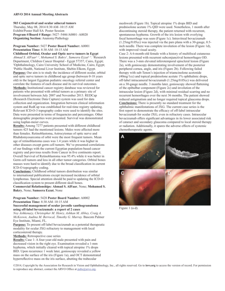
Results: Case 1: A four-year-old male presented with pain and
decreased vision in the right eye. Examination revealed a 1-mm
hyphema, which initially cleared with topical atropine 1% drops
BID. Upon recurrence 1 week later, gonioscopy revealed a yellow
mass on the surface of the iris (Figure 1a), and OCT demonstrated
hyperreflective mass on the iris surface, abutting the trabecular
meshwork (Figure 1b). Topical atropine 1% drops BID and
prednisolone acetate 1% QID were used. Nonetheless, 1 month after
discontinuing steroid therapy, the patient returned with recurrent,
spontaneous hyphema. Growth of the iris lesion with overlying
focal hemorrhage was seen (Figure 1c). Intravitreal bevacizumab
(1.25mg/0.05cc) was injected via the pars plana with a 30-gauge, 0.5inch needle. There was complete involution of the lesion (Figure 1d),
with improved visual acuity.
Case 2: A 6-month-old female with a history of multifocal cutaneous
lesions presented with recurrent subconjunctival hemorrhages OD.
There was a 3-mm elevated inferotemporal episcleral lesion (Figure
2a), with gonioscopy demonstrating involvement of the posterior
peripheral cornea, angle, and iris (Figure 2b). Following failed
therapy with sub-Tenon’s injection of triamcinolone acetonide
(40mg/1cc) and topical prednisolone acetate 1% ophthalmic drops,
off-label intracameral bevacizumab (1.25mg/0.05cc) was delivered
on a 30-gauge needle. 3 months later, gonioscopy showed flattening
of the epibulbar component (Figure 2c) and involution of the
intraocular lesion (Figure 2d), with minimal residual scarring and no
recurrent hemorrhages over the next 36 months. The patient showed
reduced astigmatism and no longer required topical glaucoma drops.
Conclusions: There is presently no standard treatment for the
ophthalmic manifestations of JXG. The current case series is the
first report to demonstrate the efficacy of off-label intraocular
bevacizumab for ocular JXG, even in refractory cases. Intraocular
bevacizumab offers significant advantages in its lower associated risk
of cataract and secondary glaucoma compared to local steroid therapy
or radiation. Additionally, it spares the adverse effects of systemic
chemotherapeutic agents.
Figure 1 (a-d).
©2014, Copyright by the Association for Research in Vision and Ophthalmology, Inc., all rights reserved. Go to iovs.org to access the version of record. For permission
to reproduce any abstract, contact the ARVO Office at pubs@arvo.org.
ARVO 2014 Annual Meeting Abstracts
of them in the pediatric population. Presumably, the deeper location
of these common skin lesions results either from embryonic rests or
from traumatic implantation of glandular epithelium. All 4 previously
reported pediatric cases were congenital and of apocrine subtype.
Two of the cases in our series were of eccrine subtype, and one was
traumatic in origin, expanding the clinical and pathological spectrum
of this entity. Clinicians should be aware that an orbital cystic lesion
in a child may represent a giant hydrocystoma.
Figure 2 (a-d).
Commercial Relationships: Noy Ashkenazy, None; Christopher
M. Henry, None; Ashkan M. Abbey, None; Craig A. McKeown,
None; Audina M. Berrocal, None; Timothy G. Murray, None
Program Number: 5429 Poster Board Number: A0003
Presentation Time: 8:30 AM–10:15 AM
Giant Orbital Hydrocystoma in Children: Report of Three Cases
Mehrdad Malihi1, Roger Turbin1, Neena Mirani2, Paul D. Langer1.
1
The Institute of Ophthalmology and Visual Sciences, New Jersey
Medical School, Newark, NJ; 2Department of Pathology, New Jersey
Medical School, Newark, NJ.
Purpose: Hydrocystoma (also known as sudoriferous cyst) is a
benign cystic proliferation of a sweat gland found commonly on
the eyelid skin of adults; we report three cases of giant orbital
hydrocystoma in children, expanding the clinical and pathological
spectrum of this entity.
Methods: Interventional case series.
Results: Case 1: An 8 year-old-boy presented with a 1 year history
of painless progressive right proptosis. Computed tomographic
and magnetic resonance imaging (MRI) revealed a well-defined,
intraorbital, extraconal cystic lesion in the lateral orbit posterior
to the globe causing bony erosion (Fig 1). Pathologic examination
following total resection via lateral orbitotomy reveals a cystic lesion
with a clear cavity lined by a smooth surface of a double layer of
cuboidal cells, consistent with eccrine hydrocystoma.
Case 2: A 13-year-old girl who had suffered blunt orbital trauma one
year earlier developed a soft, mobile, non-tender subconjunctival
mass in the temporal part of the right upper eyelid. MRI revealed a
large well-defined cystic lesion in the right anterior orbit, which was
later dissected from beneath the conjunctiva. Pathologic examination
reveals a cystic cavity with papillary projections lined by two layers
of cuboidal epithelial cells, with the innermost cells displaying
eosinophilic cytoplasm and apical “snouting” (decapitation),
characteristic of an apocrine hydrocystoma (Fig 2).
Case 3: A two-month-old infant was noted to have a non-tender
subconjunctival orbital growth visible in the medial left palpebral
aperture. Surgical excision revealed a cystic lesion with a smooth
internal surface. Microscopic evaluation revealed characteristics
similar to case 1 and consistent with eccrine hydrocystoma.
Conclusions: Periocular hydrocystoma, which typically presents
on the eyelid skin in adults, only rarely occur beneath the skin or
conjunctiva: only 8 such lesions have previously been reported, 4
Commercial Relationships: Mehrdad Malihi, None; Roger
Turbin, None; Neena Mirani, None; Paul D. Langer, None
Program Number: 5430 Poster Board Number: A0004
Presentation Time: 8:30 AM–10:15 AM
Growth rates of orbital cavernous hemangiomas: A quantitative
analysis
Liza M. Cohen1, Anupam Jayaram1, Gary Lissner1, Achilles
Karagianis2. 1Ophthalmology, Northwestern University, Chicago, IL;
2
Radiology, Northwestern University, Chicago, IL.
Purpose: Cavernous hemangiomas of the orbit are characterized by
their often asymptomatic presentation and slow growth over time.
Our purpose is to calculate the growth rate of orbital cavernous
hemangiomas in order to provide quantitative evidence in counseling
patients regarding the natural progression of these lesions.
Methods: A retrospective chart review from January 1983 to 2013,
searching by radiologic diagnoses consistent with orbital cavernous
hemangioma, identified a total of 57 lesions. Of these 57, 20 had at
least three interval CT and/or MRI scans of the orbit over a period of
at least two years follow-up. Serial imaging studies were reviewed by
a single neuroradiologist, and the size of each lesion was determined
by calculating volume from three-dimensional measurements. Rates
of change in size were computed by constructing best-fit lines for size
of the lesion versus time.
Results: The 20 patients included 13 (65%) females and 7 (35%)
males, with average age at presentation of 56.7 ± 14.9 years. The
average length of follow-up was 5.7 ± 3.3 years. The average number
of serial imaging scans was 5.9 ± 3.8. Of the 20 lesions, 10 (50%)
decreased in size and 10 (50%) increased in size over the course of
the study. Two lesions that increased in size were resected after 2.5
and 3 years. For the 10 lesions that decreased in size, the average
rate of regression was 0.34 ± 0.61 cubic mm/year (range 0.029-2.04),
equating to 2.89 years for the lesion to shrink one cubic mm. For the
eight lesions not requiring surgery that increased in size, the average
rate of growth was 0.25 ± 0.39 cubic mm/year (range 0.029-1.17),
equating to 4.00 years for the lesion to grow one cubic mm. In
total, for all 18 masses not requiring surgery that both increased and
decreased in size, the average of the absolute value of the rate of
change in size was 0.30 ± 0.51 cubic mm/year, equating to an average
3.30 years for the lesion to either grow or shrink one cubic mm. Of
©2014, Copyright by the Association for Research in Vision and Ophthalmology, Inc., all rights reserved. Go to iovs.org to access the version of record. For permission
to reproduce any abstract, contact the ARVO Office at pubs@arvo.org.
ARVO 2014 Annual Meeting Abstracts
note, one lesion requiring surgery at a size of 22.5 cubic mm grew at
a rate of 3.69 cubic mm/year over three years.
Conclusions: Orbital cavernous hemangiomas, when followed with
serial imaging over an extended period of time, tend to primarily
either increase or decrease in size at a steady slow rate. Quantifying
this rate of growth/regression can aid in confirming a diagnosis of
cavernous hemangioma and in reassuring patients as to the slowly
changing nature of these lesions.
Commercial Relationships: Liza M. Cohen, None; Anupam
Jayaram, None; Gary Lissner, None; Achilles Karagianis, None
Program Number: 5431 Poster Board Number: A0005
Presentation Time: 8:30 AM–10:15 AM
Clinicopathologic Correlation of Caruncular Lesions
Sander R. Dubovy1, 2, Antonio J. Bermudez1, 2, Jordan Thompson1,
2 1
. Bascom Palmer Eye Institute, University of Miami, Miami, FL;
2
FLorida Lions Ocular Pathology Laboratory, Miami, FL.
Purpose: Lesions of the caruncle are relatively uncommon. Herein
we report the caruncular lesions seen at the Bascom Palmer Eye
Institute by characterizing the type of lesion, relative frequency and
clinical findings from a single institution.
Methods: The case files of the Florida Lions Ocular Pathology
Laboratory at the Bascom Palmer Eye Institute were reviewed from
1997 to October 2013 searching for lesions that were designated to
have been biopsied from the caruncle. The reports were reviewed and
the histopathologic findings were correlated to the clinical findings.
Results: A total of 198 lesions of the caruncle were found in the
files from January 1997 to October 2013. Thirty different diagnostic
entities were identified. The most common were nevi (n=61, 31%),
followed by non-specific inflammation (n=26,13%), papilloma (n=16,
8%), sebaceous hyperplasia (n=13, 6.6%), oncocytoma (n=10, 5%)
and sebaceous carcinoma (n=8, 4%).
Conclusions: This case series demonstrates that benign nevi are
the most common lesions identified in the caruncle. While more
commonly benign, malignant lesions of the caruncle account for up to
14% of caruncular lesions including sebaceous carcinoma, squamous
cell carcinoma, basal cell carcinoma, lymphoma, melanoma and
intraepithelial carcinoma. A wide variety of lesions may present in
the caruncle and the clinician should be aware of the differential
diagnosis in this anatomic location.
Commercial Relationships: Sander R. Dubovy, None; Antonio J.
Bermudez, None; Jordan Thompson, None
Support: Florida Lions Eye Bank
Program Number: 5432 Poster Board Number: A0006
Presentation Time: 8:30 AM–10:15 AM
Immunohistochemical Analysis of Sebaceous Cell Carcinoma in
Comparison to Both Basal Cell Carcinoma and Squamous Cell
Carcinoma
Andre N. Ali-Ridha1, 2, Seymour Brownstein1, 2, Kailun Jiang1, 2,
Tatyana Milman3, Bruce Burns2, Paula Blanco2, James Farmer2.
1
Department of Ophthalmology, University of Ottawa, The Ottawa
Hospital, The Ottawa Hospital Research Institute, Ottawa, ON,
Canada; 2Department of Pathology and Laboratory Medicine,
University of Ottawa, The Ottawa Hospital, Ottawa, ON, Canada;
3
Departments of Ophthalmology and Pathology, New York Eye and
Ear Infirmary, New York, NY.
Purpose: Basal cell carcinoma is the most common malignancy of
the eyelid followed by sebaceous cell and squamous cell carcinoma.
The mortality rate of sebaceous cell carcinoma has been reported
as 9 to 30% and both it and squamous cell carcinoma can develop
metastatic disease. Clinically, sebaceous cell carcinoma frequently
mimics inflammatory conditions and other neoplasms of the eyelid,
including both squamous cell and basal cell carcinoma. Our study
compares the immunostaining profile of sebaceous cell carcinoma to
that of basal cell and squamous cell carcinoma.
Methods: Retrospective and prospective case series. Eight specimens
each of sebaceous cell, basal cell and squamous cell carcinoma of the
eyelid were obtained from the Ottawa Ocular Pathology Laboratory
from 2007 to 2013. We compared the immunohistochemical profile
of these specimens by staining each of them with EMA, BER-EP4,
adipophilin, androgen receptor (AR), P16, BCL-2, CK7, Ki67,
BRST1, BRST2, p53, and CK20. We compared the extent of staining
to that of normal surface and glandular epithelial tissue on each slide
as normal internal controls. The immunoreactivity data was then
analyzed using a 2-tailed Kruskal-Wallis test (p<0.05 is significant)
with a post-hoc analysis using Mann-Whitney tests.
Results: Our test results were statistically significant (p<0.05) for
positive staining of EMA, adipophilin, P16 and AR in sebaceous cell
carcinoma. This group also showed a higher percentage of positivity
for Ki67. The basal cell tumours exhibited positive staining for
CK7, BCL2, and BER-EP4, while squamous cell carcinoma showed
substantial positivity only for EMA. Adipophilin stained positive
in sebaceous cell carcinoma with intracytoplasmic lipid vesicles
in 94% of the cells in our series as compared to the nonspecific or
minimal staining of granules in basal and squamous carcinoma cells
respectively.
Conclusions: Our study helps clarify much of the controversy in the
literature concerning immunostains which overlap in reactivity for
sebaceous cell carcinoma. We have found that the most informative
panel of immunostains, from our original 12 stains, for differentiating
sebaceous cell carcinoma from other related tumors consists of P16,
adipophilin, EMA, AR, CK7 and Ki67. This diagnostically optimal
panel of immunostains may allow for earlier diagnosis and treatment
of sebaceous cell carcinoma of the eyelid.
Commercial Relationships: Andre N. Ali-Ridha, None; Seymour
Brownstein, None; Kailun Jiang, None; Tatyana Milman, None;
Bruce Burns, None; Paula Blanco, None; James Farmer, None
Program Number: 5433 Poster Board Number: A0007
Presentation Time: 8:30 AM–10:15 AM
Sebaceous gland carcinoma of the ocular adnexa – variability in
histological and immunhistochemical appearance
Eva J. Becker1, 2, Martina C. Herwig1, 2, Frank G. Holz1, Hans-Peter
Fischer3, Karin U. Loeffler1, 2. 1Ophthalmology, University Eye
Hospital Bonn, Bonn, Germany; 2Ophthalmic Pathology, University
Eye Hospital Bonn, Bonn, Germany; 3Pathology, University Bonn,
Bonn, Germany.
Purpose: To evaluate the characteristics of sebaceous gland
carcinoma (SGC) of the ocular adnexae which is - due to a high
variability in clinical, histological and immunhistochemical
characteristics – challenging to diagnose.
Methods: Records of 6 patients (1 female, 5 male) with SGC
were reviewed, who underwent surgical excision and who were
histologically diagnosed SGCs. For comparison, specimens from
4 patients with basal cell carcinoma (BCC), 1 with squamous cell
carcinoma (SCC) and 2 with indeterminate lesions were examined.
Histological and immunhistochemical analysis included stains for
HE and PAS, cytokeratins (CKpan, Cam5.2), epithelial membrane
antigen (EMA), androgen receptor (AR441), and adipophilin.
Results: SGCs were located in the upper (n=2) or lower (n=4)
eyelid and were associated with various clinical signs including
chalazion-like lesions with pyogenic granuloma (n=1), papillomatous
conjunctival tumors (n=3), a hyperkeratotic exophytic neoplasm
(n=1) and an ulcerating crusted lesion resembling chronic blepharitis
(n=1). The treatment was tumor resection, followed (if necessary)
©2014, Copyright by the Association for Research in Vision and Ophthalmology, Inc., all rights reserved. Go to iovs.org to access the version of record. For permission
to reproduce any abstract, contact the ARVO Office at pubs@arvo.org.
ARVO 2014 Annual Meeting Abstracts
by adjuvant therapy with topical Mitomycin C (n=2). Histologic
characteristics included basophilic pleomorphic cells with vacuolated
cytoplasm, prominent nucleoli, mitotic figures and in some cases
pagetoid spread (n=2). CKpan, EMA and Cam5.2 showed a strong
positive immunoreactivity in all specimens (SGC, BCC, SCC).
AR441 positivity was noted with variable intensities in almost all
lesions and in particular in pagetoid spread in contrast to non-tumor
cells. Adipophilin showed an annular staining of lipid granules in
immature sebaceous cells and was mainly found in SGC.
Conclusions: SGCs display a variety of clinical signs and may mimic
many other lesions. Tumor resection, followed by histological and
immunhistochemical analysis, leads to the diagnosis and initiation
of the proper treatment regimen. Herein, immunohistochemistry
showed an unequivocal profile in SGC and did not allow for an
exact differentiation from BCC and SCC by immunohistochemical
means only. An extended evaluation of HE stains remains essential.
However, immunohistochemistry can make relevant contributions to
the diagnosis of SGC, especially in cases of inconclusive histology,
by positive staining for adipophilin in immature sebaceous cells or by
AR441 labeling in cases of pagetoid spread.
Commercial Relationships: Eva J. Becker, None; Martina C.
Herwig, None; Frank G. Holz, None; Hans-Peter Fischer, None;
Karin U. Loeffler, None
Program Number: 5434 Poster Board Number: A0008
Presentation Time: 8:30 AM–10:15 AM
Molecular Profiling of Ocular Surface Squamous Neoplasia
Identifies Multiple DNA Copy Number Alterations Including
Recurring 8p11.22 Amplicons
Saeed AlWadani1, 3, Laura Asnaghi1, Hind Alkatan2, Hilal Al
-Hussain2, Deepak Edward1, 2, Charles Eberhart1. 1Ophthalmic
Pathology, Johns Hopkins Hospital, Baltimore, MD; 2King Khaled
Eye Specialist Hospital, Riyadh, Saudi Arabia; 3Ophthalmology, King
Saud University, Riyadh, Saudi Arabia.
Purpose: To uncover novel diagnostic biomarkers and molecular
pathways, which can be targeted using new therapies in Conjunctival
squamous cell carcinoma (cSCC). Very little is known about the
molecular pathways, which drive the formation and growth of ocular
surface squamous neoplasia.
Methods: We analyzed DNA extracted from 14 snap frozen cSCC
tumor specimens using Agilent 180K high density oligonucleotide
array-based Comparative Genomic Hybridization (aCGH), with
12 samples giving high quality hybridizations. 11 cases with
DNA remaining were used to confirm chromosomal alterations by
nanostring analysis.
Results: Of these 12 tumors, the number of clear regions of DNA
loss ranged from 1 to 24 per tumor, while gains ranged from 2 to 14
per tumor. Two of the 12 tumors were recurrent, and these had the
highest number of copy number gains/amplifications 9 and 14, and
8 and 11 losses among the cohort. These recurring aberrations were
observed in chromosome 6, where the region 6p22.1-p21.32 was lost
in 33%, in chromosome 14, where the locus 14q13.2 was lost in 42%,
and in chromosome 22, where 5 samples showed DNA loss and one
DNA gain at 22q11.22. However, the most frequent alteration was
observed in chromosome 8, where the locus 8p11.22 was amplified
in 75% and lost in 25%. This region contains a group of genes coding
for “a disintegrin and metalloprotease” (ADAM) proteins, known
to be involved in the activation of oncogenic receptors and tumor
formation. We observed the most profound DNA alterations in the
region of 8p11.22 which contains part or all of the ADAM1B, 3A,
and 5p genes. We are now investigating mRNA expression at the
8p11.22 loci with PCR.
Conclusions: Recurrent cSCC tumors were found to have significant
chromosomal alteration in region 8p11.22, suggesting that increased
numbers of DNA alterations may be associated with more aggressive
clinical behavior and/or tumor progression. In contrast, case with
fewer gains and losses was somewhat distinct microscopically, and
noted to be relatively undifferentiated with adnexal features. The
ADAM genes discovered in this study could be related to ocular
tumors and previously reported ADAM9 alterations associated with
oral mucosa neoplasia, suggest that ADAM family members may be
involved in the pathogenesis of several types of mucosal squamous
neoplasia, and could be potential therapeutic target.
Commercial Relationships: Saeed AlWadani, King Khaled Eye
Specialist Hospital (F); Laura Asnaghi, King Khaled Eye Specialist
Hospital (F); Hind Alkatan, King Khaled Eye Specialist Hospital
(F); Hilal Al -Hussain, King Khaled Eye Specialist Hospital (F);
Deepak Edward, King Khaled Eye Specialist Hospital (F); Charles
Eberhart, King Khaled Eye Specialist Hospital (F)
Support: King Khaled Eye Specialist Hospital Grant, Riyadh, Saudi
Arabia
Program Number: 5435 Poster Board Number: A0009
Presentation Time: 8:30 AM–10:15 AM
Establishment and characterization of squamous cell carcinoma
cells from the human bulbar conjunctiva
Bettina Mueller, Henning Thomasen, Klaus-Peter Steuhl, Daniel
Meller. University Duisburg Essen, Essen, Germany.
Purpose: Until now only limited information about cultivated tumor
initiating cells out of squamous cell carcinoma (SCC) from the
human bulbar conjunctiva are available. Therefore, the establishment
of a model cell line would be a useful tool for further studies.
In particular, the phenotypic and molecular characterization in
comparison to other SCC cells is of high interest. This would enable
the development of new treatment options for clinical application.
Methods: Epithelial cells were isolated from a bulbar conjunctival
SCC obtained from a 74 year old male and were named PeCaUkHb-01. For receiving a pure cell culture, epithelial cells were
harvested by stepwise trypsinisation. Furthermore, mycoplasma
contamination was tested. Cell doubling time and number of
passages were determined. STR (short tandem repeats) and karyotype
analyses were performed to verify the origin of the cells and to
analyze chromosomal heterogeneity. For further characterization
semiquantitative real-time PCR and immunofluorescence staining
were carried out to detect tumor and epithelial progenitor cell
markers. Spheroid and colony forming ability were performed.
Results: The morphology of the new cell line resembled other
epithelial SCC cell lines, furthermore STR experiments confirmed the
origin of the cells from the donor. The cells were free of mycoplasma.
They grew above passage number forty and could be frozen and
recultured. Karyotype analyses revealed a heterogeneous composition
of the cell culture and the karyogram itself showed aberrations and
changes in the chromosome numbers. PeCa-UkHb-01 cells were
able to build colonies and spheroids. Semiquantitative real-time PCR
results displayed an upregulation of SOX2 in comparison with A-431
SCC cells. Furthermore the cells showed expression of the putitative
ocular surface stem cell markers ABCG2, c Myc, Lin28, Oct4, P63,
which indicates stem-cell like characteristics.
Conclusions: PeCa-UkHb-01 cells fulfill the criteria of a cell line
which might display characteristics of cancer stem cells. They
showed similarities to established cell lines from SCCs and other
tumors. Further characterizations are needed to confirm the hint of
stem cell characteristics. This cell line will be a huge advantage for
further basic research and development of therapeutic applications
due to the fact that it is the first of its kind.
©2014, Copyright by the Association for Research in Vision and Ophthalmology, Inc., all rights reserved. Go to iovs.org to access the version of record. For permission
to reproduce any abstract, contact the ARVO Office at pubs@arvo.org.
ARVO 2014 Annual Meeting Abstracts
Commercial Relationships: Bettina Mueller, None; Henning
Thomasen, None; Klaus-Peter Steuhl, None; Daniel Meller, None
Program Number: 5436 Poster Board Number: A0010
Presentation Time: 8:30 AM–10:15 AM
Intraoperative High Dose Rate 32P Brachytherapy for Diffuse
Conjunctival Neoplasms
Brian Marr1, David H. Abramson1, Gil’ad Cohen3, Christopher
Barker2. 1Surgery, Memorial Sloan Kettering, New York, NY;
2
radiation oncology, Memorial Sloan Kettering, New York, NY;
3
medical physics, Memorial Sloan Kettering, New York, NY.
Purpose: : Malignancies occurring near the eye are often effectively
managed with radiation therapy. However, the sensitivity and small
size of the eye often limit options with teletherapy. For this reason,
we have employed a novel brachytherapy system for ophthalmic
malignancies in recent years. This study was conducted to assess the
outcome of treatment with this modality.
Methods: : With permission of the IRB, medical records of patients
treated with 32P brachytherapy for ophthalmic malignancies were
reviewed. Demographic, comorbidity, cancer and treatment related
factors were recorded. Visual acuity, intraocular pressure, grade >2
adverse events (defined and graded per CTCAE) and tumor control
were noted.
Results: : 5 patients (2 women, 3 men) underwent 6 courses of
32P ophthalmic brachytherapy. Median age was 63 (range 51-80).
Median ACE-27 comorbidity score was 1 (range 1-2). Four of 5
patients (80%) had recurrent cancer, which had failed a median of
1 prior therapy (range 1-3), including surgical excision, and topical
chemotherapy and immunotherapy. No patients had evidence of
regional or distant metastases at presentation for brachytherapy.
Doses were prescribed to 1 mm from the surface of the applicator,
and ranged from 5-17 Gy (median 15 Gy), at dose rates of 0.3250.770 Gy/minute (median 0.450), from 32P sources with activities
of 1.35-6.00 mCi (median 5.60), using custom designed applicators
1.0-6.7 cm2 (median 4.5). With a median follow-up of 17 months,
2 patients developed clinical evidence of local recurrence 11 and 4
months after brachytherapy; biopsy confirmed recurrence in only one
patient. One patient required enucleation for extensive local tumor
recurrence. No patient developed regional or distant recurrence, or
has died since brachytherapy. One grade 4 corneal ulcer occurred
1 month after brachytherapy, and one grade 3 cataract occurred 30
months after brachytherapy. Visual acuity >20/200 was preserved in 4
of 5 patients. Glaucoma was not noted in any
Conclusions: : Intraoperative high dose rate 32P brachytherapy
is feasible for ophthalmic malignancies. We have not noted major
complications with doses of ≤15 Gy. Further study with a larger
group of patients will be necessary to validate these preliminary
findings.
Commercial Relationships: Brian Marr, None; David H.
Abramson, None; Gil’ad Cohen, None; Christopher Barker, None
Program Number: 5437 Poster Board Number: A0011
Presentation Time: 8:30 AM–10:15 AM
A heterotopic model for tumor-associated (lymph)angiogenesis
in the murine cornea - feasibility of intrastromal tumor cell
injections
Konrad R. Koch, Nasrin Refaian, Deniz Hos, Mario Matthaei, Felix
Bock, Simona L. Schlereth, Martina Becker, Claus Cursiefen, Ludwig
M. Heindl. Department of Ophthalmology, University of Cologne,
Cologne, Germany.
Purpose: The avascular cornea is predestined to study neovascular
responses. First experiences with this model date back to 1972, when
Gimbrone et al heterotopically implanted tumor fragments in rabbit
corneal pockets. Different assays in rabbit, rat, and mouse corneas
have since been published including placement of (anti-)angiogenic
growth factor releasing pellets or proangiogenic corneal sutures.
Using murine corneas is surgically more intricate but advantageous
due to the well-defined genetic background and availability of
genetically modified animals. In mice, so far tumor-associated
angiogenesis has been studied by inserting pre-grown tumor
fragments into corneal pockets. Here we describe an alternative
approach, where a suspension of cultured tumor cells is directly
injected into the corneal stroma.
Methods: One ml of B16F10 melanoma cells suspended in PBS
(100.000 cells/ml) and pre-stained with FITC+ CellTracker Green was
injected into the paracentral corneal stroma of C57Bl/6 mice (n=10,
OD) using a Hamilton 33G microsyringe. After 7 days, eyes were
enucleated and fixated either in aceton for wholemount preparation,
or in formalin for paraffin-embedded sections. LYVE1+ stained
wholemounts were evaluated for the relative corneal area covered by
FITC+ tumor cells (RAT, in relation to the entire corneal area) and
the smallest distance between the tumor cell cluster and the LYVE1+
lymphatic limbus (DTL). Paraffin-embedded sections were stained
for HMB45 or Ki67. The rate of Ki67+ tumor cells was calculated.
Results: Macroscopic evaluation in 9 eyes revealed a smooth
corneal surface with localized pigmented stromal areas. One
mouse was euthanized on post-op day 2 due to corneal perforation/
endophthalmitis. All wholemounts (n=4) showed a paracentral area
of FITC+ tumor cells. RAT was 5,05% (±2.27, mean ±SD). DTL was
1.32mm (±0.34). In paraffin-embedded sections (n=5) pigmented
HMB45+ tumor cells were observed within the corneal stroma. No
tumor cells were found in the adjacent anterior chamber. Thirty-seven
percent of intrastromal tumor cells were Ki67+.
Conclusions: Intrastromal tumor cell injection into the murine cornea
appears feasible, allowing for malignant cell survival and ongoing
proliferation. Further studies are needed to assess tumor growth and
the potential manifestation of associated (lymph)angiogenesis over a
longer time period.
Commercial Relationships: Konrad R. Koch, None; Nasrin
Refaian, None; Deniz Hos, None; Mario Matthaei, None; Felix
Bock, None; Simona L. Schlereth, None; Martina Becker, None;
Claus Cursiefen, None; Ludwig M. Heindl, None
Program Number: 5438 Poster Board Number: A0012
Presentation Time: 8:30 AM–10:15 AM
New mouse model for conjunctival melanoma
Simona L. Schlereth1, Sandra Iden2, Melina Mescher2, Konrad
R. Koch1, Claus Cursiefen1, Ludwig M. Heindl1. 1Department of
Ophthalmology, University of Cologne, Cologne, Germany; 2CECAD
Cologne, University of Cologne, Cologne, Germany.
Purpose: Conjunctival melanoma is a rare but potentially aggressive
ocular cancer, affecting about 0.8 patients per million with a mortality
rate of 7-24% in five years. By now, there are no published mouse
models to study this malignancy. In this study we investigate a new
mouse model for conjunctival melanoma.
Methods: Female 6-8 weeks old black six mice were
subconjunctivally injected with different doses of 384-HGF+Cdk4
tumor cells (3 groups - first group: 1x10⁶ cells, second: 5x10⁵, third:
1x10⁵ cells). These tumor cells were isolated from dermal melanoma
of HGF-Cdk4 mice. We tested different doses and documented
findings by a clinical score (including redness, chemosis and tearing
in a grade 0 (not detectable) to 3 (severe) and exophthalmos (yes/
no)). After development of macroscopically detectable conjunctival
tumor, mice were sacrificed and immunohistochemical staining was
performed in liver, lymph nodes, spleen and bulbus including the
conjunctiva for hematoxylin&eosin and different tumor markers
©2014, Copyright by the Association for Research in Vision and Ophthalmology, Inc., all rights reserved. Go to iovs.org to access the version of record. For permission
to reproduce any abstract, contact the ARVO Office at pubs@arvo.org.
ARVO 2014 Annual Meeting Abstracts
(including Ki67, TRP1). Mice were inspected for macroscopically
visible tumors on the skin and the lung.
Results: Conjunctival melanoma was inducible in all mice receiving
1x10⁶ (group1) or 5x10⁵ HGF+cells (group 2) after only 3 days,
by darkly pigmented conjunctival swelling. Experiments had to be
stopped at day nine due to unilateral exophthalmos in 83% of the
animals. The exophthalmos was induced by complete infiltration
of tumor mass into the retroorbital space. Mice that received 1x10⁵
HGF+cells (group 3) did not develop any clinical alterations of the
eye in an observation period of 40 days.
Immunohistochemistry showed intense Ki67 and TRP1 positivity
within the conjunctival melanoma, even higher than in isolated in
vitro cells. Ki67 expression was elevated in the draining lymphnode.
Conclusions: Injection of HGF+ cutaneous melanoma cells into the
conjunctiva imitates a solid tumor growth within the conjunctiva and
may be used as a new mouse model for a better understanding and
treatment of conjunctival melanoma.
Commercial Relationships: Simona L. Schlereth, None; Sandra
Iden, None; Melina Mescher, None; Konrad R. Koch, None; Claus
Cursiefen, None; Ludwig M. Heindl, None
Support: Cologne GEROK program to SLS; DFG grant to CC: Cu
47/6-1 and LMH: HE 6743/2-1, CIO Cologne-Bonn Special Program
“Ophthalmic Oncology” to CC and LMH
Program Number: 5439 Poster Board Number: A0013
Presentation Time: 8:30 AM–10:15 AM
A new conjunctival melanoma model of primary and metastatic
tumor growth
Jinfeng Cao1, Nadine de Waard1, Aat A. Mulder1, Bruce R. Ksander2,
Martine Jager1. 1Ophthalmolgy, Leiden University Medical Center,
Warmond, Netherlands; 2Ophthalmology, Schepens Eye Research
Institute / Mass Eye & Ear, Harvard,, Boston, MA.
Purpose: There is currently no conjunctival melanoma model
available, limiting the ability to study the mechanisms of tumor
progression which would allow the development of more effective
chemotherapy and/or novel therapies, such as immunotherapy.
Methods: In order to develop a xenogeneic model of human
conjunctival melanoma in mice, cell lines derived from human
conjunctival melanoma (CRMM-1, CRMM-2, CM2005.1) were
injected orthotopically (4 x 10⁶ cells/5 μl) into the subconjunctival
space of immunodeficient NOD.Cg-Prkdcscid Il2rgtm1Wjl/SzJ (NSG)
mice. Sequential passage of in vivo grown conjunctival melanomas
was achieved by harvesting primary tumors, digestion of tumor
tissue, and injection of tumor cells into the subconjunctival space
of a new naive NSG mouse. Primary tumor growth was assessed by
slit lamp examination, H&E, and immunohistochemical staining of
conjunctival melanoma markers. Metastatic tumors derived from the
cervical lymph nodes were also analyzed.
Results: Xenogeneic human conjunctival melanomas grew
progressively within the subconjunctival space of immunodeficient
NSG mice. Primary tumors formed within two weeks in all mice
(total of 101 mice; CRMM-1 n =33, CRMM-2 n = 34, CM2005.1 n =
35). All three cell lines expressed HMB-45. CRMM-1 and CM2005.1
expressed Melan-A, while S100 was only expressed at high levels in
CM2005.1. On the primary tumors, the melanocyte markers..... were
expressed. Histological analysis demonstrated all three cell lines had
comparable growth and morphological characteristics: tumors grew
along the subconjunctival space and sclera, displayed an epithelioid
cell morphology with large nuclei, prominent nucleoli, and large
cytoplasmic compartments with vacuoles. Surprisingly, initial
primary tumors failed to metastasize in any mice, even after tumors
attained the size requiring euthanasia. However, serial in vivo passage
of primary tumors resulted in consistent metastatic tumor spread to
the cervical lymph nodes draining the tumor-containing eye.
Conclusions: Preventing metastatic spread of conjunctival melanoma
is critical for a positive prognosis. Human xenogeneic conjunctival
melanomas growing in immunodeficient NSG mice display tumor
growth and metastatic progression that is similar to the human
disease, allowing for the analysis of the protective effectiveness of
current and novel therapies.
Commercial Relationships: Jinfeng Cao, None; Nadine de Waard,
None; Aat A. Mulder, None; Bruce R. Ksander, None; Martine
Jager, None
Support: Chinese Scholarship Council
Program Number: 5440 Poster Board Number: A0014
Presentation Time: 8:30 AM–10:15 AM
Role of the Renin-Angiotensin System in Human Conjunctival
Lymphoma
Erdal Tan Ishizuka, Atsuhiro Kanda, Satoru Kase, Satoshi Kinoshita,
Saori Takashina, Yoko Dong, Kousuke Noda, Susumu Ishida.
Laboratory of Ocular Cell Biology and Visual Science, Department of
Ophthalmology, Hokkaido University Graduate School of Medicine,
Sapporo, Japan.
Purpose: The renin-angiotensin system (RAS), a known important
controller of systemic blood pressure (circulatory RAS), plays
distinct roles in inflammation and pathological vascular conditions
in various organs including the eye (tissue RAS). Conjunctival
lymphoma is one of the common malignancies found in the ocular
adnexa. In this study, we investigated the role of RAS in extranodal
marginal zone B-cell lymphoma (EMZL) of the conjunctiva.
Methods: Gene expressions of RAS components [prorenin, (pro)
renin receptor ((P)RR), angiotensinogen, angiotensin converting
enzyme, angiotensin II type 1 receptor (AT1R) and AT2R] in EMZL
tissues surgically excised from patients were analyzed using reverse
transcription PCR. Localization of AT1R and (P)RR in the EMZL
tissues were studied by immunofluorescent analyses. Expression
levels of pro-angiogenic and inflammatory genes in human
B-lymphocyte cell lines treated with angiotensin II (Ang II) were
analyzed by real-time PCR.
Results: Gene expressions of RAS components were detected in
EMZL and several human B-lymphocyte cell lines. (P)RR and AT1R
were detected in both vascular endothelial cells and the cytoplasm
of atypical lymphoid cells, co-localized with CD31 (endothelial
tissue marker) and CD20 (B lymphocyte marker), respectively.
Immunofluorescent analyses showed co-localization of prorenin
and angiotensinogen in (P)RR-positive and AT1R-positive cells,
respectively.
Real time-PCR analysis revealed that Ang II significantly upregulated
several mRNA expression levels [e.g. basigin and vascular
endothelial growth factor A], which were suppressed by pre-treatment
of valsartan (AT1R blocker), in human B lymphocyte cell line.
Conclusions: Our data using clinical samples together with in
vitro results provide evidence that tissue RAS is associated with
pathological events in human conjunctival lymphoma.
Commercial Relationships: Erdal Tan Ishizuka, None; Atsuhiro
Kanda, None; Satoru Kase, None; Satoshi Kinoshita, None;
Saori Takashina, None; Yoko Dong, None; Kousuke Noda, None;
Susumu Ishida, None
Support: Creation of Innovation Centers for Advanced
Interdisciplinary Research Areas Program, Takeda Science
Foundation, Mishima Saiichi Memorial Ophthalmic Research Japan
Foundation, and JSPS KAKENHI-24791823
©2014, Copyright by the Association for Research in Vision and Ophthalmology, Inc., all rights reserved. Go to iovs.org to access the version of record. For permission
to reproduce any abstract, contact the ARVO Office at pubs@arvo.org.
ARVO 2014 Annual Meeting Abstracts
Program Number: 5441 Poster Board Number: A0015
Presentation Time: 8:30 AM–10:15 AM
Prognosis of lymphomas developed in the ocular adnexa: A
single-center study
Yoshiko Matsumoto1, Atsushi Azumi2, Azusa Akashi1, Mari Sakamoto1,
Takayuki Nagai1, Makoto Nakamura1. 1Devision of Ophthalmology,
Kobe University Graduate School of Medicine, Kobe, Japan;
2
Ophthalmology, Kobe Kaisei hospital, Kobe, Japan.
Purpose: To demonstrate a prognostic feature of patients suffering
from lymphomas in the ocular adnexa.
Methods: A single-center retrospective study was conducted.
Patients’ information was collected from the medical records of
patients that underwent surgical resection of the lesions developed
in the ocular adnexa, the conjunctiva, lid and orbit, and were
diagnosed with lymphomas by pathological examination from
April 1992 to June 2013. In all cases, Southern blot hybridization
for immunoglobulin-gene rearrangement was performed to exclude
non-lymphoma diseases such as IgG4-related ophthalmic disease.
Flow cytometry analysis and G-banding were also done to make
precise subtype diagnoses in accordance with WHO classification.
For staging classification, systemic examinations were performed to
evaluate metastasis. Kaplan-Meier method was used for survival rate
analyses.
Results: One hundred fifteen cases were diagnosed as lymphomas
developed in the ocular adnexa. The median of patient age was 61
(range 10-87). The most frequent subtype was extranodal marginal
zone B cell lymphoma of mucosa-associated lymphoid tissue type
(MALT lymphoma), seen in 98 cases (85.2%). As for other subtypes
of lymphoma, diffuse large B-cell lymphoma was seen in 6 cases
(5.2%), follicular lymphoma in 5 (4.3%), and mantle cell lymphoma
in 4 (3.5%). Affected sites in the ocular adnexa were orbit; 61 cases
(53.0%), conjunctiva; 52 (45.2%), lid; 2 (1.7%). According to AnnArbor staging system, 75 cases (65.2%) were in stage I, 7 (6.1%) in
stage II, 2 (1.7%) in stage III, and 15 (13.0%) in stage IV.
In most of the cases, the disease was treated by either radiotherapy
or chemotherapy, or both, but some cases were just observed after
diagnosis. Systemic carcinoma development was observed in 14
cases (12.2%).
The 10-year-overall-survival rate of all lymphoma cases was 94.0%
and that of MALT lymphoma cases 94.8%. The cause-specific
survival rates were 98.6% in all cases and 100% in MALT lymphoma
cases. As for 10-year-non-recurrent rates in the cases of stage I or
II, the rates were 64.6% in all cases and 67.6% in MALT lymphoma
cases.
Conclusions: Most common subtype of lymphoma in the ocular
adnexa is MALT lymphoma, and the 10-year-cause-specific survival
rate was 100%. However, the non-recurrent rate was less than
70% and the development of cancer is not rare. The ocular adnexal
lymphomas require long-term systemic follow-up.
Commercial Relationships: Yoshiko Matsumoto, None; Atsushi
Azumi, None; Azusa Akashi, None; Mari Sakamoto, None;
Takayuki Nagai, None; Makoto Nakamura, None
Program Number: 5442 Poster Board Number: A0016
Presentation Time: 8:30 AM–10:15 AM
Prognostication in Ocular Adnexal Lymphoma – Feasibility of
the Latest Tumour, Tode, Metastasis Staging System
Peter Rasmussen2, 1, Sarah E. Coupland3, Paul T. Finger4, Enrique
O. Graue4, Hans E. Grossniklaus5, Penny McKelvie7, Kaustubh
Mulay6, Jan Ulrik Prause2, Elisabeth Ralfkiaer8, Steffen Heegaard2,
1 1
. Department of Ophthalmology, Glostrup Hospital, University
of Copenhagen, Glostrup, Denmark; 2Eye Pathology Institute,
University of Copenhagen, Copenhagen, Denmark; 3Department
of Cellular and Molecular Pathology, University of Liverpool,
Liverpool, Liverpool, United Kingdom; 4Department of Ocular
Oncology, The New York Eye Cancer Center, New York City, NY;
5
Emory Eye Center, Section of Ocular Oncology, Emery University,
Atlanta, GA; 6Prasat Eye Institute, Kallam Anji Reddy Campus,
Hyderabad, India; 7The Orbital Plastic and Lacrimal Clinic, The
Royal Victorian Eye and Ear Hospital, Melbourne, VIC, Australia;
8
Department of Pathology, Rigshospitalet, University of Copenhagen,
Copenhagen, Denmark.
Purpose: To evaluate the prognostic utility of the tumour, node,
metastasis (TNM)-based staging system for primary ocular adnexal
lymphomas (i.e. lymphomas arising in the orbit, the eyelids, the
conjunctiva, the lacrimal gland and the lacrimal sac) proposed by the
American Joint Committee on Cancer.
Methods: Retrospective, multicenter study of patients with primary
ocular adnexal lymphoma collected from six eye-cancer centers from
January 1st 1980 through December 31st 2010.
Results: A total of 578 eligible patients were included in the study,
293 (51%) were females. The median age was 61 years (range 3 –
97 years). The most frequent lymphoma subtypes were: extranodal
marginal zone lymphoma (397/578, 69%), follicular lymphoma
(63/578, 11%), and diffuse large B-cell lymphoma (52/578, 9%).
The TNM-stages were: T1N0M0/conjunctiva (23%), T2N0M0/
orbit including the lacrimal gland (67%), T3N0M0/eyelid (6%) and
T4N0M0/extension beyond the orbit (2%).
For the entire study group the 5-year survival was 73%. The group
of patients with diffuse large B-cell lymphoma had a significantly
poorer survival compared with extranodal marginal zone lymphoma
and follicular lymphoma (5-year survival, 31% vs. 79% vs. 81%,
respectively) (log-rank p < 0.01). The survival was not associated
with the extension or site-specific location of the lesions as depicted
by the TNM-based stages in any of the lymphoma subtypes.
Conclusions: Lymphoma arising in the ocular adnexal region
is mainly prevalent in elderly patients. The survival is primarily
determined by histopathology rather tumour size or site-specific
location.
Commercial Relationships: Peter Rasmussen, None; Sarah E.
Coupland, None; Paul T. Finger, None; Enrique O. Graue, None;
Hans E. Grossniklaus, None; Penny McKelvie, None; Kaustubh
Mulay, None; Jan Ulrik Prause, None; Elisabeth Ralfkiaer, None;
Steffen Heegaard, None
Support: Fight for Sight Denmark, the Danish Cancer Society,
the Danish Eye Research Foundation, Synoptik Foundation, the
Danish Foundation for Cancer Research, Engineer Lars Andersens
Foundation, the A.P. Møller Foundation for the Advancement of
Medical Science and the Merchant Kjaer and Wife Kjaer, born la
Cour-Holmens Foundation, and The Eye Cancer Foundation, Inc.
©2014, Copyright by the Association for Research in Vision and Ophthalmology, Inc., all rights reserved. Go to iovs.org to access the version of record. For permission
to reproduce any abstract, contact the ARVO Office at pubs@arvo.org.
ARVO 2014 Annual Meeting Abstracts
Program Number: 5443 Poster Board Number: A0017
Presentation Time: 8:30 AM–10:15 AM
IgG4 Immunostaining in Patients with Orbital Sarcoidosis: A
Pilot Study
Kateki Vinod1, Jordan Spindle2, Tatyana Milman1, Roman Shinder2.
1
Ophthalmology, New York Eye and Ear Infirmary, New York, NY;
2
Ophthalmology, SUNY Downstate Medical Center, New York, NY.
Purpose: IgG4-related disease (IgG4-RD) is an emerging entity
characterized histopathologically by a lymphoplasmacytic infiltrate
with increased numbers of IgG4-positive plasma cells (IgG4+ PC).
In the last decade, IgG4-RD has been described in multiple organs,
including the lung and orbit. Several case reports have appeared in
recent pulmonary literature demonstrating evidence of concurrent
sarcoidosis and IgG4-RD. The purpose of this study is to determine
the strength of association between IgG4-RD and clinically
documented and orbital biopsy-confirmed sarcoidosis.
Methods: The databases of orbital biopsies performed at the
New York Eye and Ear Infirmary (NYEEI) and the SUNY
Downstate Medical Center between 1990 and 2013 were searched
for “sarcoidosis” and/or “granulomatous inflammation” and/or
“granuloma.” Patients with orbital biopsies suggestive of sarcoidosis
(i.e. non-caseating granulomas), who also had clinical, serologic,
and/or radiographic evidence of systemic sarcoidosis, were included.
Paraffin-embedded tissue blocks were sectioned and immunostained
with IgG4 and IgG antibodies. Histopathology and the degree of
IgG4+ PC infiltration were evaluated by 1 ophthalmic pathologist.
This study is NYEEI Institutional Review Board approved.
Results: Nine patients with orbital biopsies consistent with
sarcoidosis and clinical diagnosis of sarcoidosis were identified.
Histopathologic findings included discrete non-necrotizing
granulomas in 9/9 (100%) patients, mild focal non-storiform
fibrosis in 5/9 (56%), moderate focal non-storiform fibrosis in 4/9
(44%), obliterative phlebitis in 1/9 (11%), and mild to moderate
lymphoplasmacytic infiltrate in 8/9 (89%). One patient had moderate
to severe lymphoplasmacytic infiltrate with elevated IgG4+ PC count
>50 per high power field (HPF), IgG4:IgG ratio >90%, elevated
total serum IgG and IgG1, 2, and 3 subsets, but normal IgG4. The
remaining patients had an average IgG4+ PC count of 5.3 per HPF
(range 0-21), and IgG4:IgG ratio of 6.6% (range 0-22%).
Conclusions: These results confirm prior data indicating an
infrequent association of sarcoidosis with IgG4-RD. These
observations are limited by the small sample size, lack of reliable
diagnostic criteria for orbital IgG4-RD, and lack of consensus on
IgG+ and IgG4+ PC quantification algorithms. Further research is
needed to elucidate whether elevated tissue and serum levels of IgG4
in patients with sarcoidosis have clinical significance.
Commercial Relationships: Kateki Vinod, None; Jordan Spindle,
None; Tatyana Milman, None; Roman Shinder, None
Program Number: 5444 Poster Board Number: A0018
Presentation Time: 8:30 AM–10:15 AM
IgG4-related ophthalmic disease: A clinicopathological study of
40 cases
Shunichiro Ueda1, 2, Hiroshi Goto1, Yoshihiko Usui1, Keisuke Kimura1,
Kazuhiko Umazume1, Jun Matsubayashi2, Toshitaka Nagao2.
1
Ophthalmology, Tokyo Medical University, Tokyo, Japan; 2Anatomic
Pathology, Tokyo Medical University, Tokyo, Japan.
Purpose: The aim of this study is to clarify the clinical and
histopathological features of IgG4-related ophthalmic disease (IgG4ROD).
Methods: We reviewed the medical records of patients with IgG4ROD diagnosed at Tokyo Medical University Hospital between
2002 and 2013. The age and sex of the patients as well as the IgG4
serum levels and the findings of imaging studies, histopathological
assessments, flow cytometric studies, and immunoglobulin heavy
chain rearrangement assessments were investigated. IgG4-ROD was
diagnosed based on (1) the presence of a swelling, enlargement, or
mass in any ocular adnexal tissue detected by imaging studies and (2)
IgG4 serum levels > 135 mg/dl or (3) >40% of IgG-positive plasma
cells being IgG4-positive and >50 cells/field when a biopsy sample
was observed using a high-powered microscope.
Results: Based on the above criteria, 40 patients were diagnosed
with IgG4-ROD, of which 20 (50%) were women. The mean age
of the patients was 58.4 years (age range, 27–81 years). Imaging
studies showed infiltrative lesions in both (n = 30) or one (n = 3)
lacrimal gland, eyelid and extraocular muscles on both sides (n =
2), extraocular muscles on both sides (n = 2), the caruncles on both
sides (n = 1), and as a unilateral orbital mass (n = 2). The average
IgG4 serum levels were 613 mg/dL (range, 57–1,920 mg/dL).
Twenty-seven patients underwent biopsy. Although histological
examination revealed lymphoid follicles and mild fibrosis in many
cases, strong fibrosis and unclear lymphoid follicles were also
observed in a few cases. Immunohistochemically, average 61.5%
of the IgG-positive plasma cells were IgG4-positive. No evidence
of light chain restriction was observed in the flow cytometric study.
Immunoglobulin heavy chain rearrangement was not detected but one
patient.
Conclusions: The findings of the present study suggest that IgG4ROD occurs in various regions of the ocular adnexa.
Commercial Relationships: Shunichiro Ueda, None; Hiroshi
Goto, None; Yoshihiko Usui, None; Keisuke Kimura, None;
Kazuhiko Umazume, None; Jun Matsubayashi, None; Toshitaka
Nagao, None
Program Number: 5445 Poster Board Number: A0019
Presentation Time: 8:30 AM–10:15 AM
Primary Epithelial Malignancies of the Lacrimal Gland: Trends
in Survival
Maxwell Elia1, Charles Tuggle2, Javier Servat1, Flora Levin1.
1
Ophthalmology, Yale University School of Medicine, New Haven,
CT; 2Plastic and Reconstructive Surgery, Yale University School of
Medicine, New Haven, CT.
Purpose: To determine the trends in incidence, treatment, and
survival of primary epithelial malignancies of the lacrimal gland
in the United States from 1988 to 2010 using a systemic review of
the National Cancer Institute Surveillance, Epidemiology and End
Results (SEER) database.
Methods: One hundred and thirty-two cases of primary epithelial
malignancy of the lacrimal gland were identified in the Surveillance,
Epidemiology, and End Results (SEER) program database in the
United States from 1988 to 2010. Survival rates were calculated by
the Kaplan-Meier method and significance was determined using
chi-squared testing.
Results: There were 132 cases of primary epithelial lacrimal gland
tumors with histopathologic confirmation in the SEER database. The
most common tumor types were adenoid cystic carcinoma (51.5%),
mucoepidermoid carcinoma (17.4%), adenocarcinoma in situ (15.1%
). The majority of tumors (61%) presented as locally invasive disease.
The remainder, were confined to a tumor capsule (35%) or metastatic
at presentation (4%). Most tumors were 2-4 cm in size at diagnosis
(68%), with 19% less than 2cm and 13% greater than 4cm (13%).
Surgery was the treatment of choice in 90% of patients, with 58%
receiving radiation therapy (RT). There was a statistically significant
improvement in survival among patients undergoing surgery versus
those without surgical treatment (8.24 years vs. 1.0 year; p< 0.0001).
Those undergoing radiation therapy fared poorer (2.25 years vs. 8.94
©2014, Copyright by the Association for Research in Vision and Ophthalmology, Inc., all rights reserved. Go to iovs.org to access the version of record. For permission
to reproduce any abstract, contact the ARVO Office at pubs@arvo.org.
ARVO 2014 Annual Meeting Abstracts
years; p = 0.02). Patients with advanced disease were significantly
more likely to be treated with radiation therapy alone (70% regionally
invasive or metastatic vs. 45% locally invasive; p<0.01). Patients
with a history of prior malignancy had worse survival (5.3 years vs.
9.6 year; p = 0.014). There was no significant gender, age, or race
predilection. There has been no significant improvement in survival
between the 1988-1997 group and the 2004-2010 group (9.6 years vs.
8.36 years, p=0.77).
Conclusions: Surgery confers a statistically significant improvement
in survival among patients diagnosed with primary epithelial tumors
of the lacrimal gland and should remain an important tool in disease
management. The SEER data demonstrates that there has been no
significant improvement in survival among patients diagnosed with
primary epithelial malignancies of the lacrimal gland during the
period of data collection.
Purpose: Fluorescein-guided resection of tumors is a relatively
new modality being used in neurosurgery and urology, with recent
literature describing the ability to obtain clearer margins. However,
current knowledge on direct staining characteristics in various tissue
types is limited at best. Herein, we investigate the staining properties
of vital dyes to advance our knowledge for potential use during in
vivo resection of tumors.
Methods: 4 vital dyes were prepared into aqueous solutions of
2 concentrations: either 1 mg/ml and 0.5 mg/ml concentrations
(fluorescein, lissamine green, and rose bengal) or 0.1% and 0.06%
(trypan blue.) Sprague-Dawley rats were used to harvest periorbital
tissues of interest: muscle, adipose, optic nerve, brain and meninges.
Tissue samples were stained with the dyes for 2 minutes then washed
with 3 saline baths (3 x 5 mins) to determine the amount of residual
staining. Color photographs were taken prior to staining, after
staining, and after saline washes.
Results: Muscle, adipose, nerve and meninges appeared to stain
relatively strongly with each dye, with visibly significant retention
of stain after saline washing. Brain had variable but often minimal
preliminary staining with the dyes; however, it appeared to have
little to no retention of stain. All of the vital dyes appeared to stain
meninges quite strongly, with little appreciable loss of dye. Of the
4 vital stains, lissamine green was the poorest at both initial and
retained staining. Rose bengal appeared to be the most resilient stain
of the 4, with minimal dissipation of dye after washing of the tissues.
Conclusions: With respect to vital dye utility for in vivo tumor
resection, the most promising finding of our investigation is the
visible disparity in staining between brain and meningeal tissue, with
minimal uptake of the vital dyes in brain tissue. This predilection for
staining in meningeal tissue would be of most interest for resection
of meningiomas, potentially offering clear delineation between brain
and meningioma, allowing for maximal visualization of tumor and
ideally greater preservation of healthy brain during surgery.
Brain and dura prior to staining with vital dye, after staining, and
after saline washes.
Above: 0.5 mg/ml fluorescein
Below: 0.5 mg/ml lissamine green
Commercial Relationships: Maxwell Elia, None; Charles Tuggle,
None; Javier Servat, None; Flora Levin, None
Program Number: 5446 Poster Board Number: A0020
Presentation Time: 8:30 AM–10:15 AM
Selective Uptake of Vital Stains in Orbital and Periorbital Tissues
Nicole Nikolic1, 2, James Qiao1, Ping Bu1, 2, David K. Yoo1, 2.
1
Ophthalmology, Loyola University Stritch School of Medicine,
Chicago, IL; 2Ophthalmology, Hines Veterans Affairs Hospital,
Hines, IL.
©2014, Copyright by the Association for Research in Vision and Ophthalmology, Inc., all rights reserved. Go to iovs.org to access the version of record. For permission
to reproduce any abstract, contact the ARVO Office at pubs@arvo.org.
ARVO 2014 Annual Meeting Abstracts
Brain and dura prior to staining with vital dye, after staining, and
after saline washes.
Above: 0.5 mg/ml rose bengal
Below: 0.06% trypan blue
Commercial Relationships: Nicole Nikolic, None; James Qiao,
None; Ping Bu, None; David K. Yoo, None
Support: Illinois Society for the Prevention of Blindness - AU
514230
©2014, Copyright by the Association for Research in Vision and Ophthalmology, Inc., all rights reserved. Go to iovs.org to access the version of record. For permission
to reproduce any abstract, contact the ARVO Office at pubs@arvo.org.


