Detecting HFE Mutations in
Human Genomic DNA:
Student Laboratory Manual
I.
I.
Purpose .................................................................................................................1
II.
Introduction ...........................................................................................................1
III.
Protocol for PCR Amplification of Fragments from the HFE Gene ..................3
IV.
Protocol for Detecting RFLPs Associated with Hemochromatosis .................5
Purpose
!
This student manual
is available online
only. Reproduction for noncommercial educational
purposes only. Copyright
2008 Promega
Corporation. All Rights
Reserved.
Protocols prepared by:
The purpose of this laboratory is to screen human genomic DNA samples for mutations in
HFE gene that are linked to hereditary hemochromatosis.You will use PCR to amplify exons
2 and 4 from the HFE gene and look for restriction fragment length polymorphisms associated with the mutations. The mutations analyzed include the C187G change in exon 2,
which eliminates an MboI restriction site, and G845A change in exon 4, which creates a
novel RsaI restriction site.
Patricia Bates, PhD
Trista Schagat, PhD
Michele Arduengo, PhD
II. Introduction
Hereditary hemochromatosis is an autosomal recessive disorder of iron metabolism in
which the body accumulates too much iron, leading to iron overload. In 1996 two mutations were identified in the HFE gene, that when present in the homozygote or compound
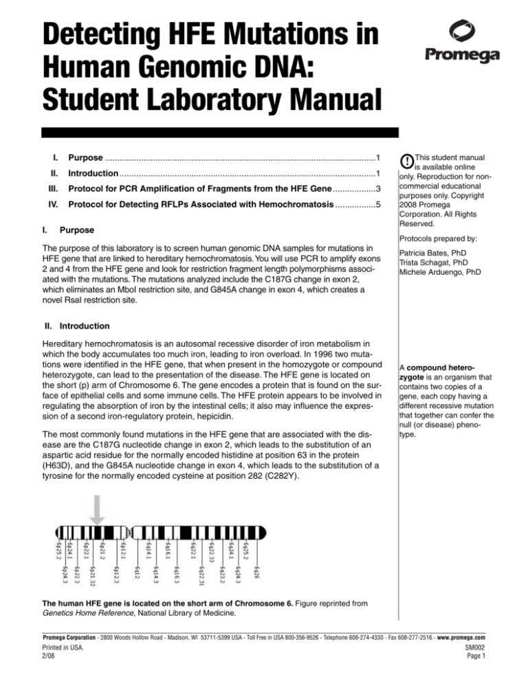
heterozygote, can lead to the presentation of the disease. The HFE gene is located on
the short (p) arm of Chromosome 6. The gene encodes a protein that is found on the surface of epithelial cells and some immune cells. The HFE protein appears to be involved in
regulating the absorption of iron by the intestinal cells; it also may influence the expression of a second iron-regulatory protein, hepicidin.
The most commonly found mutations in the HFE gene that are associated with the disease are the C187G nucleotide change in exon 2, which leads to the substitution of an
aspartic acid residue for the normally encoded histidine at position 63 in the protein
(H63D), and the G845A nucleotide change in exon 4, which leads to the substitution of a
tyrosine for the normally encoded cysteine at position 282 (C282Y).
A compound heterozygote is an organism that
contains two copies of a
gene, each copy having a
different recessive mutation
that together can confer the
null (or disease) phenotype.
The human HFE gene is located on the short arm of Chromosome 6. Figure reprinted from
Genetics Home Reference, National Library of Medicine.
Promega Corporation · 2800 Woods Hollow Road · Madison, WI 53711-5399 USA · Toll Free in USA 800-356-9526 · Telephone 608-274-4330 · Fax 608-277-2516 · www.promega.com
Printed in USA.
2/08
SM002
Page 1
Homozygosity for the mutation that results in the C282Y substitution is considered the
most common genotype of hereditary hemochromatosis. Approximately 1 in 10 caucasians in the United States are carriers of an HTE mutation, and 4.4 out of 1,000 caucasians in the United States are homozygotes.
The penetrance of a
phenotype refers to the
percent of individuals with
a particular genotype who
also exhibit the associated
phenotype. In this case not
all individuals who are
homozygous for one of the
two mutations (or compound heterozygotes) will
actually develop disease
symptoms. Hematochromatosis is said to show
incomplete penetrance.
However the actual penetrance of the disease is not as high as the genotype frequencies
would predict. Additionally, males are almost twice as likely to present with symptomatic
disease as females. The difference between disease prevalance and genetics is thought
to be due in part to environmental factors such as menstruation, pregnancy, frequent
blood donation, alcohol consumption, diet and other accompanying disease, such as
viral hepatitis.
Symptoms of hereditary hemochromatosis generally present in adults aged 30–60, with
presentation occurring later in affected females. In the disease, excess iron is deposited
in the liver, pancreas, heart, joints and endocrine glands. Early symptoms can include
hyperpigmentation, enlarged liver, impotence, testicular atrophy, joint swelling and tenderness and fatigue. The disease is managed in its early and intermediate stages by
phlebotomy (blood letting).
References
1. Emery, J. et al. (2007) Genetics and preventive health care. Australian Family
Physician 36, 808–11.
2. Whitlock, E.P. et al. (2006) Screening for hereditary hemochromatosis: a systematic review for the U.S. preventive services task force. Annals of Internal
Medicine. 145, 209–23.
3. Brissot, P. and de Bels, F. (2006) Current approaches to the management of
hemochromatosis. Hematology 2006 36–41.
4. National Library of Medicine (2006) HFE. Genetics Home Reference.
http://ghr.nlm.nih.gov/gene=hfe (accessed 2/13/2008).
Promega Corporation · 2800 Woods Hollow Road · Madison, WI 53711-5399 USA · Toll Free in USA 800-356-9526 · Telephone 608-274-4330 · Fax 608-277-2516 · www.promega.com
SM002
Page 2
Printed in USA.
2/08
III. Protocol for PCR Amplification of Fragments from the HFE Gene
!
Contamination of pipets and laboratory surfaces with DNA is a significant issue with
PCR. Wear gloves and use sterile, barrier pipet tips. Use sterile, thin-walled reaction
tubes for the PCR, and store the Master Mixes in sterile tubes as well.
!
Wear gloves, lab coats, closed-toe shoes and protective eyewear whenever you are
working in a laboratory.
Materials Required
•
•
•
•
•
•
•
•
•
vortex mixer
primer pair mix I for exon 2
primer pair mix II for exon 4
Nuclease-Free Water (Cat.# P1193)
dNTP Mix (Cat.# U1511)
GoTaq® Hot Start DNA Polymerase
(Cat.# M5001)
5X Green GoTaq® Flexi Buffer
(included with enzyme)
25 mM MgCl2 (included with enzyme)
genomic DNA homozygous for C187G
mutation
•
•
•
•
•
•
•
•
genomic DNA from compound heterozygote for C187G and G845A
wildtype genomic DNA for C187 and
G845
genomic DNA homozygous for G845A
mutation
Mineral Oil (Cat.# DY1151)
ice bucket
Barrier pipet tips (Cat.# A1491,
A1521 and A1541)
thin-walled PCR tubes
thermal cycler
Protocol for PCR
1.
Thaw, vortex and briefly centrifuge all reagents (except for the DNA polymerase) to collect liquid at the bottom of the tubes. Place reagents on ice.
Note: Do not vortex the DNA polymerase.
2.
Label eight 0.5 ml PCR tubes. (Label the tubes 1–8 plus your group letter
and the letter “P” for PCR. For example: 1AP, 2AP, etc.).
3.
Prepare master mixes I and II by combining the components listed in Tables
1 and 2 in the order listed. Prepare enough of each master mix for 10 reactions to allow for pipetting errors (use the volumes in the third column of the
tables to prepare your master mixes). Be sure to clearly label the master
mix tubes so that you will easily be able to tell which tube contains master
mix I and which one contains master mix II.
Table 1. PCR Master Mix I Components.
Volume Per Individual
Reaction
Volume for 10 Reactions
(Master Mix I)
1.0 µl
10.0 µl
10.0 µl
100 µl
dNTP Mix
5.0 µl
1.0 µl
50 µl
10.0 µl
GoTaq® Hot Start DNA Polymerase
0.25 µl
2.5 µl
Nuclease-Free Water
final volume
7.75 µl
25 µl
77.5 µl
250 µl
Components
Exon 2 primer pair
5X
GoTaq®
Green Flexi Buffer
MgCl2 Solution, 25 mM
Promega Corporation · 2800 Woods Hollow Road · Madison, WI 53711-5399 USA · Toll Free in USA 800-356-9526 · Telephone 608-274-4330 · Fax 608-277-2516 · www.promega.com
Printed in USA.
2/08
SM002
Page 3
Protocol for PCR (continued)
Table 2. PCR Master Mix II Components.
Volume Per Individual
Reaction
1.0 µl
Volume for 10 Reactions
(Master Mix II)
10.0 µl
5X GoTaq® Green Reaction Buffer
10.0 µl
100.0 µl
MgCl2 Solution, 25 mM
dNTP Mix
GoTaq® Hot Start DNA Polymerase
0.25 µl
1.0 µl
0.5 µl
2.5 µl
10.0 µl
5.0 µl
Nuclease-Free Water
final volume
7.75 µl
25 µl
77.5 µl
250 µl
Components
Exon 4 primer pair
4.
Add the components in the order listed in Table 3 to the labeled 0.5 ml PCR
amplification tubes.
Note: When adding the DNA sample, place the pipet tip at the bottom of the
tube so that the tip is submerged unter the water level. Centrifuge the tubes
briefly to collect liquid at the bottom of the tube. The final total volume for
each reaction will be 50 µl.
Table 3. Components and Volumes for Individual PCR Amplifications and Controls.
Reaction Nubmer
Nuclease-Free
Water
DNA Sample*
(Coriell Cat.#) PCR Master Mix I
1
(negative control)
25.0 µl
—
25.0 µl
—
2
(negative control)
25.0 µl
—
—
25.0 µl
3
(exon 2)
24.0 µl
1.0 µl
(NA14656)
25.0 µl
—
4
(exon 2)
24.0 µl
1.0 µl
(NA14690)
25.0 µl
—
5
(exon 2)
24.0 µl
1.0 µl
(NA14620)
25.0 µl
—
6
(exon 4)
24.0 µl
1.0 µl
(NA14656)
—
25.0 µl
7
(exon 4)
24.0 µl
1.0 µl
(NA14650)
—
25.0 µl
8
(exon 4)
24.0 µl
1.0 µl
(NA14686)
—
25.0 µl
PCR Master Mix II
*Note: Make sure that your genomic DNA concentration is 50 ng/µl. The final concentration of
each component in the reaction is: 1X Reaction Buffer, 1.5 mM MgCl2, 0.5 units GoTaq® DNA
polymerase, 200 µM each dNTP, 50 pmol of each PCR primer, and 50 ng of human genomic DNA.
5.
Add one drop of mineral oil the side of each reaction tube unless you are
using a thermal cycler with a heated lid. Store your reactions on ice until
they are placed into the thermal cycler.
Promega Corporation · 2800 Woods Hollow Road · Madison, WI 53711-5399 USA · Toll Free in USA 800-356-9526 · Telephone 608-274-4330 · Fax 608-277-2516 · www.promega.com
SM002
Page 4
Printed in USA.
2/08
6.
Process the amplifications using the profile below. The cycling profile will
take approximately 1.5–2 hours to complete.
Table 4. PCR Cycling Profile.
95 °C for 2 mintues.
1 cycle
94 °C for 30 seconds; 60 °C for 30 seconds; 72 °C for 30 seconds
35 cycles
68 °C for 7 minutes
1 cycle
4 °C soak
1 cycle
IV. Protocol for Detection of Hemochromatosis RFLPs
Materials Required
• MboI Restriction Enzyme
(Cat.# R6711)
• RsaI Restriction Enzyme
(Cat.# R6371)
• 10X Restriction Enzyme Buffer C
• BenchTop PCR Markers
(Cat.# G7531)
• TAE Buffer, 10X (Cat.# V4271)
• 5.0 µg/ml ethidium bromide
• staining trays
• Agarose, LMP, Preparative Grade for
Small Fragments (10 to 1,000 bp)
(Cat.# V3841)
• Agarose, LE, Analytical Grade
(Cat.# V3121)
• ultraviolet light box, camera and film
and/or digital imager
• power supplies for agarose gel electrophoresis
Warning: Ethidium bromide is a carcinogen. Please consult the Material Safety Data Sheet or your
institution’s chemical safety officer for appropriate usage and disposal instructions.
IV.A. Restriction Digestion
1.
Label ten 1.5 ml tubes. (Label the tubes 1–10 plus your label group letter
and the letter “D” for digests. For example: 1AD, 2AD).
2.
Assemble the restriction enzyme digests according to Table 5 below. If you
used mineral oil in your PCR, be sure to remove the aliquot of PCR product
from BELOW the mineral oil.
3.
Vortex mix and then briefly centrifuge the reactions to collect the liquid at
the bottom of the tube. Incubate reactions at 37 °C for 3 hours.
Promega Corporation · 2800 Woods Hollow Road · Madison, WI 53711-5399 USA · Toll Free in USA 800-356-9526 · Telephone 608-274-4330 · Fax 608-277-2516 · www.promega.com
Printed in USA.
2/08
SM002
Page 5
Table 5. Restriction Enzyme Digestions of Exons 2 and 4 from the HFE Gene.
Restriction
Digest
Reaction
PCR Product
Volume of
Volume of 10X
Volume of
Number
to Use
PCR Product
Buffer C
Mbo I
Volume of
RsaI
1D
#1
26 µl
3 µl
1 µl
—
2D
#2
26 µl
3 µl
—
1 µl
3D (uncut
control)
#3
8 µl
—
—
—
4D
#3
26 µl
3 µl
1 µl
—
5D
#4
26 µl
3 µl
1 µl
—
6D
#5
26 µl
3 µl
1 µl
—
7D (uncut
control)
#6
8 µl
—
—
—
8D
#6
26 µl
3 µl
—
1 µl
9D
#7
26 µl
3 µl
—
1 µl
10D
#8
26 µl
3 µl
—
1 µl
Agarose Gel Electrophoresis
Note: If you used the GoTaq® Green Flexi Buffer you will not need to add any loading dye to your samples because this buffer contains loading dyes. The BenchTop
PCR markers are also ready-to-use and do not need additional loading dyes.
1.
Prepare a 3.0 % agarose gel in 1X TAE buffer (2.5% LMP Preparative
Agarose for Small Fragments and 0.5% Agarose, LE, Analytical Grade).
Note: The gel running buffer is 1X TAE. Be sure to dilute any TAE stock to a
1X working solution.
2.
Lane 1
Load 12 µl of each reaction and markers in the following order:
2
3
4
5
6
7
8
9
10
11
BenchTop
Rxn Rxn Rxn Rxn Rxn Rxn Rxn Rxn Rxn Rxn
PCR Marker 1
2
3
4
5
6
7
8
9
10
12
BenchTop
PCR Marker
3.
Run the agarose gel at the voltage recommended by the maker of the
power supply and gel apparatus that you are using. Allow the yellow dye to
migrate to 5 mm from the bottom of the gel. The yellow dye migrates at
approximately 50 bp.
4.
Stain the gel in 5.0 µg/ml ethidium bromide for 10 minutes. Destain for
10 minutes in water.
Note: The destaining water will contain ethidium bromide and should be
disposed of properly.
5.
View the stained gel on a UV light box and photograph.
© 2008 Promega Corporation. All rights reserved.
Products may be covered by pending or issued patents or may have certain limitations. Please visit our Web site for
more information.
Promega Corporation · 2800 Woods Hollow Road · Madison, WI 53711-5399 USA · Toll Free in USA 800-356-9526 · Telephone 608-274-4330 · Fax 608-277-2516 · www.promega.com
SM002
Page 6
Printed in USA.
2/08

