Product Information
Revised: 11–September–2003
FluoCells ® Prepared Microscope Slides
Quick Facts
Storage upon receipt:
• FluoCells slides #1, #2 and #6: ≤25ºC
• FluoCells slides #3 and #4: ≤–20ºC
• Protect from light
Abs/Em:
See Table 1
Introduction
In response to requests from educators and instrument manuufacturers, Molecular Probes offers FluoCells® prepared microscope slides, which contain multilabeled cell preparations for
observation by epifluorescence or confocal microscopy. The
multicolor staining in these cell preparations results in stunning,
publication-quality images. The slides are especially useful for
setting up microscopes and camera systems and for assessing the
capabilities of optical filter sets.
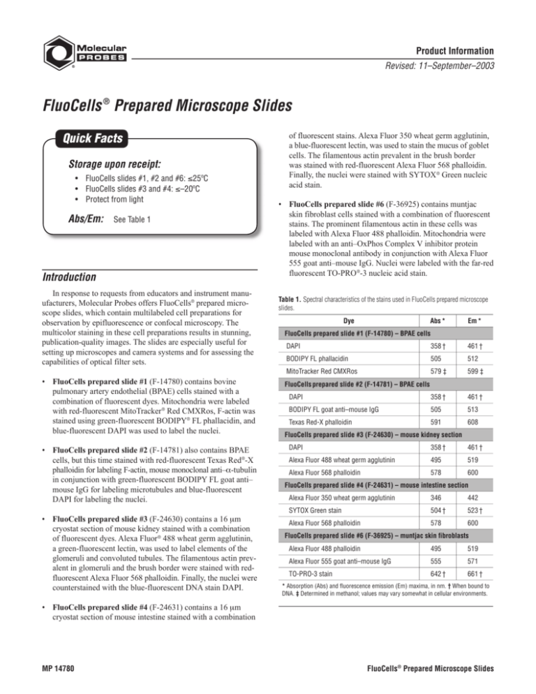
• FluoCells prepared slide #1 (F-14780) contains bovine
pulmonary artery endothelial (BPAE) cells stained with a
combination of fluorescent dyes. Mitochondria were labeled
with red-fluorescent MitoTracker ® Red CMXRos, F-actin was
stained using green-fluorescent BODIPY® FL phallacidin, and
blue-fluorescent DAPI was used to label the nuclei.
• FluoCells prepared slide #2 (F-14781) also contains BPAE
cells, but this time stained with red-fluorescent Texas Red ®-X
phalloidin for labeling F-actin, mouse monoclonal anti–α-tubulin
in conjunction with green-fluorescent BODIPY FL goat anti–
mouse IgG for labeling microtubules and blue-fluorescent
DAPI for labeling the nuclei.
• FluoCells prepared slide #3 (F-24630) contains a 16 µm
cryostat section of mouse kidney stained with a combination
of fluorescent dyes. Alexa Fluor ® 488 wheat germ agglutinin,
a green-fluorescent lectin, was used to label elements of the
glomeruli and convoluted tubules. The filamentous actin prevalent in glomeruli and the brush border were stained with redfluorescent Alexa Fluor 568 phalloidin. Finally, the nuclei were
counterstained with the blue-fluorescent DNA stain DAPI.
of fluorescent stains. Alexa Fluor 350 wheat germ agglutinin,
a blue-fluorescent lectin, was used to stain the mucus of goblet
cells. The filamentous actin prevalent in the brush border
was stained with red-fluorescent Alexa Fluor 568 phalloidin.
Finally, the nuclei were stained with SYTOX ® Green nucleic
acid stain.
• FluoCells prepared slide #6 (F-36925) contains muntjac
skin fibroblast cells stained with a combination of fluorescent
stains. The prominent filamentous actin in these cells was
labeled with Alexa Fluor 488 phalloidin. Mitochondria were
labeled with an anti–OxPhos Complex V inhibitor protein
mouse monoclonal antibody in conjunction with Alexa Fluor
555 goat anti–mouse IgG. Nuclei were labeled with the far-red
fluorescent TO-PRO ®-3 nucleic acid stain.
Table 1. Spectral characteristics of the stains used in FluoCells prepared microscope
slides.
Dye
Abs *
Em *
FluoCells prepared slide #1 (F-14780) – BPAE cells
DAPI
358 †
461 †
BODIPY FL phallacidin
505
512
MitoTracker Red CMXRos
579 ‡
599 ‡
FluoCells prepared slide #2 (F-14781) – BPAE cells
DAPI
358 †
461 †
BODIPY FL goat anti–mouse IgG
505
513
Texas Red-X phalloidin
591
608
FluoCells prepared slide #3 (F-24630) – mouse kidney section
358 †
DAPI
461 †
Alexa Fluor 488 wheat germ agglutinin
495
519
Alexa Fluor 568 phalloidin
578
600
FluoCells prepared slide #4 (F-24631) – mouse intestine section
Alexa Fluor 350 wheat germ agglutinin
346
442
SYTOX Green stain
504 †
523 †
Alexa Fluor 568 phalloidin
578
600
FluoCells prepared slide #6 (F-36925) – muntjac skin fibroblasts
Alexa Fluor 488 phalloidin
495
519
Alexa Fluor 555 goat anti–mouse IgG
555
571
TO-PRO-3 stain
642 †
661 †
* Absorption (Abs) and fluorescence emission (Em) maxima, in nm. † When bound to
DNA. ‡ Determined in methanol; values may vary somewhat in cellular environments.
• FluoCells prepared slide #4 (F-24631) contains a 16 µm
cryostat section of mouse intestine stained with a combination
MP 14780
FluoCells® Prepared Microscope Slides
FluoCells slides #3 and #4. Store at –20°C or below, protected from light. The FluoCells slides #3 and #4 can be stored
for short periods of time (a few days) at 2–25°C without harm.
Materials
Contents
FluoCells prepared slides are packaged individually, one slide
per package.
Storage
FluoCells slides #1, #2 and #6. Store at 25°C or below, protected from light.
Short-term exposure of any FluoCells slide to dim light
(e.g., room lighting) will not cause damage. When stored properly,
these permanently mounted specimens will retain their bright,
specific staining patterns for at least six months from the date
of purchase.
Product List Current prices may be obtained from our Web site or from our Customer Service Department.
Cat #
Product Name
F-14780
F-14781
FluoCells® prepared slide #1 *BPAE cells with MitoTracker ® Red CMXRos, BODIPY® FL phallacidin, DAPI*.................................................
FluoCells® prepared slide #2 *BPAE cells with mouse anti-α-tubulin, BODIPY® FL goat anti-mouse IgG, Texas Red®-X
phalloidin, DAPI* ............................................................................................................................................................................................
FluoCells® prepared slide #3 *mouse kidney section with Alexa Fluor ® 488 WGA, Alexa Fluor ® 568 phalloidin, DAPI* .................................
FluoCells® prepared slide #4 *mouse intestine section with Alexa Fluor ® 350 WGA, Alexa Fluor ® 568 phalloidin, SYTOX® Green* ...............
FluoCells® prepared slide #6 *muntjac cells with mouse anti-OxPhos Complex V inhibitor protein, Alexa Fluor ® 555 goat anti-mouse IgG,
Alexa Fluor ® 488 phalloidin, TO-PRO®-3* .......................................................................................................................................................
F-24630
F-24631
F-36925
Unit Size
each
each
each
each
each
Contact Information
Further information on Molecular Probes' products, including product bibliographies, is available from your local distributor or directly from Molecular Probes. Customers in
Europe, Africa and the Middle East should contact our office in Leiden, the Netherlands. All others should contact our Technical Assistance Department in Eugene, Oregon.
Please visit our Web site — www.probes.com — for the most up-to-date information.
Molecular Probes, Inc.
Molecular Probes Europe BV
29851 Willow Creek Road, Eugene, OR 97402
Phone: (541) 465-8300 • Fax: (541) 335-0504
Poortgebouw, Rijnsburgerweg 10
2333 AA Leiden, The Netherlands
Phone: +31-71-5233378 • Fax: +31-71-5233419
Customer Service: 6:00 am to 4:30 pm (Pacific Time)
Phone: (541) 335-0338 • Fax: (541) 335-0305 • order@probes.com
Toll-Free Ordering for USA and Canada:
Customer Service: 9:00 to 16:30 (Central European Time)
Phone: +31-71-5236850 • Fax: +31-71-5233419
eurorder@probes.nl
Order Phone: (800) 438-2209 • Order Fax: (800) 438-0228
Technical Assistance: 9:00 to 16:30 (Central European Time)
Technical Assistance: 8:00 am to 4:00 pm (Pacific Time)
Phone: (541) 335-0353 • Toll-Free (800) 438-2209
Fax: (541) 335-0238 • tech@probes.com
Phone: +31-71-5233431 • Fax: +31-71-5241883
eurotech@probes.nl
Molecular Probes’ products are high-quality reagents and materials intended for research purposes only. These products must be used by, or directly under the
supervision of, a technically qualified individual experienced in handling potentially hazardous chemicals. Please read the Material Safety Data Sheet provided for
each product; other regulatory considerations may apply.
Several of Molecular Probes’ products and product applications are covered by U.S. and foreign patents and patents pending. Our products are not available for resale
or other commercial uses without a specific agreement from Molecular Probes, Inc. We welcome inquiries about licensing the use of our dyes, trademarks or technologies. Please submit inquiries by e-mail to busdev@probes.com. All names containing the designation ® are registered with the U.S. Patent and Trademark Office.
Copyright 2003, Molecular Probes, Inc. All rights reserved. This information is subject to change without notice.
FluoCells® Prepared Microscope Slides
2

